Comparative evaluation by scanning confocal Raman spectroscopy and transmission electron microscopy of therapeutic effects of noble metal nanoparticles in experimental acute inflammation

Adriana Filip *a, Monica Potarab, Adrian Floreac, Ioana Baldeaa, Diana Olteanua, Pompei Bolfad, Simona Clichicia, Luminita Davide, Bianca Moldovane, Liliana Olenicf and Simion Astileanb
aDepartment of Physiology, “Iuliu Hatieganu” University of Medicine and Pharmacy, 1-3 Clinicilor Street, Cluj-Napoca, Romania. E-mail: gabriela.filip@umfluj.ro; adrianafilip33@yahoo.com; Fax: +40-264-597257; Tel: +40-745-268704
bNanobiophotonics and Laser Microspectroscopy Center, Interdisciplinary Research Institute in Bio-Nano-Sciences and Faculty of Physics, Babes-Bolyai University, M. Kogalniceanu Str 1, 400084 Cluj-Napoca, Romania
cDepartment of Cell and Molecular Biology, “Iuliu Hatieganu” University of Medicine and Pharmacy, Cluj Napoca, Romania
dDepartment of Biomedical Sciences, Ross University School of Veterinary Medicine Basseterre, St. Kitts, West Indies
eFaculty of Chemistry and Chemical Engineering, Babes-Bolyai University, Cluj-Napoca, Romania
fNational Institute for Research and Development of Isotopic and Molecular Technologies, Cluj-Napoca, Romania

Received 1st June 2015 , Accepted 24th July 2015

First published on 24th July 2015


Abstract

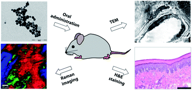
Finding appropriate experimental designs and analysis methods in order to gain insight into the mechanisms of efficiency and toxicity of nanomaterials is a major focus in today’s research in nanomedicine. In this paper, we demonstrate the ability of scanning confocal Raman spectroscopy to emphasize the molecular changes in terms of inflammation resolution after administration of a single dose of metal nanoparticles functionalized with natural extracts, in experimental inflammation. Five experimental groups of Wistar rats were used, treated with one dose of gold nanoparticles (AuNPs–CM), one dose of silver nanoparticles (AgNPs–CM), Cornus mas (CM) extract, and vehicle, before intraplantar injection of 100 μL 1% carrageenan, and one group of untreated animals. The paw tissues were harvested and used for transmission electron microscopy (TEM) and evaluation of the metal content 4 and 24 hours after the induction of inflammation and, after 24 hours, Raman spectroscopy, histopathology and prostaglandin (PG) E2 level assessment were performed. TEM revealed varying degrees of alterations in dermo-epidermal junctions and capillaries, especially in tissues treated with AgNPs–CM and vehicle, in parallel with the increase in PGE2 levels. Besides ultrastructural changes highlighted by TEM, meaningful information about the molecular changes is provided by multivariate Raman spectral images. Indeed, thorough Raman spectral analysis shows that AuNPs–CM and CM restored the normal composition of unsaturated fatty acids while the specimens treated with AgNPs–CM were dominated by the protein component. Our results suggest that the Raman spectral analysis has real potential to be used in tandem with standard methods for monitoring the subtle molecular effects induced by the administration of nanoparticles.


Introduction

In recent years, research in the fields of nanomaterials and nanotechnology has seen exponential growth due to the multiple applications in different areas such as molecular imaging, drug delivery, engineering technology and the development of materials and medical devices for diagnosis and treatment.1–3 Various biomedical applications rely on attractive properties such as small size, large surface area to volume ratio, high reactivity to living cells, flexibility in functionalization and a wide range of target delivery.4 Nevertheless, the behavior of nanoparticles inside cells is still an enigma, and not all the complexity of the interactions between these particles and cells is completely understood. Therefore, finding appropriate experimental designs and analysis methods will allow us to understand the mechanisms of efficiency and toxicity of nanomaterials, so that nanomaterials can be safely used in medicine.

Usually, the changes in the chemical compositions of tissue sequences are early events in pathology, and their detection by modern techniques is useful in early therapeutic intervention. For this purpose, Raman spectroscopy has been shown to have the ability to distinguish between normal and pathologically altered skin samples, making it a useful technique for early diagnosis. The gold standards in the diagnosis of skin pathology are biopsies and histological investigations. These processes are more invasive and time-consuming, need specially qualified technicians and allow detection of lesions after their occurrence. Optical imaging systems are ideally suited for the early detection of epithelial diseases, including most cancers, and for the assessment of tumor margins and therapy response. Raman spectroscopy is an optical technique based on the vibrational activity of chemical bonds and has been successfully applied in pathology to differentiate a variety of lesions. This technique is able to identify the unknown substances in a tissue, based on the spectral signature of the mode of vibration for each molecule from a sample. In recent decades, Raman spectroscopy has been used to characterize the molecular composition and hydration of normal skin and to analyse the conformational structures of proteins, water and lipids in mummified skin.5–8 Several studies found changes of protein and lipid structures in basal cell carcinoma or actinic keratosis compared to normal skin, changes which allowed discrimination between the tissues and identification of the tumour margins.9–11 Moreover, based on high wave number Raman bands it is possible to separate the perilesional and normal tissue.10 Such techniques are particularly relevant to inflammation, where the detection of subclinical, early disease states, could facilitate early diagnosis and targeted therapies.

It is known that inflammation is a complex biological response involved in the pathogenesis of a large variety of diseases.12 Generally, in the acute inflammatory response, numerous cytokines such as tumor necrosis factor (TNF-α), interleukin (IL)-1, IL-6 and pro-inflammatory enzymes (cyclooxygenase-2 [COX]-2, prostaglandin E synthase, inducible nitric oxide synthase [iNOS]) are released, inducing vasodilatation, edema, cellular metabolic stress and even tissue necrosis.13,14 Eicosanoids such as prostaglandin E2, derived from polyunsaturated fatty acids, promote angiogenesis, hinder apoptosis and stimulate the inflammation as well as the cell proliferation and growth of tumor tissue.15 Consequently, the ability to detect and treat inflammation is critical for the treatment and prevention of inflammatory conditions. Moreover, the use of nanoparticles for the detection of vascular activation and cellular recruitment at subclinical levels has proven effective in practice.

Gold nanoparticles (AuNPs) have been widely used as therapeutic agents in experimental gene therapy and drug delivery of anticancer agents, antibiotics, amino-acids and peptides, antioxidants or isotopes.16–21 Silver nanoparticles (AgNPs) are used to impregnate various textile materials for odor reduction, in creams for acne treatment or in healing dressings.22 Besides the antimicrobial action, it was shown that AgNPs exerted cytotoxic and pro-apoptotic effects mediated via reactive oxygen species (ROS), both in normal as well as in tumour cell lines.23–27 Furthermore, some researchers have suggested that silver ions released from AgNPs in aqueous media can induce an inflammatory response, may interact with nucleic acids and can lead to focal inflammation 14 days after topical treatment.25,28,29 Although some studies have shown that AuNPs are biologically inert, there is other research which attests that they generate inflammation, especially small nanoparticles, as a response of the host to foreign particles.30,31 The inflammatory response is transient and accompanied by an increase in macrophage phagocytosis and apoptosis.32

The phytochemical synthesis of metal nanoparticles with polyphenols from natural extracts could be an option to counteract the adverse reactions as they can reduce ROS production and protect the cellular proteins and lipids. It is crucially important to investigate the in vivo effects of such compounds before approving any potential therapeutic applications. Cornelian cherry (Cornus mas) could be a candidate for the “green synthesis” of metal nanoparticles. It was known as a medicinal plant for the treatment of inflammation in an ancient system of medicine.33

The aim of the present study was to evaluate the potential of Raman spectroscopy as a technique capable of providing the diagnosis of skin inflammatory conditions and to differentiate the effect of treatments with silver and gold nanoparticles conjugated with natural extracts compared to the extract alone. Vibrational data collected from the samples are presented in conjunction with the conventional histopathological investigation, transmission electron microscopy (TEM) and assessment of prostaglandin E2 (PGE2) levels.

Experimental

Materials

Silver nitrate (AgNO3), NaOH and EDTA–Na2 were obtained from Merck KGaA Darmstadt (Germany) and absolute ethanol and n-butanol from Chimopar (Bucharest). Glutaraldehyde for TEM was from EMS (Hatfield, USA), osmium tetroxide and λ-carrageenan type IV were from Sigma-Aldrich (St. Louis, USA), and Epon 812 resin was from Fluka GmbH (Buchs Switzerland). ELISA tests for the evaluation of PGE2 levels were obtained from Blue Gene (China). Standard gold solution 1000 mg L−1 in HCl 2 mol L−1 and standard silver solution 1000 mg L−1 in HNO3 0.5 mol L−1 were purchased from LGC Standards GmbH (Wesel, Germany).

Vegetal material and preparation of the extract

Samples of Cornus mas L. (CM) fruits were collected in September 2013 from Cluj-Napoca (altitude 360 m), Romania. The species of the fruits were identified by the Botanical Department of the Faculty of Pharmacy Cluj-Napoca and voucher specimens were deposited in the Herbarium of the Faculty of Pharmacy, “Iuliu Hatieganu” University of Medicine and Pharmacy, Cluj-Napoca. Natural extracts were obtained according to the method of David et al.34 The total anthocyanins content was determined using the pH differential method and the total phenolic content was assessed using the Folin–Ciocalteu method.35,36

Synthesis of AgNPs–CM and AuNPs–CM nanomaterials and their characterization

AgNPs–CM and AuNPs–CM were prepared by the reduction of the metallic salts with a natural extract solution of Cornus mas using a method described before.34,37 The pH of the natural extract solution is brought to 7.5 with 1 M NaOH solution. The concentrations of the solutions used were: 0.03 M HAuCl4·3H2O, 0.06 M AgNO3 and natural extract with 25 × 10−3 mM total anthocyanins. 16.6 mL Cornus mas extract and 6.6 mL solution of metallic ions were added to 200 mL double distilled water with vigorous stirring for 10 minutes, until a brown solution was obtained. The metallic nanoparticles were kept in a refrigerator until used.

The colloids were stable for 30 days after which the metallic nanoparticles aggregated very slightly. UV-visible-NIR extinction spectra were measured in a 2 mm quartz cell using a Jasco V-670 spectrometer with 1 nm spectral resolution. The morphology and size distribution of the nanoparticles were examined using a TEM JEOL-JEM 1010 instrument (JEOL Inc). The zeta potential of the gold and silver nanoparticles was determined by a laser Doppler micro-electrophoresis technique using a Malvern Zetasizer Nano ZS-90. The Nano ZS contains a He–Ne laser operating at a wavelength of 633 nm and an avalanche photodiode detector. The measurements were performed at a temperature of 25 °C. For biological applications, besides physicochemical property characterization, the tissue toxicity and biocompatibility evaluation of nanoparticles is important. Therefore, we tested the acute systemic toxic effects of both metallic nanoparticles on Wistar rats (see ESI file section S1).

Model of carrageenan-induced inflammation in Wistar rats

The study was performed on an experimental model of inflammation induced by the administration of carrageenan in the footpad of male Wistar rats. The animals (130–150 g) were obtained from the Animal Department of “Iuliu Hatieganu” University of Medicine and Pharmacy, Cluj-Napoca, Romania and were acclimatized under the following conditions: 12 h light/12 h dark cycles, 35% humidity, free access to water and feeding with a normocaloric standard diet (VRF1). Paw edema was induced by an injection of 100 μL of freshly prepared 1% carrageenan (λ-carrageenan, type IV, Sigma) diluted in normal saline in the right hind footpad.38 The left hind footpad was injected with the same volume of saline solution. One dose of nanoparticle suspension functionalised with the Cornus mas extract was administered orally 1 day prior to the injection of carrageenan, in a volume not exceeding 0.25 mL. We used 5 groups, each consisting of 8 animals divided as follows: group 1 – normal, control tissue; group 2 – animals with inflammation pre-treated with 0.9% NaCl solution as a positive control group; group 3 – inflammation pre-treated with AuNPs–CM 0.3 mg per b.w.; group 4 – inflammation pre-treated with AgNPs–CM 0.3 mg per b.w.; group 5 – inflammation pre-treated with 15 mg per b.w. per day Cornus mas extract. 24 hours after carrageenan administration, soft paw tissues were sampled and used for Raman spectroscopy and histopathological investigations. For the quantification of metal content in the samples, transmission electron microscopy and evaluation of prostaglandin E2 levels by ELISA, the paw tissues were sampled after 4 and 24 hours. All procedures for in vivo experiments were approved by the Ethics Committee on Animal Welfare of the “Iuliu Hatieganu” University, in accordance with the European Convention for the Protection of Vertebrate Animals used for Experimental and other Scientific Purposes, Council of Europe no. 123, Strasbourg 1985.

Sample preparation

For Raman analysis fresh biopsies from the paws of sacrificed rats were immediately immersed in Jung Tissue Freezing Medium (Leica Biosystems). They were cryo-sectioned within a maximum of 2 hours after sampling at a thickness of 10 μm, using a Leica CM 1850 Cryotome set at −20 °C. One section from each animal from each group was placed on normal histology glass slides then placed in an incubator for drying and better adherence at 37 °C for 1 hour. Samples were then stored in a freezer at −20 °C until tissue analysis using confocal Raman imaging. Following Raman spectroscopy analysis of the paw, the same sections were photographed unstained, then stained with H&E and photographed again using a BX51 microscope, DP 25 digital camera and Olympus Cell B software. Each microscopic section evaluated had a surface of 330.68 × 421.42 μm. For ELISA investigations the soft tissue homogenates were prepared in 50 mM TRIS–10 mM EDTA buffer (pH 7.4) and homogenized with a Polytron homogenizer (Brinkman Kinematica, Switzerland) as previously described.34

Determination of Au and Ag levels by atomic absorption spectrometry

The analysis of gold and silver nanoparticle content in the paw tissues was conducted by atomic absorption spectrometry using an AVANTA PM atomic absorption spectrometer (GBC Scientific Equipment, Braeside, Australia) equipped with a graphite furnace (GF 3000) and programmable auto-sampler (PAL 3000, GBC Scientific Equipment, Braeside, Australia). Additionally, pyrolytic graphite furnaces (P 301, Photron Ltd, Victoria, Australia), gold cathode lamps (P 821 HCl), silver and deuterium lamps were used. Calibration was performed using the “Linear Least Squares” method and an excellent correlation was obtained for 0–50 μg L−1 gold (R = 0.9997) and 1–6 μg L−1 silver (R = 0.9996). Prior to the determination, the biological samples were mineralized in 1 mL aqua regia for gold and nitric acid for silver, at room temperature. Each sample was measured three times. The corresponding calculated concentrations of metal nanoparticles were: 1.96 μg g−1 tissue for AuNPs–CM after 4 hours, 2.95 μg g−1 tissue after 24 hours, 1.62 μg g−1 tissue for AgNPs–CM after 4 hours and 1.96 μg g−1 tissue after 24 hours.

Raman spectroscopy

The Raman spectra and images presented in this work were recorded using a Confocal Raman Microscope (alpha 300 R from WITec GmbH, Ulm, Germany) equipped with a Nd-YAG laser working at 532 nm and an ultrahigh throughput spectrometer (UHTS 300) for Raman light analysis. A 100× dry microscope objective with a numerical aperture (NA) of 0.9 was used to perform all measurements from tissues. The Raman backscattered light collected through the objective was passed through a holographic edge filter, before being focused into a multimode optical fiber of 100 μm diameter which provides the optical pinhole for the confocal measurement. The light emerging from the output optical fiber was analyzed using an ultrahigh throughput spectrometer equipped with a back-illuminated deep-depletion 1024 × 128 pixel CCD camera (DV401-BV, Andor) operating at −60 °C. The tissues placed on glass slides were mounted on a piezoelectric scanning stage and scanned through the laser focus in a raster pattern. The spectroscopic imaging was performed over a 150 × 150 μm2 area which was first divided into four squares of 75 × 75 μm2 (75 × 75 pixels) selected from the unstained glass slide sections. Each square was imaged with an integration time of 0.5 s for each spectrum to prevent sample degradation at the focal point. The measurements were conducted at a power incident on the samples of 10 mW. WITec Project Plus software was used for spectral analysis and image processing. Reflected-light bright-field optical images of tissues were captured with a color video camera attached to the eyepiece out-port of the same microscope using a super-bright white LED source for illumination.

Transmission electron microscopy

For transmission electron microscopy the paw tissues were fixed for 1.5 hours with 2.7% glutaraldehyde, washed four times with 0.1 M phosphate buffer (pH 7.4), post fixed for 1.5 hours with 1.5% osmium tetroxide, dehydrated in an acetone series, and embedded in Epon 812 resin as previously described.34 The ultrathin sections obtained with glass knives using a Bromma 8800 ULTRATOME III (LKB, Stockholm, Sweden) were collected on 300 mesh copper grids (Agar Scientific Ltd, Stansted, UK). The samples were examined on a JEOL JEM 1010 transmission electron microscope (JEOL Ltd, Tokyo, Japan) at an 80 kV acceleration voltage, and photographed with a Mega VIEW III camera (Olympus, Soft Imaging System, Münster, Germany).

Prostaglandin (PG) E2 levels

PGE2 contents of the tissue homogenates were measured using an ELISA system (Blue Gene, China) according to the manufacturer’s instructions. Results are expressed as pg per mg protein.

Statistical analysis

Experimental data were analyzed by one-way analysis of variance (ANOVA) followed by the Tukey’s multiple comparisons post test using GraphPad Prism 5.0 software (GraphPad, San Diego, Ca., SUA). All values are expressed as mean ± standard deviation, with a limit of statistical significance of p < 0.05.

Results and discussion

Characterization of nanomaterials

Detailed characterization of the nanoparticles’ morphology was obtained by TEM measurements. Fig. 1A and B illustrate representative TEM pictures of the obtained nanoparticles, which clearly reveal that the products as prepared are mainly composed of spherical nanoparticles dispersed in solution with a particle diameter of 13–52 nm for AuNPs–CM and 20–100 nm for AgNPs–CM.
image file: c5ra10376b-f1.tif
Fig. 1 Transmission electron micrographs of: (A) AuNPs–CM and (B) AgNPs–CM. (C) Normalized absorption spectra of: (a) AgNPs–CM and (b) AuNPs–CM.

The optical absorption spectra of the as prepared nanoparticles in Fig. 1C exhibit a dominant plasmonic band centered at 409 nm for AgNPs–CM (curve a) and at 525 nm for AuNPs–CM (curve b), which represents the typical signature of the dipolar plasmon resonance of individual spherical silver and gold nanoparticles, respectively.39

The performance of the colloidal nanoparticles in different applications strongly depends on their chemical stability. The stability of colloidal systems is defined in terms of the charge on their surface. The zeta potential distribution of the prepared nanoparticles is relatively narrow and centered at −19.7 and −22.4 mV for AuNps–CM and AgNps–CM, respectively, indicating a good stability of the nanoparticles in colloidal suspension (result not shown). In view of the next step toward applications we further investigated the sample stability in simulated physiological conditions by mixing 500 μL of colloidal solution with 100 μL of a solution of PBS/PBS containing 10% fetal bovine serum. We found that the absorption spectra of the mixed samples remained stable after 4 h of incubation (more details in the ESI file, section S2). The acute systemic toxicity test performed on the Wistar rats revealed a transient toxicity in the liver, induced especially by high doses of AgNPs–CM nanoparticles (see ESI file, section S3 and S4).

Confocal Raman microscopy

As stated above, confocal Raman microscopy is a powerful spectral imaging tool for the investigation of molecular composition in a non-destructive and label-free manner. We used Raman spectral micro-imaging combined with multivariate statistical analysis to detect and visualize subtle biochemical changes in tissues associated with pathologically altered skin and to evaluate the effect of different therapeutic agents.

The acquired two-dimensional Raman spectral maps were processed with a K-means cluster algorithm that allows the classification of the skin tissue histological organization based on the intrinsic biochemical composition. K-means cluster analysis enables the partition of the spectra within a Raman image into a predefined number of groups of similar biochemical characteristics and the construction of color-coded spectral images. A mean spectrum of each cluster within an image dataset was derived and overlaid for spectral comparison. This method was previously described.40

Fig. 2 shows representative multivariate Raman spectral images of normal soft paw tissue (A), inflamed tissue treated with vehicle (B) and tissues treated with AuNPs–CM (C), AgNPs–CM (D) and CM (E). The presented Raman images facilitate the identification of the main histological features of the soft paw tissue and enable the visual discrimination between healthy, inflamed and treated tissues. Indeed, the distribution of proteins (red cluster), lipids (blue cluster) and unsaturated fatty acids (green cluster) regions highlights obvious visual differences between the analyzed skin tissues. To identify the origin of the visual differences and hence the biochemical alterations we compared the corresponding extracted spectra of tissue samples presented in Fig. 2F–J. The Raman spectrum of the normal paw tissue in Fig. 2F shows vibrational bands at 862, 926, 1093, 1125, 1248, 1316, 1447, 1581, 1664, 1748, 2854, 2888, 2927 and 3009 cm−1. The assignment of Raman fingerprint of the tissue is given in Table 1 which is in good agreement with other similar data published in the literature.41,42


image file: c5ra10376b-f2.tif
Fig. 2 Raman maps obtained by multivariate data analysis algorithms (K-means cluster analysis) of: (A) normal skin tissue; (B) inflamed skin tissue and tissues treated with AuNPs–CM (C), AgNPs–CM (D) and CM extract (E). The scale bar is 10 μm. Corresponding extracted spectra from distinctly colored areas in images: (F) normal skin tissue; (G) inflamed skin tissue and tissues treated with AuNPs–CM (H), AgNPs–CM (I) and CM (J).
Table 1 Assignments of the major Raman bands of the normal skin tissue
Raman (cm−1) Tentative assignment40,41
862 Proline, collagen (CC stretching)
926 Ribose-phosphate (CO and CC stretching)
1093 Phospholipids (symmetric stretching PO2, stretching CC, stretching COC) glycosidic link
1125 Glycogen (CO stretching from CH2OH of carbohydrates); carotenoids (CC stretching)
1248 Amide III, α-helix, collagen, tryptophan (stretching CN, bending NH); phospholipids (asymmetric stretching PO2); nucleic acids (ring stretching)
1316 β-D-Glucose, D-(+) dextrose (CH2 and CH2OH deformations); amide III, α-helix (NH and CH bands)
1447 Lipids (CH2 scissoring, CH2 deformation); phospholipids (δ(CH2) scissoring); collagen (δ(CH2), δ(CH3))
1581 Tryptophan, proteins (NH bending amide II, C–N stretching)
1664 Amide I/α-helix (H-bonded CO stretching); unsaturated bonds of lipids (C[double bond, length as m-dash]C stretching)
1748 Lipids (C[double bond, length as m-dash]O stretching)
2854 Fatty acids, lipids (CH stretching, CH2 symmetric stretching)
2888 Fatty acids, lipids (CH, CH2 asymmetric stretching)
2926 Fatty acids, lipids, polypeptide (CH2 asymmetric stretching); proteins (CH stretching, CH3 symmetric stretching)
3009 Fatty acids, lipids (H–C[double bond, length as m-dash]C stretching)


The biochemical differences between the analyzed samples appeared to show a strong association with the spectral region of 2800–3100 cm−1 originating from the vibrations of the lipids and unsaturated fatty acids (green cluster). Previous studies have demonstrated that the vibration characteristics from unsaturated fatty acids play a leading role in distinguishing between cancerous and noncancerous human tissues.41,43 In accordance with these findings, in the first step we carefully analyzed the distributions of oleic acid, linoleic acid, γ-linoleic acid and arachidonic acid. The Raman profile of the normal soft paw tissue in the region of 2800–3100 cm−1 is dominated by the vibrational bands at 2854, 2888, 2926 and 3009 cm−1 (Fig. 2F, green curve). The positions and the ratio between the intensities of these bands are very similar to the Raman signature of oleic acid and linoleic acid, according to the literature.41,43 In contrast, the Raman profile of the inflamed tissue in the region of 2800–3100 cm−1 (Fig. 2G, green curve) shows noticeable differences related to the vibrational bands at 2854 and 2926 cm−1. Specifically, a significant decrease of the intensity of the peak at 2854 cm−1 with a concomitant increase of the intensity of the peak at 2926 cm−1 is clearly visible in the case of the inflamed tissue. The comparison with the Raman spectra of the unsaturated fatty acids shows that the inflamed paw soft tissue is dominated by the Raman signature of γ-linoleic acid and arachidonic acid.41,43 In addition, the corresponding multivariate Raman images show that the normal tissue contains a markedly higher concentration of unsaturated fatty acids as compared to that of inflamed skin and subcutaneous tissue (Fig. 2A and B, green cluster).

It is known that oleic acid is a common monounsaturated acid found in various animal and vegetable fats while polyunsaturated fatty acids are precursors of eicosanoids released from phospholipid cell membranes as a result of phospholipase A2 action. Eicosanoids including prostaglandin (PG), prostacyclin (PGI) and thromboxanes (TX), synthesized under the cyclooxygenase (COX) activity and leukotrienes (LT), are involved in inflammation and allergic reactions as well as in cell proliferation and growth of tumor tissue.44–46 Arachidonic acid is a key inflammatory intermediate which is released immediately after the induction of inflammation, before the recruitment of leukocytes and the infiltration of immune cells at inflammatory site. Arachidonic acid is very susceptible to lipid peroxidation and generates cytotoxic compounds following its action on the macromolecules of cells such as proteins, nucleic acids and lipids. The compounds which are formed in turn amplify the inflammation.46

Colloidal gold and silver nanoparticles can selectively accumulate into pathologically altered tissues by a phenomenon termed the enhanced permeability and retention (or EPR) effect and represent an alternative for developing reliable delivery systems for targeting drugs at a specific site in a desired concentration.47 As observed in Fig. 2C (green cluster), the oral administration of AuNPs–CM leads to an increase in the concentration of unsaturated fatty acids. The effect of AuNPs–CM is associated with marked biochemical changes in the spectral region of 2800–3100 cm−1. Specifically, a decrease of the intensity of the band at 2926 cm−1 with a concomitant slight increase of the intensity of the band at 2854 cm−1 is clearly shown after the oral administration of the AuNPs–CM (Fig. 2H, green curve). The comparison of the Raman signature of normal and inflamed skin tissue shows that the spectral profile of the treated skin tissue tends to reproduce the Raman spectrum of the normal skin tissue. In contrast, after the oral administration of AgNPs–CM, there is no amount of detectable unsaturated fatty acids (Fig. 2D – the lack of green cluster). The corresponding Raman profile in the region of 2800–3100 cm−1 (Fig. 2I) confirms that the vibrations originating from unsaturated fatty acids are no longer discernible after the treatment with AgNPs–CM. It appears that this result could be surprising due to the lack of a Raman signal from unsaturated fatty acids. However, this can be explained by the fact that the Raman bands in the spectral region of 2800–3100 cm−1 originate not only from the vibrations of unsaturated fatty acids, but also from lipids and proteins. It is therefore conceivable that the inflamed skin tissue treated with AgNPs–CM contains a lower concentration of unsaturated fatty acids and a markedly higher concentration of proteins or lipids as compared to that of the inflamed tissue. This, in turn masks the spectral signature of the unsaturated fatty acids. Indeed, the multivariate Raman images of the inflamed skin tissue treated with AgNPs–CM in Fig. 2D clearly shows a noticeable contribution from proteins (red cluster). It was previously reported that the distribution of proteins plays a crucial role in distinguishing between normal and pathologically altered skin. As can be seen in Fig. 2A (red cluster) the normal skin tissue contains a much lower concentration of proteins in comparison with the tissue treated with AgNPs–CM. This finding was previously explained by the increased synthesis of large amounts of proteins in inflammatory and proliferating tissues.41,43 A detailed inspection of the red cluster in Fig. 2A–E shows that this overgrowth of protein concentration occurs only in the inflamed skin tissue treated with AgNPs–CM. Moreover, the normal skin tissue in Fig. 2A is characterized by well-differentiated regions associated with the distribution of proteins (red cluster), lipids (blue cluster) and unsaturated fatty acids (green cluster). In contrast, the inflamed skin tissue treated with AgNPs–CM is devoid of this organization and is characterized by only two spectral zones. The blue cluster in Fig. 2D is associated with the occurrence of the vibrational band at 1635 cm−1. This spectral feature was previously assigned to a conformational change in the protein secondary structure.48 Interestingly, the zones corresponding to the protein denaturation are highly localized and are observed only in the inflamed skin tissue treated with AgNPs–CM (Fig. 2D, blue cluster). Overall the above results indicate that AgNPs–CM induce inflammatory and cytotoxic effects on the skin tissue after oral administration, while AuNPs–CM exhibit moderate anti-inflammatory properties. As observed in Fig. 2E (green cluster), the treatment with CM extract leads to a slight increase in the concentration of unsaturated fatty acids. The biochemical changes in the spectral region of 2800–3100 cm−1 are very similar to those induced by AuNPs–CM (Fig. 2H and J, green curve). We can assume that the anti-inflammatory effect of the CM extract is lower than that of AuNPs–CM. CM was reported to possess antioxidant and anti-inflammatory effects and diminished the structural changes in tissues induced by inflammation.49,50 By analyzing the spectral region of 2800–3100 cm−1 our results suggest that CM exhibits only a low anti-inflammatory effect after oral administration. A possible explanation for the limited efficiency is the low concentration of the CM extract at the site of inflammation.

Histopathological analysis and transmission electron microscopy

Oral delivery is the most common method for drug administration. It is known that, under normal, healthy conditions, nanoparticles administered orally cross the mucus barrier of the gastrointestinal tract, capillary endothelium and extracellular matrix (ECM) with difficulty to reach the target cells. However, in inflammation, endothelial cells lose their cellular integrity due to the activation of proinflammatory cytokines, increased angiogenesis and over-expression of cell receptors, factors which facilitate the accumulation of nanoparticles at the target site, called the enhanced permeability and retention (EPR) effect.51–54 This ability in conjunction with the antioxidant and anti-inflammatory effects of natural compounds used for functionalization are exploited in the present work.

To assess the ultrastructural paw tissue changes after the oral administration of metal nanoparticles in acute inflammation, conventional histology and TEM were used. In Fig. 3 the left panel shows the hematoxylin–eosin stained sections of the paw tissues. In group 1 we observed a section of normal paw tissue containing a keratinized squamous epithelium covered by a homogenous thick orthokeratin layer, rete ridges, dermal papilla, loose irregular connective tissue (papillary dermis) and dense irregular connective tissue with closely packed collagen fibers (reticular dermis). Some blood vessels are also visible in the dermis. On this background, in groups 3 and 5 a minimum amount of inflammatory cells (macrophages and a few neutrophils) was observed in the dermis accompanied by minimal edema, mainly perivascular. In group 4 (AgNPs–CM) perivascular edema was more prominent and more inflammatory cells were observed infiltrating the dermis. Similar but more severe changes (increased cellularity in the dermis as well as perivascular and interstitial edema) were observed in rats from group 2 (vehicle treatment). Histologically, with H&E staining, edema is observed as clear spaces, occupied by interstitial fluid, whereas macrophages are seen as round to oval cells with basophilic nuclei. For all images, the unstained tissue section (Fig. 3, right column) shows similar architecture, with the collagen fibers being more prominent (dark fibrillar appearance) and the squamous epithelial cells being paler.


image file: c5ra10376b-f3.tif
Fig. 3 Parallel evaluation of H&E-stained (left column) and unstained (right column) paw biopsies from the 5 groups. Each section has a surface of 330.68 × 421.42 μm. Different degrees of perivascular and interstitial edema as well as varying intensities of inflammatory cells in the dermis, mainly macrophages with a few neutrophils, was observed. These changes were more intense in groups 2 and 4, and less intense in groups 3 and 5. A normal histological aspect was observed in group 1. The dotted squares represent the 4 areas of 75 × 75 μm used for Raman analysis (E = epidermis; D = dermis). Scale bar is 100 μm.

The ultrastructure of the papillary region of the dermis was investigated in all the experimental groups using transmission electron microscopy (TEM), with regard to its interface with the basal membrane and with the keratinocytes from the stratum basale of the epidermis, the aspect of blood capillaries, the number and distribution of fibroblasts and other cells, and the aspect and density of the collagen network. In the untreated, control group (group 1), a normal ultrastructure of the dermis was observed. The superficial region of the papillary dermis was immediately subjacent and tightly attached to a thin and continuous basement membrane underlying the cells from the stratum basale. This region contained relatively densely packed collagen bundles, crossing in all directions and a few extensions of fibroblasts (Fig. 4A).


image file: c5ra10376b-f4.tif
Fig. 4 TEM images of the rat papillary dermis from negative controls (group 1, A–C), positive controls – carrageenan induced inflammation after 4 h (group 2, D–F), and positive controls after 24 h (group 2, G–I). (A): Normal ultrastructure of the epidermis–dermis interface; (D and G): the abnormal ultrastructure of the epidermis–dermis interface; (B): capillaries with a normal aspect of epithelium; (E and H): modified aspect of the capillaries; (C): normal ultrastructure of fibroblasts; (F and I): fibroblasts with changed ultrastructure and function. sb-stratum basale; c-collagen bundles; f-fibroblasts; ec-endothelial cells; m-mitochondria; rer-rough endoplasmic reticulum; p-pericytes; arrowheads-regions of interruption in the basement membranes; long arrows-pinocytosis vesicles; short arrows-extensions of epithelial cells into the lumen.

Capillaries were surrounded by pericytes (not shown) and endothelial cells had elongated, euchromatic nuclei with regular outlines. They also presented flat prolongations and a relatively low number of transcytosis vesicles dispersed into their cytoplasm (Fig. 4B). Capillaries were closely surrounded by the collagen fibers network, and rare extensions of fibroblasts. The fibroblasts showed big, euchromatic nuclei, with more or less irregular contours, and prominent nucleoli. In their cytoplasm, many profiles of rough endoplasmic reticulum were present, as well as several round, or oval mitochondria, and sometimes Golgi apparatus was observed (Fig. 4C). The collagen fibers were in tight contact with the fibroblasts.

In the group with inflammation pre-treated with vehicle important ultrastructural changes were identified after 4 hours. The basal layer of the epidermis was disorganized, as well as several layers of keratinocytes in the stratum spinosum. The basement membrane appeared discontinued in some images, and the density of collagen fibers adjacent to it was reduced (Fig. 4D). Capillaries had a thin endothelium, and sometimes the endothelial cells were prominent in the lumen (not shown). Many granulocytes were observed within the capillaries, and also in their immediate vicinity (Fig. 4E). The collagen network around the capillaries had a generally reduced density, even though some more densely packed bundles were identified. The fibroblasts still showed euchromatic nuclei and prominent nucleoli, but the perinuclear space was distended. Vacuolated rough endoplasmic reticulum and swollen mitochondria were observed in their cytoplasm (Fig. 4F). The fibroblasts were surrounded by large electron-lucent halos due to the absence of collagen fibers, mainly in regions close to the epidermis. In the same group, the ultrastructural changes were still important after 24 hours, but without the same amplitude as after 4 hours. Thus, a very low density of collagen fibers was observed, both at the interface with the epidermis (Fig. 4G), and deeper in the dermis (Fig. 4H and I).

The most representative ultrastructural feature of the fibroblasts in this group was the large volume of rough endoplasmic reticulum that sometimes appeared as electron-transparent vesicles of various shapes and sizes (Fig. 4I). Swollen mitochondria were also observed. The collagen fibers were denser and consequently the halos around the fibroblasts were smaller. The large halos around the fibroblasts could be explained either by an interruption of collagen synthesis due to the toxic effect of the inflammatory mediators, or to a migration of fibroblasts in areas devoid of collagen in order to repair the lesions.

It was reported that carrageenan injection causes paw edema in rats in two phases: an initial phase up to 6 hours and a late phase extending up to 24 hours. The early phase is characterized by the release of histamine and serotonin, both responsible for vasodilatation and increased permeability of capillaries, and the late phase is due to the action of bradykinin, prostaglandins, neutrophil-derived ROS, nitric oxide, cytokines, proteases and lysosomal enzymes, compounds which amplify the acute tissue inflammation.55–57 Besides these, in the rat, a soft tissue edema fluid accumulates and neutrophil extravasations appear; changes which amplify the halos around the fibroblasts and induce the swelling of mitochondria and endothelial cells.58,59

In the experimentally treated groups, the ultrastructural aspect of the dermis ranged between that of the control group and of the positive group, treated with vehicle. In group 3 (with inflammation which received treatment from AuNPs–CM) ultrastructural changes of low amplitude were identified 4 h after the induction of inflammation. Thus, at the dermoepidermal interface, rare and localized lesions in the basement membrane were noted, associated with a certain degree of disorganization of the basal layer, and of the collagen layer underlying the basement membrane (Fig. 5A). Capillaries displayed endothelial cells with fine extensions into the lumen and low number of transcytosis vesicles (Fig. 5B). Granulocytes were not observed, either inside or outside the capillaries. The collagen bundles had a reduced density around the capillaries (Fig. 5A) and left small halos around the fibroblasts (Fig. 5A–C). The fibroblasts showed normal ultrastructure, except the mitochondria, which were swollen, and had a rarefied matrix (Fig. 5C). After 24 hours we noticed the lowest level of ultrastructural changes among all the treated groups. In most of the regions of the dermis, the collagen bundles were more densely packed (Fig. 5D–F), even though some of the fibroblasts were partially surrounded by thin clear zones, devoid of collagen (Fig. 5D and F). As compared to the same group after 4 hours, the fibroblasts contained a significantly higher amount of rough endoplasmic reticulum, also with a larger volume. Some mitochondria were swollen and contained a rarefied matrix (Fig. 5F).


image file: c5ra10376b-f5.tif
Fig. 5 TEM images of rat papillary dermis showing the low amplitude of ultrastructural changes after carrageenan induced inflammation and treatment with AuNPs–CM after 4 h (group 3, A–C), and even lower after 24 h treatment (group 3, D–F). (A and D): Epidermis–dermis interface; (B and E): ultrastructure of capillaries; (C and F): ultrastructure of fibroblasts. sb-stratum basale; c-collagen bundles; f-fibroblasts; ec-endothelial cells; n-nucleus; nu-nucleolus; m-mitochondria; rer-rough endoplasmic reticulum; b-basophils; e-eosinophils; n-neutrophils; p-pericytes; arrowhead-interruption in the basement membranes; long arrows-pinocytosis vesicles.

In group 4, with inflammation treated by AgNPs–CM, lesions are produced 4 hours after carrageenan injection, both in the most superficial layer of collagen (Fig. 6A), and in the capillaries (Fig. 6B). Granulocytes were observed in the proximity of the capillaries (Fig. 6B) as well as at different distances from the blood vessels (not shown). Fibroblasts had low amounts of rough endoplasmic reticulum, swollen mitochondria and many vacuoles (Fig. 6C). Collagen fibers were dispersed, having reduced density around the capillaries (Fig. 6B). They were also present in small amounts next to the fibroblasts (Fig. 6A–C), but without forming well delimited halos. These structural changes could be explained by the cellular uptake of the nanoparticles and the ability to induce an oxidative reaction by the release of silver ions and dysfunction of the mitochondria, with the generation of free radicals.60 They are extremely reactive; attack biological molecules situated within the diffusion range and modulate intracellular calcium concentrations, activate transcription factors and induce cytokine production which in turn lead to the production of reactive oxygen and nitrogen species and the initiation of inflammation.61,62


image file: c5ra10376b-f6.tif
Fig. 6 TEM images of the rat papillary dermis presenting important ultrastructural changes after carrageenan induced inflammation and treatment with AgNPs–CM for 4 h (group 4, A–C), which are more accentuated after 24 h treatment (group 4, D–F). (A and D): Epidermis–dermis interface; (B and E): ultrastructure of capillaries; (C and F): ultrastructure of fibroblasts. sb-stratum basale; c-collagen bundles; f-fibroblasts; ec-endothelial cells; n-nucleus; m-mitochondria; b-basophils; p-pericytes; arrowhead-interruption in the basement membranes; long arrow-pinocytosis vesicles.

Highlighting the direct accumulation of particles is difficult, firstly due to their extremely low concentration in tissue after oral administration. The results obtained from ICP-MS confirmed these features. Furthermore, the discrimination between the metal nanoparticles and the structural components of the paw tissue was extremely difficult in a grey-scale background. It would probably be necessary to examine the nanoparticles in tissues using a microscope operating at higher voltage and higher magnification or by analytical microscopic methods such as energy filtered TEM (EFTEM).63,64

After 24 hours, the lesions were more accentuated, with the presence of small amounts of collagen under the basement lamina of the stratum basale, an area very rich in fibroblasts (Fig. 6D). Granulocytes were present outside the capillaries, surrounded by discontinuous collagen bundles (Fig. 6E). Fibroblasts observed as intact were strongly affected, while others underwent extensive damage, with plasma membrane breakdown, dilatation of perinuclear space and vacuolation of cytoplasm (Fig. 6F). Swollen mitochondria, with a rarefied matrix also contributed to this appearance. Superoxide anions produced in mitochondria were reported to trigger inflammasome formation in a specific specialized subdomain of the endoplasmic reticulum (ER) membrane that regulates ER–mitochondria communications called the mitochondria associated ER membrane (MAMs).65,66 TEM analysis revealed mitochondrial swelling and membrane damage after AgNPs–CM administration, and also granulocyte extravasations, markers of inflammation.

In group 5, with inflammation treated by CM extract, after 4 hours, the general ultrastructure of the dermis showed similarities with that observed in the group treated with AuNPs–CM. The basement membrane at the interface between the dermis and epidermis had small interruption zones, and the superficial collagen bundles had a relatively high density (Fig. 7A). Capillaries presented a continuous endothelium with an intense traffic of endocytosis vesicles, but also with fine extensions towards the lumen. They were surrounded by dense bundles of collagen (Fig. 7B). Fibroblasts preserved in general an almost unaffected ultrastructure, except for rare swollen mitochondria (Fig. 7C). Many of the fibroblasts also had halos of various sizes around them. After 24 hours, in the same group, the dermis resembled more that of the animals treated with vehicle after 24 hours, presenting relatively extensive ultrastructural lesions. However, the density of the collagen bundles was increased next to the epidermis interface (Fig. 7D) and around the capillaries (Fig. 7E). The endothelial cells, containing many transcytosis vesicles, extended long prolongations into the lumen, and often endothelial cells or fragments of endothelial cells were found in the capillary lumen (Fig. 7E). Fibroblasts appeared with normal ultrastructure, but many were surrounded by large clear halos (Fig. 7F).


image file: c5ra10376b-f7.tif
Fig. 7 TEM images of the rat papillary dermis presenting some ultrastructural changes after carrageenan induced inflammation and treatment with CM extract for 4 h (group 5, A–C), and extensive ultrastructural lesions after 24 h treatment (group 5, D–F). (A and D): Epidermis–dermis interface; (B and E): ultrastructure of capillaries; (C and F): ultrastructure of fibroblasts. sb-stratum basale; c-collagen bundles; f-fibroblasts; ec-endothelial cells; rbc-red blood cell; n-nucleus; m-mitochondria; rer-rough endoplasmic reticulum; long arrows-pinocytosis vesicles; short arrows-extensions of epithelial cells into the lumen.

Quantitative estimation of PGE2 levels by ELISA

In order to evaluate the biological impact of the gold and silver nanoparticle administration on arachidonic acid metabolite generation we quantified the PGE2 levels in the paw tissue homogenate. The induction of inflammation with carrageenan increased PGE2 levels in the paw tissue, both after 4 and 24 hours, compared to the control group, but was statistically insignificant (p > 0.05) (Fig. 8). Preadministration of AgNPs–CM induced secretion of PGE2 in soft tissue, soon after injection of carrageenan, compared to the control, untreated group (p < 0.01) (Fig. 8). The same pattern was maintained after 24 hours. AuNPs–CM reduced the inflammation (25% inhibition) but insignificantly compared to vehicle (p > 0.05) (Fig. 8). The results obtained confirmed the structural changes induced in fatty acids by silver nanoparticle administration. It is known that arachidonic acid is metabolized by the sequential actions of prostaglandin synthase or by cyclooxygenase (COX) in prostaglandin E2 (PGE2), prostacyclin (PGI2), prostaglandin D2 (PGD2), and prostaglandin F2α (PGF2α), known lipid mediators which act as autocrine and paracrine factors to maintain local homeostasis. PGE2 is the most representative of PGs and acts locally through binding of 1 or more of its 4 cognate receptors, termed E prostanoid receptor (EP)1–EP4.67 PGE2 mediates augmentation of arterial dilatation and increases microvascular permeability, regulates the function of macrophages, dendritic cells (DCs) and T and B lymphocytes and secretion of proinflammatory cytokines.68
image file: c5ra10376b-f8.tif
Fig. 8 Prostaglandin E2 levels in the paw tissue homogenates 4 and 24 hours after the induction of inflammation with carrageenan in animals pretreated with AuNPs–CM, AgNPs–CM and CM extract. AgNPs administered orally both 4 and 24 hours after carrageenan injection gave significantly increased PGE2 secretion compared to the control, untreated group. Values are means ± SD. Statistical analysis was done by a one-way ANOVA, with Tukey’s multiple comparisons posttest (**p < 0.01; ***p < 0.001).

Formation of eicosanoids via the metabolism of arachidonic acid in inflammation and the important role of fatty acids in this pathology were the reasons that led us to investigate their spectroscopic properties.

Conclusions

We provide here the proof of concept for employing Raman technique in examining the inflamed tissue from rats and demonstrated its applicability for monitoring the effect of nanoparticle administration on inflammation. Specifically, we have shown that AgNPs–CM induced inflammatory and cytotoxic effects on the skin tissue after oral administration, increased PGE2 levels and induced varying degrees of alteration of capillaries, cellularity, and dermo-epidermal junctions using conventional microscopy and TEM. The administration of AuNPs–CM exhibited moderate anti-inflammatory effects while CM extract tends to reproduce the Raman spectrum of the normal skin tissue.

In conclusion, our results demonstrate that Raman spectroscopy can achieve high diagnostic accuracy and, combined with microscopy, is very powerful for imaging biological samples. The ultrastructural and histological changes in tissue in combination with the biochemical results support the importance of Raman scanning for early detection of molecular changes in vivo and monitoring the efficiency of the treatment. The promising results obtained create perspectives for further progress in the early treatment of inflammatory conditions based on natural extract-functionalized gold nanoparticles.

Acknowledgements

This work was supported by the Ministry of Education, Research, Youth and Sports, Romania as a part of the research project no. 147/2012 PN-II-PT-PCCA-2011-3-1-0914. Monica Potara gratefully acknowledges the financial support of the Sectoral Operational Programme for Human Resources Development 2007–2013, co-financed by the European Social Fund, under the project POSDRU/159/1.5/S/137750 – “Doctoral and postdoctoral programs – support for increasing research competitiveness in the field of exact Sciences”. Special thanks are given to Dr Ute Schmidt for assisting in cluster analysis.

References

  1. W. H. de Jong and P. J. A. Borm, Int. J. Nanomed., 2008, 3, 133–149 CrossRef CAS.
  2. M. C. Garnett and P. Kallinteri, Occup. Med., 2006, 56, 307–311 CrossRef CAS PubMed.
  3. A. Nel, T. Xia, L. Mädler and N. Li, Science, 2006, 311, 622–627 CrossRef CAS PubMed.
  4. C. S. Yah, Biomed. Res., 2013, 24, 400–413 CAS.
  5. P. J. Caspers, G. W. Lucassen and G. J. Puppels, Biophys. J., 2003, 85, 572–580 CrossRef CAS.
  6. L. Knudsen, C. K. Johansson, P. A. Philipsen, M. Gniadecka and H. C. Wulf, J. Raman Spectrosc., 2002, 33, 574–579 CrossRef CAS PubMed.
  7. S. Peterson, N. O. Faurskov, D. H. Christensen, H. G. M. Edwards, D. W. Farwell, R. David, P. Lambert, M. Gniadecka and H. C. Wulf, J. Raman Spectrosc., 2003, 34, 375–379 CrossRef PubMed.
  8. S. Mert and M. Culha, Appl. Spectrosc., 2014, 68, 617–624 CrossRef CAS PubMed.
  9. J. Choi, J. Choo, H. Chung, D. G. Gweon, J. Park, H. J. Kim, S. Park and C. H. Oh, Biopolymers, 2005, 77, 264–272 CrossRef CAS PubMed.
  10. A. Nijssen, T. C. B. Schut, F. Heule, P. J. Caspers, D. P. Hayes, M. H. A. Neumann and G. J. Puppels, J. Invest. Dermatol., 2002, 119, 64–69 CrossRef CAS PubMed.
  11. T. R. Hata, T. A. Scholz, I. V. Ermakov, R. W. McClane, F. Khachik, W. Gellermann and L. K. Pershing, J. Invest. Dermatol., 2000, 115, 441–448 CrossRef CAS PubMed.
  12. G. Brevetti, G. Giugliano, L. Brevetti and W. R. Hiatt, Circulation, 2010, 122, 1862–1875 CrossRef PubMed.
  13. R. Medzhitov, Nature, 2008, 454, 428–435 CrossRef CAS PubMed.
  14. G. Ren, X. Zhao, L. Zhang, J. Zhang, A. L’Huillier, W. Ling, A. I. Roberts, A. D. Le, S. Shi, C. Shao and Y. Shi, J. Immunol., 2010, 5, 2321–2328 CrossRef PubMed.
  15. H. Tapiero, G. N. Ba, P. Couvreur and K. D. Tew, Biomed. Pharmacother., 2002, 56, 215–222 CrossRef CAS.
  16. D. Pissuwan, T. Niidome and M. B. Cortie, J. Controlled Release, 2011, 149, 65–71 CrossRef CAS PubMed.
  17. G. E. Craig, S. D. Brown, D. A. Lamprou, D. Graham and N. J. Wheate, Inorg. Chem., 2012, 51, 3490–3497 CrossRef CAS PubMed.
  18. R. T. Tom, V. Suryanarayanan, P. G. Reddy, S. Baskaran and T. Pradeep, Langmuir, 2004, 20, 1909–1914 CrossRef CAS.
  19. C. A. Simpson, K. J. Salleng, D. E. Cliffel and D. L. Feldheim, Nanomedicine, 2013, 9, 257–263 CrossRef CAS PubMed.
  20. P. C. Chen, S. C. Mwakwari and A. K. Oyelere, Nanotechnology, 2008, 1, 45–66 CAS.
  21. N. G. Bastus, E. Sanchez-Tillo, S. Pujals, C. Farrera, M. J. Kogan, E. Giralt, A. Celada, J. Lloberas and V. Puntes, Mol. Immunol., 2009, 46, 743–748 CrossRef CAS PubMed.
  22. H. J. Johnston, G. Hutchinson, F. M. Christensen, S. Peters, S. Hankin and V. Stone, Crit. Rev. Toxicol., 2010, 40, 328–346 CrossRef CAS PubMed.
  23. J. S. Kim, E. Kuk, K. N. Yu, J. H. Kim, S. J. Park, H. J. Lee, S. H. Kim, Y. K. Park, Y. H. Park, C. Y. Hwang, Y. K. Kim, Y. S. Lee, D. H. Jeong and M. H. Cho, Nanomedicine: Nanotechnology, Biology and Medicine, 2007, 3, 95–101 CrossRef CAS PubMed.
  24. B. Carlson, S. Hussain, A. Schrand, L. Braydich-Stolle, K. L. Hess, R. L. Jones and J. J. Schlager, J. Phys. Chem., 2008, 112, 13608–13619 CrossRef PubMed.
  25. M. E. Shamberg, S. J. Oldenburg and N. A. Monteiro-Riviere, Environ. Health Perspect., 2010, 118, 407–413 Search PubMed.
  26. P. V. Asharani, M. P. Hande and S. Valiyaveettil, BMC Cell Biol., 2009, 10, 65,  DOI:10.1186/1471-2121-10-65.
  27. E. J. Park, E. Bae, J. Yi, Y. Kim, K. Choi, S. H. Lee, J. Yoon, B. C. Lee and K. Park, Environ. Toxicol. Pharmacol., 2010, 30, 162–168 CrossRef CAS PubMed.
  28. M. Ahamed, M. S. AlSalhi and M. K. Sidduqui, Clin. Chim. Acta, 2010, 14, 1841–1848 CrossRef PubMed.
  29. M. Ahmad, M. Karns, M. Goodson, J. Rowe, S. M. Hussain, J. J. Schlager and Y. Hong, Toxicol. Appl. Pharmacol., 2008, 233, 404–410 CrossRef PubMed.
  30. D. Mahl, C. Greulich, W. Meyer-Zaika, M. Koller and M. Epple, J. Mater. Chem., 2010, 20, 6176–6181 RSC.
  31. A. H. Khan, M. A. K. Abdelhalim, A. S. Alhomida and M. S. Al-Ayed, BioMed Res. Int., 2013, 590730,  DOI:10.1155/2013/590730.
  32. W. S. Cho, M. Cho, J. Jeong, M. Choi, H. Y. Cho, B. S. Han, S. H. Kim, H. O. Kim, Y. T. Lim, B. H. Chung and J. Jeong, Toxicol. Appl. Pharmacol., 2009, 236, 16–24 CrossRef CAS PubMed.
  33. K. U. Yilmaz, S. Ercisli, Y. Zengin, M. Sengul and E. Y. Kafkas, Food Chem., 2009, 114, 408–412 CrossRef CAS PubMed.
  34. L. David, B. Moldovan, A. Vulcu, L. Olenic, M. Perde-Schrepler, E. Fischer-Fodor, A. Florea, M. Crisan, I. Chiorean, S. Clichici and G. A. Filip, Colloids Surf., B, 2014, 1(122), 767–777 CrossRef PubMed.
  35. M. M. Giusti and R. E. Wrolstad, in Current Protocols in Food Analytical Chemistry, ed. R. E. Wrolstad, Wiley, New York, 2001, ch. F1.2 Search PubMed.
  36. V. L. Singleton, R. Orthofer and R. M. Lamuela-Raventós, Methods Enzymol., 1999, 299, 152–178 CAS.
  37. M. Crisan, L. David, B. Moldovan, A. Vulcu, S. Dreve, M. Schrepler-Perde, C. Tatomir, A. G. Filip, P. Bolfa, M. Achim, I. Chiorean, I. Kacso, C. Berghian Grosan and L. Olenic, J. Mater. Chem. B, 2013, 1, 3152–3158 RSC.
  38. C. R. Patil, A. R. Gadekar, P. N. Patel, A. Rambhade, S. J. Surana and M. H. Gaushal, Homeopathy, 2009, 98, 88–91 CrossRef PubMed.
  39. S. A. Maier, Plasmonics: Fundamentals and Applications, Springer, New York, 2007 Search PubMed.
  40. T. Dieing and W. Ibach, Software Requirements and Data Analysis in Confocal Raman Microscopy, in Confocal Raman microscopy, Springer series in optical sciences 158, ed. T. Dieing, O. Hollricher and J. Toporski, Springer-Verlag, Berlin, Heidelberg, 2010 Search PubMed.
  41. J. Surmacki, J. Musial, R. Kordek and H. Abramczyk, Mol. Cancer, 2013, 12, 48 CrossRef PubMed.
  42. D. I. Ellis, D. P. Cowcher, L. Ashton, S. O’Hagana and R. Goodacre, Analyst, 2013, 138, 3871–3884 RSC.
  43. B. Brozek-Pluska, J. M. R. Kordek, E. Bailo, T. Dieing and H. Abramczyk, Analyst, 2012, 137, 3773–3780 RSC.
  44. L. Zhou and A. Nilsson, J. Lipid Res., 2001, 42, 1521–1542 CAS.
  45. P. Benatti, G. Peluso, R. Nicolai and M. Calvani, J. Am. Coll. Nutr., 2004, 23, 281–302 CrossRef CAS PubMed.
  46. D. N. Granger, E. Senchenkova and J. Granger, Inflammation and the Microcirculation, New York, San Rafael, CA, Morgan and Claypool Life Sciences, 2010 Search PubMed.
  47. L. A. Dykman and N. G. Khlebtsov, Chem. Rev., 2014, 114, 1258–1288 CrossRef CAS PubMed.
  48. M. Verdonck, N. Wald, J. Janssis, P. Yan, C. Meyer, A. Legat, D. E. Speiser, C. Desmedt, D. Larsimont, C. Sotirioud and E. Goormaghtigh, Analyst, 2013, 138, 4083–4091 RSC.
  49. N. P. Seeram, R. Schutzki, A. Chandra and M. G. Nair, J. Agric. Food Chem., 2002, 50, 2519–2523 CrossRef CAS PubMed.
  50. D. G. Kang, M. K. Moon, A. S. Lee, T. O. Kwon, J. S. Kim and H. S. Lee, Biol. Pharm. Bull., 2007, 30, 1796–1799 CAS.
  51. H. F. Galley and N. R. Webster, Br. J. Anaesth., 2004, 93, 105–113 CrossRef CAS PubMed.
  52. J. Shaji and M. Lal, Asian J. Pharm. Clin. Res., 2013, 6, 3–12 CAS.
  53. B. Semete, L. I. Booysen, L. Kalombo, J. D. Venter, L. Katata, B. Ramalapa, J. A. Verschoor and H. Swai, Toxicol. Appl. Pharmacol., 2010, 249, 158–165 CrossRef CAS PubMed.
  54. C. Heneweer, J. P. Holland, V. Divilov, S. Carlin and J. S. Lewis, J. Nucl. Med., 2011, 52, 625–633 CrossRef CAS PubMed.
  55. M. Valko, C. J. Rhodes, J. Moncol, M. Izakovic and M. Mazur, Chem.-Biol. Interact., 2006, 160, 1–40 CrossRef CAS PubMed.
  56. G. A. Fitzgerald and C. Patrono, N. Engl. J. Med., 2001, 345, 433–442 CrossRef CAS PubMed.
  57. P. J. W. Naude, A. D. Cromarty, E. Constance and R. V. Jansen, Inflammaopharmacology, 2010, 18, 33–39 CrossRef CAS PubMed.
  58. V. A. Chatpalliwar, A. A. Johrapurkar, M. M. Wanjari, R. R. Chakraborty and V. T. Kharkar, Indian Drugs, 2002, 39, 543–545 Search PubMed.
  59. J. Guay, K. Bateman, R. Gordon, J. Mancini and D. Riendeau, J. Biol. Chem., 2004, 279, 24866–24872 CrossRef CAS PubMed.
  60. C. Sioutas, R. J. Delfino and M. Singh, Int. J. Environ. Health Res., 2005, 113, 947–955 Search PubMed.
  61. L. Risom, P. Moller and S. Loft, Mutat. Res., 2005, 592, 119–137 CrossRef CAS PubMed.
  62. D. M. Brown, K. Donaldson, P. J. Borm, R. P. Schins, M. Dehnhardt, P. Gilmour, L. A. Jimenez and V. Stone, Am. J. Physiol.: Lung Cell. Mol. Physiol., 2004, 286, L344–L353 CrossRef CAS PubMed.
  63. B. Baroli, M. G. Ennas, F. Loffredo, M. Isola, R. Pinna and M. A. Lopez-Quintela, J. Invest. Dermatol., 2007, 127, 1701–1712 CAS.
  64. B. M. Rothen-Rutishauser, S. Schurch, B. Haenni, N. Kapp and P. Gehr, Environ. Sci. Technol., 2006, 40, 4353–4359 CrossRef CAS.
  65. M. T. Sorbara and S. E. Girardin, Cell Res., 2011, 21, 558e60 CrossRef PubMed.
  66. D. Arnoult, F. Soares, I. Tattoli and S. E. Girardin, EMBO Rep., 2011, 12, 901e10 CrossRef PubMed.
  67. C. E. Trebino, J. L. Stock, C. P. Gibbons, B. M. Naiman, T. S. Wachtmann, J. P. Umland, K. Pandher, J. M. Lapointe, S. Saha, M. L. Roach, D. Carter, B. A. Thomas, B. A. Durtschi, J. D. McNeish, J. E. Hambor, P. J. Jakobsson, T. J. Carty, J. R. Perez and L. P. Audoly, Proc. Natl. Acad. Sci. U. S. A., 2003, 100, 9044–9049 CrossRef CAS PubMed.
  68. E. Ricciotti and G. A. Fitzgerald, Arterioscler., Thromb., Vasc. Biol., 2011, 31, 986–1000 CrossRef CAS PubMed.

Footnotes

Electronic supplementary information (ESI) available. See DOI: 10.1039/c5ra10376b
These authors contributed equally to this work.

This journal is © The Royal Society of Chemistry 2015