DOI:
10.1039/C5RA09715K
(Paper)
RSC Adv., 2015,
5, 60114-60120
Synthesis of graphene/α-Fe2O3 composites with excellent electromagnetic wave absorption properties†
Received
23rd May 2015
, Accepted 6th July 2015
First published on 6th July 2015
Abstract
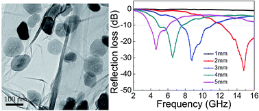
A novel three-dimensional (3D) composite based on reduced graphene oxide (rGO)/Fe2O3 was prepared by a one-pot hydrothermal method. Fe2O3 nanoparticles were either attached on the surface of graphene sheets or coated uniformly in the graphene sheets. The resulting composite is found to self-assemble to form a 3D network via hydrothermal treatment. Our results indicate that the as-prepared Fe2O3 nanoparticles show a porous morphology, which results in the rGO/Fe2O3 composites exhibiting excellent microwave absorbing properties in the range of 2–16 GHz and are therefore expected to be a promising candidate as microwave absorbing materials.
Introduction
With the rapid development of electromagnetic devices, electromagnetic (EM) wave radiation has become a pollution problem, which not only influences the performance of electronic devices, but may also be harmful to the health of human beings.1,2 For this reason, EM wave absorbing materials are of great interest for EM wave pollution. In this regard, extensive efforts have been made to develop EM wave absorption materials that are lightweight, with strong absorption and a wide absorption frequency range. Iron and carbon are abundant and nontoxic with a low cost. Meanwhile, carbon based and iron based composite materials have good EM absorption properties and they can meet the strict requirements of EM wave absorption materials.3–6 Therefore, iron oxides composited with carbon can become a promising candidate for lightweight microwave absorption materials.
Graphene, a new kind of carbon material, has been considered the ‘mother of all carbon forms’ as a building block.7 Low cost graphene can be produced in large-scale through chemical oxidation and reduction process using graphite as a raw material. Generally, the graphene product obtained by the above method is also known as the reduced graphene oxide (rGO). Due to the unique properties of graphene, it is promising for the applications in wide areas including energy storage and conversion,8–11 chemical catalysts,12 biomedicine,13 and etc.
Recently, scientists have reported that graphene and their composites have good microwave absorption properties and can be used as wave absorbing materials. Wang et al. have studied the microwave absorption properties of rGO and reported that rGO exhibited better microwave absorption performance compared with graphite and carbon nanotubes.14 Yu et al. have prepared graphene/polyaniline nanorods and found that the graphene had a strong dielectric loss.15 In addition, various iron based composites have been fabricated, for instance, Sun et al. have developed a facile method to synthesize hierarchical dendritic-like Fe3O4, γ-Fe2O3 and Fe and found that the composites exhibited excellent microwave absorbability in low or middle frequency (2–9 GHz).16 Wang et al. fabricated GO/CNT–Fe3O4 composites by using a one-pot co-precipitation in-situ growth route and found that the composite took on both dielectric loss and magnetic loss.17 The microwave absorption property of the porous Fe3O4-decorated graphene was reported by Dan et al., and they found that the porous, flower-like structured GN-Fe3O4 exhibited high magnetic loss in the low-frequency range and unique dielectric loss in the high-frequency range.18 The novel rGO composites with hematite nanoparticles embedded in rGO layers and wrapped by rGO sheets are found to show dramatically improved electromagnetic absorbing performance.19 Zhang et al. synthesized hybrid 3D rGO/Fe2O3 composite hydrogel by a hydrothermal method with GO and Fe3O4 nanoparticles as the raw materials and found the composite exhibited both wider absorption band and a larger reflection loss.20 M. Mishra et al. have demonstrated the presence of rGO plays a crucial role in enhancing the dielectric losses.21 Although there exist some work on carbon-based and iron-based absorbents, until now there have been only few reports on the microwave absorption performance of the composites filled with porous Fe2O3. Porous Fe2O3 nanoparticles have a low gravity and multiform framework. Therefore, it is expected that porous rGO/Fe2O3 hybrids can exhibit the excellent microwave absorption properties.
Herein, we report the in situ one-step synthesis of rGO/Fe2O3 composites without using any surfactants. The as-prepared Fe2O3 nanoparticles are poriferous, and show crystal structures with relatively uniform sizes. Our method provides operational simplicity and the capability for the large-scale production of graphene-based composites. Moreover, the microwave absorption properties of the rGO/Fe2O3 are studied, and the results indicate that as-prepared rGO/Fe2O3 composites show excellent microwave absorption performance.
Experimental
Materials
Graphite powder (200 mesh, Alfa Aesar, Johnson Matthey Company). KMnO4, K2S2O8, P2O5, H2O2, FeCl3·6H2O, NaOH (Beijing Chemical Works). All chemicals were analytical grade and used directly without further purification.
Preparation of porous Fe2O3 nanoparticles
Porous Fe2O3 nanoparticles were synthesized by a hydrothermal method. In a typical preparation procedure, 0.5 g FeCl3·6H2O was dissolved in 35 mL deionized (DI) water. The mixture was transferred into a 50 mL Teflon-sealed autoclave and hydrothermally treated at 180 °C for 12 h. The Teflon-sealed autoclave was washed by alkaline solution in advance. After that, the as-prepared samples were freeze-dried overnight.
Preparation of rGO/Fe2O3 composites
Graphene oxide (GO) was synthesized from natural graphite flakes by a modified Hummers method.22,23 Exfoliation was carried out by ultrasonicating the GO dispersion under ambient conditions. For the preparation of rGO/Fe2O3, 35 mL of a 1 mg mL−1 GO suspension was ultrasonicated for 0.5 h, in which 0.5 g FeCl3·6H2O was slowly added. The mixture was transferred into a 50 mL Teflon-sealed autoclave which was first washed by alkaline solution and then hydrothermally treated at 180 °C for 12 h. Next, the as-prepared sample was freeze-dried overnight.
Characterization
The morphology of the samples was investigated by field-emission scanning electron microscope (SEM, FEI, Quanta 200F) and transmission electron microscope (TEM, FEI, F20). Raman spectra were recorded on a Renishaw inVia confocal Raman microscope system using green (532 nm) laser excitation. The X-ray photoelectron spectrum (XPS) was conducted on a Thermo Fisher K-Alpha spectrometer. X-ray diffraction (XRD) patterns were carried out on a Bruker D8 Advance X-ray diffractometer. Thermogravimetric analysis (TGA) was carried out by a STA7200 (HITACHI) with a heating rate of 20 °C min−1 under flowing oxygen. The Fourier transform infrared (FT-IR) spectra were collected with a Magna-IR 560 E.S.P spectrometer.
EM absorption measurement
EM absorption measurement of the samples was prepared by mixing rGO/Fe2O3 with paraffin with 30 wt% of the composites. The mixtures were then pressed into toroidal-shaped samples (Φout = 7.00 mm and Φin = 3.04 mm). The complex permittivity and permeability values were measured in the 2–16 GHz range with the coaxial line method by an Agilent N5224A vector network analyzer.
Results and discussion
The preparation process of rGO/Fe2O3 is illustrated in Fig. 1. Firstly, Fe3+ cations from FeCl3 can favourably bind with oxygen-containing on GO sheets via electrostatic interactions. Secondly, the hydrolysis of Fe3+ leads to the formation of FeO(OH) anchored on the surface of the GO sheets due to the heating. Thirdly, 2D GO sheets with a uniform decoration of FeO(OH) act as a building block and self-assemble into 3D monolithic networks by hydrothermal treatment. The GO is simultaneously transformed into rGO, and the FeO(OH) is transformed into porous Fe2O3 after dehydration during this step. The formation mechanism of porous Fe2O3 is similar to the cases reported in other literatures.24,25 Finally, black rGO/Fe2O3 composites are obtained after the freeze drying process.
 |
| Fig. 1 A schematic illustration of the synthesis process of rGO/Fe2O3 composites. | |
Characterization of rGO/Fe2O3 composites
The structures and morphologies of the porous rGO/Fe2O3 composites were characterized by SEM and TEM. A SEM image shown in Fig. 2(a) reveals that rGO/Fe2O3 hybrid composite has the interconnected 3D network microstructure, which indicates efficient assembly between the particles and graphene sheets during the hydrothermal treatment. It also can be observed that some Fe2O3 nanoparticles anchored uniformly on the graphene sheets. Fig. 2(b) shows that some Fe2O3 nanoparticles are attached on the surface of the graphene sheets, and there is significant portion of the Fe2O3 nanoparticles encapsulated inside the graphene sheets, which can prevent the aggregation of particles efficiently. Such a geometric confinement of Fe2O3 nanoparticles within graphene layers can enhance their interface contact and prevent the dissolution and agglomeration of nanoparticles, which can be observed in Fig. 3(a). TEM images in Fig. 3(b) and (c) reveal that all the formed Fe2O3 nanoparticles show the nanoscale porous morphology with size distribution about 110 nm. The size of Fe2O3 nanoparticles become much bigger in the absence of GO compared with the case with GO (Fig. S2, ESI†). Their pore size distributions are in the range of several nanometers, as shown in Fig. 3(d), in which the weak contrast regions represent the pores, and the dark parts represent the Fe2O3 walls. The high-resolution TEM (HRTEM) image of Fe2O3 nanoparticles (Fig. S1, ESI†) demonstrates the crystalline nature of Fe2O3 nanoparticles with the lattice fringes distance of 0.252 nm, which can be assigned to the (110) plane. The result of HRTEM is well consistent with XRD result of porous rGO/Fe2O3 composites, as shown in Fig. 4(a) in which the 2θ angles at 24.1°, 33.2°, 35.6°, 40.8°, 49.5°, 54.1°, 62.4°, and 63.9° can be assigned to the (012), (104), (110), (113), (024), (116), (214) and (300) planes of Fe2O3 (JCPDS No. 33-0664). No obvious graphite peak appeared at around 26° suggests that the rGO sheets have been homogenously dispersed on the surface of the Fe2O3 particles.26 In addition, the TGA characterization has been used to quantify the amount of rGO in the composites. From the TG curve in Fig. 4(b), the significant weight loss is observed at approximately 450 °C and a constant weight is observed above 550 °C. The percentage of Fe2O3 nanoparticles in the hybrid composite is estimated to be 70% according to the TG curve. The slight mass loss below 300 °C could be attributed to the evaporation of adsorbed water and the removal of oxygen containing groups on the surface of GO sheets. The major weight loss from 300 to 550 °C was due to the combustion of graphene.
 |
| Fig. 2 (a) A SEM image of rGO/Fe2O3 composites shows the interconnected 3D network microstructure. (b) An enlarged SEM image indicates the Fe2O3 nanoparticles are either attached on the surface or encapsulated inside the graphene sheets. | |
 |
| Fig. 3 (a) A TEM image showing that the Fe2O3 nanoparticles are uniformly distributed on the graphene sheets. (b) and (c) TEM images of Fe2O3 nanoparticles with porous structure. (d) An enlarged TEM image showing the pore morphology (indicated by arrows) in a Fe2O3 nanoparticle. | |
 |
| Fig. 4 (a) The XRD pattern of rGO/Fe2O3 composites. (b) A TGA curve of rGO/Fe2O3 composites. | |
Fig. 5 shows the Raman spectra of the GO and rGO/Fe2O3 composites. There are two characteristic peaks located at around 1350 and 1597 cm−1 which are attributed to the D and G bands of GO. Compared with pure GO, the observed peaks at 219, 279, 395, 480, 593 cm−1 for the rGO/Fe2O3 are due to the existence of Fe2O3.27,28 The intensity ratio of the D band to the G band for GO and rGO/Fe2O3 is 1.15 and 1.05, respectively, indicating that the formation of Fe2O3 nanoparticles on rGO sheets leads to the disordered stacking of graphene sheets.29 FT-IR transmittance spectra of the as-prepared samples are presented in Fig. 5(b). Three intense absorption bands are observed at 3429, 2917 and 1627 cm−1, which are assigned to the asymmetrical stretching vibration, symmetrical stretching vibration and deformation vibration of physically adsorbed H2O molecules and the hydroxy groups on the GO in the sample. For bare α-Fe2O3, the bands at 574 and 480 cm−1 can be assigned to the stretching vibrations of the Fe3+–O2− bond in the FeO6 octahedron and FeO4 tetrahedron, respectively.30 There are also intense bands observed at 538 and 451 cm−1, corresponding to the stretching vibration of the Fe3+–O2− bond in the FeO6 octahedron and FeO4 tetrahedron, that are shifted to lower wavenumbers compared to that of 574 and 480 cm−1, which further confirms the existence of Fe2O3 nanoparticles and Fe2O3 is bound to the rGO surface.
 |
| Fig. 5 (a) Raman spectra and (b) FT-IR spectra of GO and rGO/Fe2O3 composites. | |
Moreover, XPS measurements have been performed to determine the composition of the as-produced rGO/Fe2O3 composites. The XPS survey spectrum in Fig. 6(a) shows the predominant C1s peak at 285 eV and typical characteristic peaks of Fe2O3 at 711 eV and 725 eV, corresponds to the Fe2p3/2 and Fe2p1/2,16,26 respectively. The Fe2p3/2 and Fe2p1/2 main peaks are clearly accompanied by two satellite peaks on their high binding-energy side (at ∼8 eV), as shown in Fig. 6(b), which are the characteristic peaks of Fe2O3.27
 |
| Fig. 6 (a) XPS spectra of rGO/Fe2O3 composite. (b) High resolution XPS spectrum of Fe 2p. | |
EM absorption properties of rGO/Fe2O3 composites
We have further investigated the electromagnetic parameters (relative complex permittivity, εr = ε′ − jε′′) of the Fe2O3, porous Fe2O3 and rGO/Fe2O3 composites in order to study their microwave absorbing properties.
Fig. 7 shows the real (ε′) and imaginary (ε′′) parts of the complex permittivity for the solid Fe2O3, porous Fe2O3, and rGO/Fe2O3 composites in the range of 2–16 GHz. It can be seen that the value of ε′ for solid Fe2O3 and porous Fe2O3 both are almost about 2.6 in the whole measurement range, as seen in Fig. 7(a). By comparison, when the rGO is added in the composite, the value of ε′ shows a dramatic increase, and the value of ε′ for rGO/Fe2O3 composite decreased from 11.1 to 6.25 with increasing frequency in the range of 2 to 16 GHz. Similarly, as shown in Fig. 7(b), the values of ε′′ of solid Fe2O3 and porous Fe2O3 remain almost unchanged as the frequency increases, and their average values were 0.052 and 0.053, respectively, indicating a weak dielectric loss. It can be seen that the value of ε′′ increases from zero to 3.66 for the rGO/Fe2O3 composite. It is obvious that the relative complex permittivity of the composites depends on the existence of rGO which consists of lots of dipolar polarization and electric polarization at microwave frequency.26 Fig. 7(c) shows the dielectric loss tangent (tan
δE = ε′′/ε′), which indicates that the rGO/Fe2O3 composites have very strong and efficient dielectric loss in the range of 2–16 GHz. This result demonstrates that the good EM wave absorption property may be due to dielectric loss. In the cases of Fe2O3 and porous Fe2O3, the values of dielectric loss are close to about 0.02 and keep constant throughout the whole frequency range.
 |
| Fig. 7 Frequency dependence of real part (a) and imaginary part (b) of relative complex permittivity, dielectric loss tangent (c) of solid Fe2O3, porous Fe2O3 and rGO/Fe2O3 composites. | |
In order to evaluate the EM absorption properties, the reflection loss (RL) of the EM waves can be calculated using transmission theory:
|
 | (1) |
|
 | (2) |
where
Zin is the normalized input impedance of the microwave absorption layer,
εr and
μr are the relative permittivity and permeability of the materials,
j is the microwave frequency,
d is the thickness of the absorber and
c is the velocity of light in free space.
The calculated theoretical RL of the composites-wax with different thicknesses in the range of 2–16 GHz with a loading of 30 wt% is shown in Fig. 8. As shown in Fig. 8(a) and (b), The maximum RL values for the pure Fe2O3 with a thickness of 1–5 mm is −4.6 dB, which demonstrates that the pure Fe2O3 has a very weak ability to absorb EM waves. The maximum RL value for the porous Fe2O3 with a thickness of 1–5 mm is −7.8 dB. Obviously, the maximum RL value of the porous Fe2O3 is higher than that of Fe2O3, with enhancement by about 69.5%. These results give us the evidence that the porous structure of Fe2O3 can effectively enhance the property of absorbing EM waves.
 |
| Fig. 8 The reflection loss of solid Fe2O3 nanoparticles (a), porous Fe2O3 nanoparticles (b), and rGO/Fe2O3 composites (c) measured with different thickness from 1 to 5 mm. | |
As shown in Fig. 8(c), the RL value of rGO/Fe2O3 is significantly improved. It is found that the thickness of the sample is one of major factors affecting both the intensity of the reflection loss peak and the position of the frequency at the reflection loss minimum. The minimum RL is −38 dB when the porous Fe2O3 weight ratio is 70% with thickness is 2 mm at 14.78 GHz, better than the case of reported novel rGO/α-Fe2O3.20 The bandwidths of RL values below −10 dB (90% of EM wave absorption) exceeds 5.8 GHz when the porous Fe2O3 weight ratio is 70% with thickness of 2 mm. With the increase of the absorber thickness, the minimum RL corresponding to the maximum absorption gradually decreases. The calculated results above demonstrate that the as-prepared rGO/Fe2O3 composites perform a better EM wave absorption than those of pure Fe2O3, dendritic Fe3O4,16 dendritic γ-Fe2O3,16 dendritic Fe16 and graphene-wrapped ZnO hollow spheres.3 and 2D Co3O4@C@PGC nanosheets.31 Compared to conducting ferrofluid composite from which the RL can reach 41 dB, and the bandwidth is 4 GHz.21 In our work, the RL can reach 38 dB and the bandwidth exceeds 5.8 GHz.
We have also evaluated the EM wave absorption properties of samples with different loading amounts of Fe2O3 (78% and 61%) which have been confirmed by TG curves (Fig. S3, ESI†). Our measurements indicate that rGO/Fe2O3 composites with different loading amounts of Fe2O3 show good EM wave absorption properties (Fig. S4, ESI†). The data show that the minimum RL is about −22 dB when the Fe2O3 weight ratio is 78% with thickness of 5 mm at 6.78 GHz. The minimum RL is about −39 dB when the Fe2O3 weight ratio is 61% with thickness of 2 mm at 7.9 GHz and 12.85 GHz, but the bandwidth of RL value below −10 dB is only about 4.5 GHz. Interestingly, the EM wave absorption property of rGO/Fe2O3 composites (70%) is better than that of rGO/Fe2O3 (78%) and rGO/Fe2O3 (61%). The reason for above phenomenon is possible due to that the dielectric loss of rGO/Fe2O3 (78%) is too small from frequency dependence of real part and imaginary part of relative complex permittivity, dielectric loss tangent (Fig. S5, ESI†) and the impedance match of rGO/Fe2O3 (61%) is not good. For rGO/Fe2O3 (70%) composites, which contains suitable ratio α-Fe2O3, a kind of semiconductor material, can adjust impedance match which is related to the microwave absorption. The too high permittivity of absorber is harmful to the impedance match, yielding a strong reflection and weak absorption.
The possible microwave absorbing mechanism could be explained by strong dielectric loss, conduction loss and multiple reflections in the porous structure of Fe2O3, as schematically shown in Fig. 9. According to the electromagnetic theory, the dielectric loss is related to the intrinsic physical properties of the composites and the nanostructures. Firstly, there are residual oxygen functional groups and defects in the thermal rGO which can enhance the electromagnetic energy absorption. These functional groups and defects act as polarized centers, which give rise to higher ε′ values. Secondly, as shown in Fig. 2, the interface between graphene sheets and the exterior Fe2O3 nanoparticles can be clearly observed, which causes interfacial polarization (called as the Maxwell–Wagner effect) and the associated relaxation. The reason for this phenomenon is possibly due to the formation of large dipoles and the accumulation of charge at interface.32–35 Moreover, Debye dipolar relaxation is an important mechanism by which dielectric loss materials absorb microwave radiation. What is more, due to the high thermal conductivity of the composites, EM energy might be dissipated rapidly by interference and by thermal energy form.
 |
| Fig. 9 Schematic diagram for possible microwave absorbing mechanism of rGO/Fe2O3 composites. | |
Conclusions
In summary, porous rGO/Fe2O3 composites with enhanced EM wave absorption property has been successfully synthesized via in situ one step synthesis without using any surfactant. The as-prepared porous Fe2O3 nanoparticles with relatively uniform size distribution attached onto the surface of rGO or wrapped by rGO to form 3D framework. The rGO/Fe2O3 composites exhibit remarkably improved dielectric properties and EM wave absorbing performance in 2–16 GHz. The minimum RL reaches −38 dB when the porous Fe2O3 weight percentage is 70% with thickness of 2.0 mm at 14.78 GHz and the effective absorption bandwidth exceeds 5.8 GHz. Additionally, the EM wave absorption property can be adjusted easily by varying the thickness of the samples and the weight percentage of rGO. It is believed that porous rGO/Fe2O3 composites could be a potential kind of excellent microwave absorbing material with lightweight, strong absorption and wide absorption bandwidth.
Acknowledgements
We gratefully thank for the National Natural Science Foundation of China (No. 21322609 and 21202203), the Science Foundation Research Funds Provided to New Recruitments of China University of Petroleum, Beijing (No. 2462014QZDX01, YJRC-2013-31), the National Basic Research Program of China (No. 2012CB933102), and Thousand Talents Program.
Notes and references
- R. C. Che, L. M. Peng, X. F. Duan, Q. Chen and X. L. Liang, Adv. Mater., 2004, 16, 401–405 CrossRef CAS PubMed.
- J. W. Liu, R. C. Che, H. J. Chen, F. Zhang, F. Xia, Q. S. Wu and M. Wang, Small, 2012, 8, 1214–1221 CrossRef CAS PubMed.
- M. K. Han, X. W. Yin, L. Kong, M. Li, W. Y. Duan, L. T. Zhang and L. F. Cheng, J. Mater. Chem. A, 2014, 2, 16403–16409 CAS.
- L. Kong, X. W. Yin, X. Y. Yuan, Y. J. Zhang, X. M. Liu, L. F. Cheng and L. T. Zhang, Carbon, 2014, 73, 185–193 CrossRef CAS PubMed.
- T. S. Wang, Z. H. Liu, M. M. Lu, B. Wen, Q. Y. Ouyang, Y. J. Chen, C. L. Zhu, P. Gao, C. Y. Li, M. S. Cao and L. H. Qi, J. Appl. Phys., 2013, 113, 024314 CrossRef PubMed.
- X. C. Zhao, Z. M. Zhang, L. Y. Wang, K. Xi, Q. Q. Cao, D. H. Wang, Y. Yang and Y. W. Du, Sci. Rep., 2013, 3, 3421 Search PubMed.
- A. K. Geim and K. S. Novoselov, Nat. Mater., 2007, 6, 183–191 CrossRef CAS PubMed.
- H. L. Wang, H. S. Casalongue, Y. Y. Liang and H. J. Dai, J. Am. Chem. Soc., 2010, 132, 7472–7477 CrossRef CAS PubMed.
- S. B. Yang, X. L. Feng, S. Ivanovici and K. Müllen, Angew. Chem., Int. Ed., 2010, 49, 8408–8411 CrossRef CAS PubMed.
- H. B. Yang, C. X. Guo, G. H. Guai, Q. L. Song, S. P. Jiang and C. M. Li, ACS Appl. Mater. Interfaces, 2011, 3, 1940–1945 CAS.
- Y. J. Chen, Q. S. Wang, C. L. Zhu, P. Gao, Q. Y. Ouyang, T. S. Wang, Y. Ma and C. W. Sun, J. Mater. Chem., 2012, 22, 5924–5927 RSC.
- Z. Y. Zhang, F. Xiao, J. B. Xi, T. Sun, S. Xiao, H. R. Wang, S. Wang and Y. Q. Liu, Sci. Rep., 2014, 4, 4053 Search PubMed.
- X. J. Fan, G. Z. Jiao, L. Gao, P. F. Jin and X. Li, J. Mater. Chem. B, 2013, 1, 2658–2664 RSC.
- C. Wang, X. J. Han, P. Xu, X. L. Zhang, Y. C. Du, S. R. Hu, J. Y. Wang and X. H. Wang, Appl. Phys. Lett., 2011, 98, 072906 CrossRef PubMed.
- H. L. Yu, T. S. Wang, B. Wen, M. M. Lu, Z. Xu, C. L. Zhu, Y. J. Chen, X. Y. Xue, C. W. Sun and M. S. Cao, J. Mater. Chem., 2012, 22, 21679–21685 RSC.
- G. B. Sun, B. X. Dong, M. H. Cao, B. Q. Wei and C. W. Hu, Chem. Mater., 2011, 23, 1587–1593 CrossRef CAS.
- L. N. Wang, X. L. Jia, Y. F. Li, F. Yang, L. Q. Zhang, L. P. Liu, X. Ren and H. T. Yang, J. Mater. Chem. A, 2014, 2, 14940–14946 CAS.
- D. P. Sun, Q. Zou, G. Q. Qian, C. Sun, W. Jiang and F. S. Li, Acta Mater., 2013, 61, 5829–5834 CrossRef CAS PubMed.
- D. Z. Chen, G. S. Wang, S. He, J. Liu, L. Guo and M. S. Cao, J. Mater. Chem. A, 2013, 1, 5996–6003 CAS.
- H. Zhang, A. J. Xie, C. P. Wang, H. S. Wang, Y. H. Shen and X. Y. Tian, J. Mater. Chem. A, 2013, 1, 8547–8552 CAS.
- M. Mishra, A. P. Singh, B. P. Singh, V. N. Singh and S. K. Dhawan, J. Mater. Chem. A, 2014, 2, 13159–13168 CAS.
- W. S. Hummers and R. E. Offeman, J. Am. Chem. Soc., 1958, 80, 1339 CrossRef CAS.
- Y. Y. Liang, D. Q. Wu, X. L. Feng and K. Müllen, Adv. Mater., 2009, 21, 1679–1683 CrossRef CAS PubMed.
- Y. Wang, S. Wang, H. Zhang, X. Gao, J. Yang and L. Wang, J. Mater. Chem. A, 2014, 2, 7935–7943 CAS.
- J. Wang, L. Li, C. Wong, L. Sun, Z. Shen and S. Madhavi, RSC Adv., 2013, 3, 15316–15326 RSC.
- H. L. Xu, H. Bi and R. B. Yang, J. Appl. Phys., 2012, 111, 07A522 Search PubMed.
- A. Nasibulin, S. Rackauskas, H. Jiang, Y. Tian, P. Mudimela, S. Shandakov, L. Nasibulina, S. Jani and E. Kauppinen, Nano Res., 2009, 2, 373–379 CrossRef CAS PubMed.
- C. T. Cherian, J. Sundaramurthy, M. Kalaivani, P. Ragupathy, P. S. Kumar, V. Thavasi, M. V. Reddy, C. H. Sow, S. G. Mhaisalkar, S. Ramakrishna and B. V. R. Chowdari, J. Mater. Chem., 2012, 22, 12198–12204 RSC.
- L. Li, Z. P. Guo, A. J. Du and H. K. Liu, J. Mater. Chem., 2012, 22, 3600–3605 RSC.
- D. Z. Chen, W. Wei, R. N. Wang, J. C. Zhu and L. Guo, New J. Chem., 2012, 36, 1589–1595 RSC.
- F. S. Wen, H. Hou, J. Y. Xiang, X. Y. Zhang, Z. B. Su, S. J. Yuan and Z. Y. Liu, Carbon, 2015, 89, 372–377 CrossRef CAS PubMed.
- L. Kong, X. W. Yin, Y. J. Zhang, X. Y. Yuan, Q. Li, F. Ye, L. F. Cheng and L. T. Zhang, J. Phys. Chem. C, 2013, 117, 19701–19711 CAS.
- J. K. Nelson and C. F. John, Nanotechnology, 2004, 15, 586 CrossRef CAS.
- P. K. Mandal, A. Lapanik, R. Wipf, B. Stuehn and W. Haase, Appl. Phys. Lett., 2012, 100, 073112 CrossRef PubMed.
- X. F. Zhang, X. L. Dong, H. Huang, Y. Y. Liu, W. N. Wang, X. G. Zhu, B. Lv, J. P. Lei and C. G. Lee, Appl. Phys. Lett., 2006, 89, 053115 CrossRef PubMed.
Footnote |
† Electronic supplementary information (ESI) available: Additional data. See DOI: 10.1039/c5ra09715k |
|
This journal is © The Royal Society of Chemistry 2015 |
Click here to see how this site uses Cookies. View our privacy policy here.