DOI:
10.1039/C5RA09172A
(Paper)
RSC Adv., 2015,
5, 68621-68631
Bioactive prodigiosin-impregnated cellulose matrix for the removal of pathogenic bacteria from aqueous solution
Received
16th May 2015
, Accepted 15th July 2015
First published on 15th July 2015
Abstract
The increase in concern over safe water for human consumption demands disinfection of water. Conventional chemical disinfection methods release counter ions into the treated water and they impair human health significantly. The present investigation was focused on the disinfection of pathogenic bacteria via a bio-disinfection route using a prodigiosin-impregnated cellulose column reactor (PICCR). Escherichia coli and Bacillus cereus were chosen as model pathogens to validate the efficiency of the PICCR for pathogen removal from water. The pathogen removal efficiency was evaluated by the pour plate method, regrowth ability in nutrient broth and quantitative estimation of live and dead cells using fluorescent microscopy. The PICCR showed effective reduction of E. coli by 97.31% and B. cereus by 97.33%. Further, bacterial cell membrane damage by bio-disinfection was verified through analysis of the residual protein and nucleic acid in the treated water using UV-visible spectroscopy, a trans-illuminometer and SDS PAGE. The proposed PICCR was found to be effective for the removal of pathogens from water and this may be regarded as a viable purification technique for drinking water.
Introduction
The treatment of water/wastewater draws worldwide attention to alleviate increased water scarcity. One of the main drawbacks to the reuse of water/wastewater is the waterborne diseases caused by pathogens. This has been considered as a major public health concern in developing countries, as it has caused millions of deaths due to occurrence of frequent outbreaks.1 There is evidence that human and cattle populations have been frequently infected by microorganisms, such as bacteria, moulds and viruses, present in contaminated water.2 There are many research reports on the steady increase in persistent antibiotic-resistant microorganisms and their continuous effect on healthcare. There have been many methods reported for the removal of pathogenic microorganisms from water/wastewater.3–7 The application of chemical disinfectants for the disinfection of microorganisms in water is accompanied by the release of counter ions into treated water while removing the bacteria. Beyond a certain concentration, the increased counter ions and byproducts are considered to be pollutants in the treated water/wastewater.8 Thus, there has been constant research into antimicrobial agents such as chemical surfactants and nanoparticles for the disinfection of pathogens.9–14 The present investigation was focused on developing a device using bioactive prodigiosin for the removal of pathogens without release of counter ions. Prodigiosin is a secondary metabolite produced by many types of bacteria. Prodigiosin has an excellent antibacterial activity towards Gram positive and Gram negative bacteria.15,16 Prodigiosin could impart an instantaneous antibacterial effect and prevent the formation of bacterial biofilms.17 Prodigiosin acts as an antibacterial agent due to its strong affinity towards DNA and its subsequent inactivation of bacterial cells.18 Prodigiosin is efficient in the disinfection of waterborne pathogens such as fungi, algae, mosquito larvae and plankton, besides bacteria.19–22 However, bioactive prodigiosin has not yet been used in water/wastewater disinfection applications because of its hydrophobicity. Moreover, the carrier matrix needed for the immobilization of prodigiosin is yet to be developed. The cellulose matrix is an effective material for filtration purposes compared to other filtration materials. It is an effective matrix for the immobilization of prodigiosin and is highly stable in acidic and alkaline solutions.23–25 In the present investigation, prodigiosin was immobilized onto a cellulose matrix and fabricated as a stacked column reactor for the disinfection of water under the continuous mode. This is the first report on the application of prodigiosin-impregnated cellulose matrix for the disinfection of pathogenic bacteria in water.
Materials and methods
Chemicals
The chemicals and solvents used in the present study were of analytical grade. Silica gel, SYTO 9, propidium iodide, ethidium bromide and solvents were ordered from Merck chemicals, India. Microbiological ingredients were obtained from HiMedia, India.
Production, extraction and purification of prodigiosin
The red pigmented strain Serratia marcescens was isolated from self-fermented limed animal fleshing, the proteinaceous substrate generated from the leather industry. The isolated strain was inoculated in nutrient broth and incubated at 30 °C for 24 h and was named as the mother culture.26 The fermentation medium was prepared by mixing 30 g solid substrate (wheat bran, 22 g and tannery fleshing, 8 g) with a mineral solution of 15 ml volume (Na2HPO4–7H2O, 33.90 g; KH2PO4, 15 g; NaCl, 2.5 g; NH4Cl, 5 g per liter), which were transferred into an Erlenmeyer flask of 250 ml volume. The flask was plugged with cotton and was sterilized in an autoclave at a temperature of 121 °C and pressure of 15 lbs per inch2 for 20 min. The flask with the fermentation medium was cooled to room temperature and the medium was inoculated with 5 ml of inoculum collected from the mother culture. The flasks were incubated at 30 °C for 72 h. After the colour of the medium had changed to red (indicative of prodigiosin), the contents of the flask were extracted with acidified methanol. The extracted prodigiosin was dried in a rotary vacuum evaporator at 40 °C. The resultant prodigiosin was scraped and stored under refrigerated conditions until used in further experiments. It was pre-analyzed using Thin Layer Chromatography for the initial purification of the prodigiosin, using methanol
:
ethyl acetate (6
:
4, v/v) as the mobile solvent mixture. The separated compounds were identified under UV light. The identified compounds were purified by column chromatography using silica gel (mesh size 80–100) as the solid adsorbent and methanol
:
ethyl acetate (6
:
4 v/v) as the mobile solvent mixture and the eluted compound was identified using thin layer chromatography. The purified prodigiosin was further characterized using UV-visible and FT-IR spectrophotometers.
Design and fabrication of prodigiosin-impregnated cellulose column reactor
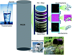
Prodigiosin (1, 5 or 10 mg) was dissolved in 30 ml methanol to obtain the prodigiosin solution. Cellulose matrix (diameter, 5 cm and thickness, 1 cm) was immersed in the prodigiosin solution under agitation at an optimized speed of 60 rpm for 30 min at 30 °C. The prodigiosin-impregnated cellulose matrix was placed in a hot air oven for 30 min at 50 °C for stabilization of the bonding. The resulting material was named as prodigiosin-impregnated cellulose (P1IC, P5IC and P10IC) matrix. The presence of prodigiosin in PIC was confirmed using a solid diffuse reflectance spectrophotometer (Varian TCA, Cary 300) in the wavelength range of 200–800 nm using barium sulphate as a reference compound. Further, the stability of the immobilized prodigiosin was checked by soaking the PIC in 100 ml of distilled water for 24 h under agitation at 60 rpm, following which the concentration of prodigiosin in the residual solution was determined. The PIC matrices were stacked one over the other with interleaved nylon mesh cloth (thickness, 2 mm) to act as a spacer and support. The stacked PIC was housed in an acrylic column (diameter, 5 cm and length, 20 cm) with a 1.5 l capacity as shown in Fig. 1 and the resulting configuration was named as a prodigiosin-impregnated cellulose column reactor (P1ICCW, P5ICCW and P10ICCW).
 |
| Fig. 1 Schematic diagram of prodigiosin-impregnated cellulose column reactor. | |
Selection of pathogens and preparation of model infected water
Escherichia coli (MTCC-2939), Gram-negative bacteria, and B. cereus (MTCC-8372), Gram-positive bacteria, were selected as the model pathogens in the present investigation for their removal using the PIC matrix. The above listed pathogens were considered in the present investigation for the reason that ground water contaminated with these pathogens was reported to cause a severe health hazard. The selected pathogens were inoculated in autoclaved nutrient broth medium of 100 ml volume and incubated under agitation at 80 rpm and a temperature of 37 °C. The matured culture was harvested after 24 h and the growth was measured by absorbance at λ600 nm using a UV-visible spectrophotometer. The bacteria were separated from the nutrient broth by centrifuging at 5000 rpm for 20 min; they were resuspended in deionized water and the colony forming unit of the resuspended solution was determined using the pour plate method. This bacteria-inoculated water was named as model infected water (MIW). The MIW, with known bacterial count, was passed through the PICCR using a peristaltic pump at a flow rate of 1 ml min−1.
Bactericidal activity
The PICCR-treated water (PICCW) was analyzed and compared with cellulose (without prodigiosin) column reactor-treated water (CCW), serving as a control for the evaluation of bacterial removal efficiency. The bacterial concentrations (CFU ml−1) of the MIW, PICCW and CCW were evaluated at different time intervals by inoculating 1 ml of water onto agar plates, which were incubated at 37 °C for 24 h. The regrowth of pathogens after different treatments was evaluated in nutrient broth and the efficiency was determined by measuring absorbance at λ600 nm. Also, the bacterial removal efficiencies of CCW and PICCW were assessed by the fluorescent dye uptake capacity of the cells. CCW and PICCW samples of 1 ml volume were separately mixed with 0.05 ml (1 mg ml−1) of propidium iodide and SYTO 9 and then incubated at room temperature for 5 min. The cells were separated by centrifugation at 5000 rpm for 10 min and the pellets were resuspended in 1 ml of phosphate buffer (pH, 7.0). Then, 20 μl of the suspension was transferred onto a microscope slide and its fluorescence images captured using a fluorescence microscope (Olympus BX-61) with a green filter (excitation/emission, 440–480 nm/515–540 nm) and a red filter (540–560 nm/630–660 nm). Viable and lysed cells were viewed in the same microscopic field with different fluorescence filter sets. The cell viability was assayed by counting green (live) and red (dead) cells.27 The pathogen removal efficiency, in percentage, was calculated using the following mathematical expression:
Further, the bacterial cell damage was quantified by analyzing the protein content and nucleic acid content in the CCW and PICCW samples. CCW and PICCW samples of 1 ml volume were each centrifuged at 5000 rpm, and the supernatants were immediately frozen at 10 °C. The protein content was determined using the Lowry method. The content of nucleic acids and derivative compounds, such as purines and pyrimidines, was determined by their absorption in a UV-visible spectrophotometer at λ260 nm.
Results and discussion
Production and extraction of prodigiosin
A solid state fermentation process was performed for different incubation periods. The prodigiosin production started after an induction period of 24 h and gradually increased up to 96 h (Fig. 2a). The high yield of prodigiosin observed in the present investigation may be attributed to the high amino acid content of the proteinaceous solid waste (tannery animal fleshing) used as the substrate.28 Tannery fleshing serves as the nitrogen source for the effective growth of bacterial biomass and subsequent formation of prodigiosin. The prodigiosin was extracted from the broth using petroleum ether in a separating funnel at a 1
:
2 (v/v) ratio. The extracted pigment was analyzed through thin layer column chromatography. The thin layer chromatography showed three bands corresponding to Rf values of 0.8, 0.9, 0.98. Amongst them, the band at Rf of 0.9 corresponds to prodigiosin, with reference to the standard prodigiosin pigment.29 The same solvent system was used in column chromatography for the purification of the prodigiosin.30
 |
| Fig. 2 Characterization of prodigiosin: (a) UV visible spectrophotometric profile of prodigiosin production at different time intervals and (b) FT-IR spectrum of prodigiosin. | |
Characterization of prodigiosin
The purified prodigiosin was lyophilized to obtain it in powder form for the characterization using UV-visible and Fourier transform-infra red (FT-IR) spectroscopy. The purified prodigiosin was scanned in a UV-visible spectrophotometer in a wavelength range of λ200 to 800 nm. Fig. 2a shows a sharp single peak at λ532 nm which corresponds to the characteristic peak of prodigiosin.31,32 Fig. 2b shows the FT-IR spectrum of the purified prodigiosin pigment. The strong stretching peaks at 1218 cm−1 and 2928 cm−1 are attributed to the presence of aromatic –CH and –CH2– stretching in prodigiosin. Another stretching at 1630 cm−1 is due to the presence of conjugated aromatic C
C bonds in prodigiosin. The N–H stretching at 3421–3540 cm−1 corresponds to the pyrrole ring in the prodigiosin molecular structure. The C–C stretching was observed at 3295 cm−1. The strong peaks near 2928 cm−1 and at 2800 cm−1 (C–O–CH3) may be attributed to the presence of methyl groups in the prodigiosin molecular structure. The (–C–O–C–)–O–CH3 group attached to the aromatic pyrrole ring in the prodigiosin molecular structure was confirmed by the strong band at 1133–1060 cm−1. The fingerprint region was characterized by medium peaks at 1734 cm−1 (C
O), 1452 cm−1 (C–O), 1133 cm−1 (C–N), 986 cm−1 & 817 cm−1 present in the prodigiosin.33
Characterization of prodigiosin-impregnated cellulose matrix
The immobilization of prodigiosin onto the cellulose matrix was confirmed by the increase in intensity of the red color. Fig. 3 illustrates that the increase in the mass of prodigiosin loading from 1 g to 10 g in the cellulose matrix significantly increased the intensity of the red color in the cellulose matrix. The PIC was further characterized using UV diffuse reflectance spectroscopy. Fig. 4a shows a sharp deflection peak at λ532 nm, which is due to the presence of the chromophore structure of the O–C group in prodigiosin.34 The peak intensity increased with the increase in concentration of prodigiosin in the cellulose matrix. The energy band gap values of the prodigiosin-impregnated matrices (P1IC, P5IC and P10IC) were calculated from the UV-DRS spectra (Fig. 4). The calculated energy band gaps for P1IC, P5IC and P10IC were found to be 3.01 eV, 2.6 eV and 2.4 eV, respectively. The energy band gap values of the PIC matrices suggest that they are semiconducting in nature. The required activation energy for the impregnation was observed to decrease with an increase in concentration, which confirmed the quick immobilization of prodigiosin molecules on the cellulose matrix (bonding between prodigiosin and cellulose matrix). The energy band gap decreased with an increase in concentration of prodigiosin molecules in the impregnation and inversely increased the number of actives sites (free electrons) in the matrix. The increase in free electron density in the impregnated matrix enhances the antimicrobial property of the PIC matrix. Fig. 4b shows the thermogravimetric analysis of the cellulose matrix and PIC matrix. The maximum weight loss was observed in the temperature range from 250 °C to 400 °C for the cellulose matrix. However, in the case of the prodigiosin-impregnated cellulose matrix, the maximum weight loss extended from 350 °C to 550 °C, which confirms the impregnation of prodigiosin molecules into cellulose matrix. Also, the prodigiosin molecules enhanced the thermal stability of the cellulose matrix significantly. The FT-IR spectra of the cellulose matrix and PIC were recorded as shown in Fig. 4c. A strong stretching peak at 3415 cm−1 is attributed to the presence of the intra-molecular hydrogen bonding of the cellulose matrix. The stretching vibration peak at 3415 cm−1 corresponds to the O–H groups in the cellulose matrix. The sp3 hybridized C–H stretching vibration for the methylene group was identified from peaks at 2934 cm−1 and 2852 cm−1. The C–H bending vibration for the –O–C–H group was identified from peaks at 1389 cm−1 and 1035 cm−1, attributed to the stretching vibration of the –C–O–C– group. The weak stretching band peak at 844 cm−1 confirmed the presence of –C–O–C– in the cellulose matrix and the C–H deformation or isomer of the matrix was identified from a peak at 700 cm−1. The PIC showed a strong stretching vibration peak at 3424 cm−1, attributed to the OH and N–H functional groups. Two broad stretching vibration peaks at 2934 cm−1 and 2852 cm−1 indicate the presence of the C–H functional group of the methylene structure in the PIC matrix. Also, the stretching vibration at 2900 cm−1 was attributed to the presence of a methoxy group in the prodigiosin molecule. The aromatic ring was identified from a peak at 1475 cm−1. Prodigiosin has an aryl alkyl ether (C6H5–O–CH3) group and it was identified from the presence of an asymmetric band at 1271 cm−1 and a symmetric band at 1035 cm−1. The presence of the pyrrole ring in the prodigiosin molecule was identified from the stretching vibration peak at 1107 cm−1. A weak stretching vibration observed at 844 cm−1 may be attributed to the presence of –C–O–C– and C–H deformation in the cellulose matrix due to the immobilization of prodigiosin. The surface morphologies of the cellulose matrix and PIC matrix were analyzed using a scanning electron microscope. Fig. 5a shows the smooth surface of the irregular multilayered cellulose matrix porous structure. Fig. 5b shows that the PIC matrix was found to be covered with a greater number of spherical-shaped molecules. The smaller spherical shaped prodigiosin molecules were distributed evenly throughout the cellulose matrix and some spherical particles were held inside the porous structure of the cellulose matrix. This evidence clearly confirmed that the prodigiosin molecules were strongly immobilized on the cellulose matrix.
 |
| Fig. 3 Prodigiosin-impregnated cellulose matrix at different prodigiosin concentrations: (a) control (CM), (b) P1IC (c) P5IC and (d) P10IC. | |
 |
| Fig. 4 Characterization of cellulose matrix and prodigiosin-impregnated cellulose matrix using (a) UV-diffuse reflectance spectroscopy, (b) TGA analysis and (c) FT-IR spectroscopy. | |
 |
| Fig. 5 SEM images of (a) PIC and (b) PIC with bacterial cells. | |
The stability of PIC in aqueous medium was evaluated for 5 days, as shown in Fig. 6. The concentration of prodigiosin in the aqueous solution was measured every 24 h after extracting with acidified methanol and was analyzed with a UV visible spectrophotometer at λ532 nm. The concentration of prodigiosin in the PIC matrix decreased by only 1.2%, even after 5 days (Fig. 6). The results confirmed that the prodigiosin molecules have strong stability in the cellulose matrix. The hydrophobic nature of the prodigiosin results in a very low degree of removal of prodigiosin in the treated water at a 1 ml min−1 flow rate (Fig. 6). However, there is no report on the adverse health effects of prodigiosin in human/animals except that prodigiosin is efficient as an antibiotic/antioxidant compound.19–22,24 Hence, leached prodigiosin may be regarded as a harmless refractory organic substrate in the treated water.
 |
| Fig. 6 Leachability of prodigiosin from impregnated cellulose matrix at different time intervals. | |
Evaluation of bactericidal activity of the PICCR
The bactericidal activity of the PICCR was evaluated through removal of Gram-negative bacteria, E. coli, and Gram-positive bacteria, B. cereus. E. coli and B. cereus are widely distributed pathogens in water/wastewater systems. E. coli is a highly reproductive strain of bacteria and has a complex recombinant structure, making it able to survive even under high stress conditions. B. cereus mainly causes waterborne diseases, such as diarrhoea, in humans and animals. Hence, these pathogens were considered in the present investigation. The removal efficiency of the pathogens by the PICCR was examined through the bacterial concentration in treated water using the pour plate method, fluorescence imaging and residual protein concentration in PICCR-treated water.
Bactericidal and bacterial cell membrane damaging potential of the PICCR
The pathogen removal efficiency of the PICCR was examined by the antimicrobial activity of the PIC against E. coli and B. cereus. The MIW containing pathogens (E. coli, 23 × 105 and B. cereus, 12 × 104 CFU ml−1) was passed through the PICCR at a flow rate of 1 ml min−1. MIW and PICCW of 1 ml volume were added to the nutrient agar plate using the serial dilution method. In the plate count method, the P10ICCW contained E. coli, log
1(14 × 101), and B. cereus, log
1(11 × 101), after the 15th h of treatment and no bacterial growth was observed up to 6th h of treatment (Fig. 7). The positive control (CCW) contained E. coli, log
3(4 × 10³), and B. cereus, log
2(4 × 10²), bacterial concentrations. The reduction may be due to the surface attachment of pathogens onto the cellulose matrix. The P5ICCW contained log
2(17 × 102) and log
1(49 × 101) concentrations of E. coli and B. cereus, respectively, at the 15th h of treatment. The bacterial removal efficiency of the PICCR was directly proportional to the concentration of prodigiosin in the PIC matrix, and the bactericidal activity was high in the P10ICCW.35 The P10ICCW showed an effective removal of E. coli of 97.31% and B. cereus of 97.33% (Fig. 8). The results confirm that the PIC matrix could effectively eliminate the pathogens in contaminated water.
 |
| Fig. 7 Quantification of viable bacteria using the plate counting method in nutrient agar pate. Quantification of E. coli in (a) MIW, (b) P1ICCW, (c) P5ICCW and (d) P10ICCW; quantification of B. cereus in (e) MIW, (f) P1ICCW, (g) P5ICCW and (h) P10ICCW. | |
 |
| Fig. 8 Bacterial log reduction of (a) E. coli and (b) B. cereus by CCW and PICCW at different prodigiosin concentrations (P1ICCW, P5ICCW, P10ICCW and MIW) at different time intervals. | |
Live and dead cells profile
The bacterial cell viability in water was investigated by fluorescence microscopic LIVE/DEAD assay. Fig. 9 shows the fluorescence microscope bacterial images of the CCW and PICCW samples stained with SYTO 9 and propidium iodide. Fig. 9 shows that the fluorescent images of P5ICCW contained about 70–80% lysed bacteria, while the dead cell concentration increased to 95–98% in P10ICCW. The lysed bacterial cells emit red-coloured florescence, indicating that bacterial cell lysis was caused by the prodigiosin in the matrix. The CCW showed entirely green-coloured cells, concentrated with viable bacterial cells. The P10ICCW sample contained the maximum number of pathogens loaded with propidium iodide rather than SYTO 9, which was confirmed by the presence of red-colored cells. This may be due to the adhesion of the propidium iodide stain to nucleic acid functional sites in the damaged bacterial cell membranes.36 The pathogenic bacterial cells in the CCW showed only green cells, indicating that the bacteria could retain the SYTO 9 stain only on the cell wall. Further to that, propidium iodide staining did not occur because nucleic acid leakage sites were not available in the bacterial cell membranes of the pathogens in the CCW. The results confirmed that the maximum pathogen removal efficiency was observed with a high mass loading of prodigiosin (10 mg) loaded in P10ICCW.
 |
| Fig. 9 Fluorescent images of CCW, P1ICCW, P5ICCW and P10ICCW: (1) viable E. coli cells, (2) dead E. coli cells, (3) viable B. cereus cells and (4) dead B. cereus cells. | |
Evaluation of protein and nucleic acid leakages from damaged bacterial cells
The bacterial cell membrane damage was confirmed by quantitative estimation of protein and nucleic acid concentrations in CCW and PICCW. Fig. 10(1) shows that the concentration of protein was 45 μg ml−1 from E. coli and 35 μg ml−1 from B. cereus in CCW. For P10ICCW the concentrations of proteins recorded were 214 μg ml−1 and 428 μg ml−1 from E. coli and B. cereus, respectively. This could be due to cell membrane damage caused by prodigiosin owing to its antibacterial activity, leading to a significant increase in the protein and nucleic acid concentrations in PICCW.37 Furthermore, the SDS-PAGE also confirmed that the increase in mass loading of prodigiosin in the PIC increased the intensity of the protein band in SDS-PAGE (Fig. 10(2)). The results reveal that the P10ICCW contained higher protein content than CCW. This indicates that the bacterial cell damage increased with mass of prodigiosin in PIC, leading to leakage of intracellular constituents into the aqueous solution. Fig. 11(1) illustrates that the nucleic acid concentration increased with the increase in concentration of prodigiosin in PIC. The intensity of the nucleic acid concentration increased from 0.012 μg ml−1 to 0.095 μg l−1 for B. cereus and 0.018 μg l−1 to 0.083 μg l−1 for E. coli in P10ICCW. The fluorescence emission was not present in CCW, which may correlate to the absence of nucleic acids. The PICCW sample emitted fluorescence under the trans-illuminometer. The trans-illuminator showed the maximum emission for P10ICCW, which confirmed the leakage of nucleic acids at the instant of cell membrane rupture (Fig. 11(2)).38–40 These results concluded that the protein and nucleic acid concentrations in P10ICCW increased due to cell membrane damage of the pathogens by prodigiosin. The scanning electron microscopy also showed the damaged bacterial cells in PICCW and MIW. Fig. 12a shows the presence of bacterial cells in MIW with uniform structure which indicates the bacterial cells are in live conditions. The homogeneous micro structure of damaged bacterial cells are indicated in yellow arrow lines in Fig. 12b, which corresponds to leakage of protein and nucleic acid.41,42
 |
| Fig. 10 (1) Protein leakage of E. coli and B. cereus in (a) CCW, (b) P1ICCW, (c) P5ICCW and (d) P10ICCW. (2) SDS-PAGE of protein leakage in (a) CCW, (b) P1ICCW (c) P5ICCW and (d) P10ICCW. | |
 |
| Fig. 11 (1) Nucleic acid leakage of E. coli and B. cereus in (a) CCW, (b) P1ICCW, (c) P5ICCW and (d) P10ICCW. (2) Trans-illuminometer image of nucleic acid leakage in (a) initial CCW, (b) P1ICCW, (c) P5ICCW and (d) P10ICCW. | |
 |
| Fig. 12 SEM image of (a) initial MIW (live cells) and (b) cells trapped on surface of PIC (dead cells). | |
Mechanistic approach for bactericidal activity of prodigiosin
The results illustrate that prodigiosin had high toxicity towards bacterial cells. This may be explained by the mechanistic approach. Prodigiosin contains a C-6 methoxy substituent in the 4-methoxy-2,2′-bipyrrolyl ring.43 The peptide linkages in the bacterial cell membrane are ruptured by the methoxy group present in the prodigiosin molecule. The methoxy group (–OCH3) has high electron density and has a tendency to attack the nucleophilic center of the carbonyl group present in the peptide linkage, thus forming a negatively charged species in the peptide by shifting the pi-bonded electrons towards the oxygen atom. The high electron density attacks the peptide linkages, causing electrostatic cleavage of the proton from the N–H group in the peptide linkage, which shifts to the methoxy group. The negatively charged electron in the carbonyl group of the peptide linkage further shifts to form a stable carbonyl carbon by electron transfer (Fig. 13). On hydrolysis, the peptide linkage in the cell membrane is rearranged into a carboxylic group and a secondary amine. The proton at the methoxy group of prodigiosin shifts to the secondary amine and transforms it to a primary amine. The damage of the peptide bond in the cell membrane forms carboxylic and primary amine compounds while the prodigiosin molecule is regenerated, as illustrated in Fig. 13, and is responsible for the overall antimicrobial activity of prodigiosin.
 |
| Fig. 13 Proposed molecular mechanism of bacterial cell membrane damage by prodigiosin-impregnated matrix. | |
Re-growth potential of PICCR-treated water
The bactericidal activity of the PICCR on E. coli and B. cereus was examined through regrowth experiments using nutrient broth. Fig. 14 shows the regrowth of pathogens in MIW, P1ICCW, P5ICCW and P10ICCW. The pathogens were completely deactivated after passing through P10ICCW, as regrowth was absent in the nutrient broth. The E. coli in P5ICCW needed nearly 48 h for regrowth to occur, while in P1ICCW the regrowth occurred within 24 h. Similarly, in P5ICCW and P1ICCW, B. cereus also required induction periods of 60 h and 36 h. This clearly demonstrates that irreversible damage was caused to the bacterial cell wall by a high concentration of prodigiosin. The prodigiosin inhibited the growth of both Gram positive and Gram negative bacteria.44,45 The prodigiosin molecules are active enough to avoid the formation of a biofilm of the dead bacteria. Since the bacterial cell walls are ruptured and thus release the prodigiosin molecules, they are regenerated back into the matrix. Thus, the occurrence of biofouling in the prodigiosin-impregnated cellulose column reactor is presented as explained in the mechanistic approach towards the antibacterial activity of prodigiosin (Fig. 13). This corroborates the observations reported by many researchers on the antifouling property of prodigiosin.21,46 However, the prodigiosin impregnated cellulose matrix may have a tendency to adsorb protein and nucleic acids released from the damaged cells. This may warrant cleaning with an alkali solution and frequent cleaning may reduce the dimensional stability of the cellular matrix. The stabilization of the cellulose matrix with cross-linkers to increase the dimensional stability is a strategic approach.
 |
| Fig. 14 Bacterial re-growth ability of (a) E. coli in PICCW and MIW and (b) B. cereus in PICCW and MIW. | |
Conclusion
Hydrophobic antibacterial prodigiosin was produced from bacteria using tannery fleshing as the substrate. The prodigiosin was impregnated into a cellulose matrix for the removal of pathogenic bacteria from contaminated water. The pathogen removal efficiency was confirmed by the pour plate method, bacterial regrowth in nutrient broth and fluorescent microscopy. The amounts of E. coli and B. cereus were reduced by 97.31% and 97.33%, respectively, with PICCR treatment. The excess protein and nucleic acid molecules in the PICCW treated water confirmed the bacterial cell damage by the antibacterial activity of the PICCR. The proposed PICCR technology can be employed for the treatment of water contaminated with pathogens without periodic addition of disinfecting chemicals into the secondary biologically treated waste water.
Acknowledgements
The financial assistance under the CSIR-STRAIT (CSC0201) programme is highly acknowledged.
References
- M. Elimelech, J. Water Supply: Res. Technol.–AQUA, 2006, 55, 3–10 Search PubMed.
- World Health Organization (WHO), Guidelines for drinking water quality, 2006, pp. 121–144 Search PubMed.
- X. Ding, H. Wang, W. Chen, J. Liu and Y. Zhang, RSC Adv., 2014, 4, 41993–41996 RSC.
- L. Duan, W. Huang and Y. Zhang, RSC Adv., 2015, 5, 6666–6674 RSC.
- K. Rajashekhar, V. N. Bhanu Priya, S. S. Sudarson, P. Francisco, J. J. Stacy, P. Avijit, C. Suhash Reddy, T. Christine, Y. Shi, V. Aruna, S. Dhiraj and C. R. Paresh, RSC Adv., 2015, 5, 18881–18887 RSC.
- M. Gozdziewska, G. Cichowicz, K. Markowska, K. Zawada and E. Megiel, RSC Adv., 2015, 5, 58403–58415 RSC.
- S. Josset, N. Keller, M. C. Lett, M. J. Ledoux and V. Keller, Chem. Soc. Rev., 2008, 37, 744–755 RSC.
- T. A. Dankovich and D. G. Gray, Environ. Sci. Technol., 2011, 45, 1992–1998 CrossRef CAS PubMed.
- J. Cai, S. Kimura, M. Wada and S. Kuga, Biomacromolecules, 2009, 87–94 CrossRef CAS PubMed.
- M. Ferraria, S. Boufi, N. Battaglini, A. M. Botelho Do Rego and M. Reivilar, Langmuir, 2010, 26, 1996–2001 CrossRef PubMed.
- T. Maneerung, S. Tokura and R. Rujiravanit, Carbohydr. Polym., 2008, 72, 43–51 CrossRef CAS PubMed.
- F. Tang, L. Zhang, Z. Zhang, Z. Cheng and X. Zhu, J. Macromol. Sci., Part A: Pure Appl. Chem., 2009, 46, 989–996 CrossRef CAS PubMed.
- Q. Fang, C. Shang and G. H. Chen, J. Environ. Eng., 2006, 132, 13–22 CrossRef CAS.
- C. Zhu, J. Xue and J. He, J. Nanosci. Nanotechnol., 2009, 9, 3067–3074 CrossRef CAS PubMed.
- G. PatilNilam and S. B. Chincholkar, Int. J. Adv. Res., 2014, 2, 838–846 Search PubMed.
- J. W. Bennett and R. Bentley, Adv. Appl. Microbiol., 2000, 47, 1–32 CAS.
- G. Chandni, B. Sourav and D. Arijit, Malays. J. Microbiol., 2012, 8, 116–122 Search PubMed.
- M. S. Melvin, J. T. Tomlinson, G. Park, C. S. Day, G. R. Saluta, G. L. Kucera and R. A. Manderville, Chem. Res. Toxicol., 2002, 15, 734–741 CrossRef CAS PubMed.
- N. Someya, M. Nakajima, K. Hirayae, T. Hibi and K. Akutsu, J. Gen. Plant Pathol., 2001, 67, 312–317 CrossRef CAS.
- Y. Fei, W. H. Yan, L. X. Qin, L. Y. Hui, L. X. Bo, Y. L. Hong and P. Y. Pu, Biomed. Environ. Sci., 2013, 26, 148–154 Search PubMed.
- K. AnandaPriya, S. Satheesh, B. Ashokkumar, P. Varalakshmi, G. Selvakumar and N. Sivakumar, Microbiological Research In Agroecosystem Management, 2013, 2, 11–21 Search PubMed.
- C. D. Patil, S. V. Patil and B. K. Salunk, Parasitol. Res., 2011, 104, 1179–1187 CrossRef PubMed.
- G. Jissa, M. Soorej, K. K. Elyas and M. Chandrasekaran, International Journals of Biotechnology and Biochemistry, 2011, 2687–2691 Search PubMed.
- C. Gulani, S. Bhattacharya and A. Das, Malays. J. Microbiol., 2012, 8, 116–122 CAS.
- F. Alihosseini, K. Ju, J. Lango, B. D. Hammock and G. Sun, Biotechnol. Prog., 2008, 24, 742–747 CrossRef CAS PubMed.
- B. Ravindran and G. Sekaran, Waste Manag., 2011, 30, 2622–2630 CrossRef PubMed.
- T. Nakashima, M. Kurachi, Y. Kato, K. Yamaguchi and T. Oda, Microbiol. Immunol., 2005, 49, 407–415 Search PubMed.
- M. A. Jabra-Rizk, T. F. Meiller, C. E. James and M. E. Shirtliff, Antimicrob. Agents Chemother., 2006, 50, 1463 CrossRef CAS PubMed.
- C. Sumathi, D. Mohanapriya, S. Swarnalatha, M. G. Dinesh and G. Sekaran, World J. Pharm. Res., 2014, 290327 CAS.
- J. S. Lee, Y. S. Kim, S. L. Kang, M. H. Lee, S. Ray, J. M. Choi, T. K. Oh and J. H. Yoon, Appl. Environ. Microbiol., 2011, 77, 4967–4973 CrossRef CAS PubMed.
- M. H. Bharmal, N. Jahagirdar and K. Aruna, Int. J. Adv. Biotechnol. Res., 2012, 2, 671–680 Search PubMed.
- N. Shahla, G. Rahul, Z. Wan, Z. Ahmad and N. Nordiana, Int. J. Chem. Sci. Appl., 2013, 4, 116–129 Search PubMed.
- H. Y. Song, K. K. Ko, L. H. Oh and B. T. Lee, Eur. Cells Mater., 2006, 11, 58 Search PubMed.
- M. C. Goldschmid and R. P. Williams, J. Bacteriol., 1968, 96, 609–616 Search PubMed.
- A. Furstner, J. Grabowski and C. W. Lehmann, J. Org. Chem., 1999, 64, 8275–8280 CrossRef PubMed.
- H. Konno, H. Matsuya and M. Okamoto, J. Biochem., 1998, 124, 547–556 CrossRef CAS.
- S. Mo, P. K. Sydor, C. Corre, M. M. Alhamadsheh, A. E. Stanley, S. W. Haynes, L. Song, K. A. Reynolds and G. L. Challis, Chem. Biol., 2008, 15, 137–148 CrossRef CAS PubMed.
- K. Sujoy, R. Motiar, T. Parandhaman, L. Fathima, K. Arun, G. Sekarana and A. B. Mandal, Nanoscale, 2013, 5, 5549 RSC.
- J. R. Morones, J. L. Elechiguerra, A. Camacho-Bragado, K. Holt, J. B. Kouri, J. T. Ramírez and M. J. Yacaman, Nanotechnology, 2005, 16, 2346–2353 CrossRef CAS PubMed.
- R. Perez-Toma, B. Montaner, E. Llagostera and V. Soto-Cerrato, Biochem. Pharmacol., 2003, 66, 1447–1452 CrossRef.
- A. Fürstner and E. J. Grabowski, ChemBioChem, 2001, 2, 706–709 CrossRef.
- B. Montaner and S. Pe’rez-Toma, Curr. Cancer Drug Targets, 2003, 3, 57–65 CrossRef CAS.
- Q. L. Feng, J. Wu, G. Q. Chen, F. Z. Cui and T. N. Kim, J. Biomed. Mater. Res., Part A, 2000, 52, 662–668 CrossRef CAS.
- C. Hu, Y. Q. Lan, J. H. Qu, X. X. Hu and A. M. Wang, J. Phys. Chem. B, 2006, 110, 4066–4072 CrossRef CAS PubMed.
- A. M. Cerdeno, M. J. Bibb and J. L. Challis, Chem. Biol., 2001, 8, 817–829 CrossRef CAS.
- T. Sathish kumar and H. Aparna, Int. J. Curr. Microbiol. Appl. Sci., 2014, 3, 712–725 Search PubMed.
|
This journal is © The Royal Society of Chemistry 2015 |
Click here to see how this site uses Cookies. View our privacy policy here.