
A comparative analysis of a TiO2 nanoparticle dispersion in various biological extracts
Tushar Yadav,
Alka A. Mungray and
Arvind K. Mungray*
Chemical Engineering Department, Sardar Vallabhbhai National Institute of Technology, Ichchhanath, Surat-395007, Gujarat, India. E-mail: akm@ched.svnit.ac.in; Fax: +91 02612201641; Tel: +91 02612201642
First published on 23rd July 2015
Abstract
Chemical surfactants are used for efficient dispersion of nanoparticles (NPs) but they also cause a hazard to the environment. Therefore, research is required to replace such chemicals with eco-friendly ones. In the present study, the dispersion of TiO2 NPs in various biologically originating extracts was investigated and compared with that of sodium dodecyl sulphate (SDS), a chemical surfactant. The adsorption of extract components on TiO2 NPs was checked and confirmed by UV-vis spectroscopy and Fourier transform infrared spectroscopy (FT-IR). Dynamic light scattering was used to analyze the particle size distribution (PSD) and zeta potential (ZP) of the TiO2 NPs. The ANOVA test suggests that there was no difference in the PSD and ZP of the various dispersions, indicative of similar dispersion behavior. Turbiscan analysis was used to measure the stability of TiO2 NPs in biological dispersions, which was found to be equivalent to the dispersion stability in SDS. The biodegradability of dispersions was compared using chemical oxygen demand (COD) on day 1 and 15 after inoculation with sludge in both aerobic and anaerobic conditions. The maximum biodegradability achieved among the biological extracts was 92% while the SDS showed the least biodegradability (<5%) after 15 days of incubation. Our results demonstrate the suitability of biological compounds as NP stabilizers, and strongly recommend further research work for complete replacement of chemical surfactants for environmental safeguard.
Introduction
Fabricated nanoparticles (NPs) are produced to accomplish unique physicochemical properties, and they are finding utility in several commercial products. NPs differ in their surface configurations and surface interactions when compared to sub-micron sized particles. They show an exceptionally high tendency to adhere and aggregate. Therefore, it is crucial to build up a method to control the aggregation phenomenon of NPs so that they could be applied to some purposeful work.The Derjaguin–Landau–Verwey–Overbeek (DLVO) theory states that the electrostatic, steric, and van der Waal forces between the NPs determine their stability and agglomeration properties in dispersion.1,2 The DLVO theory is a valuable tool to illustrate and control the NP dispersion in aqueous media. Though, it is difficult to hold the NP suspension stability especially in higher concentration, or when it is dispersed in the organic medium. For improvement of NP dispersion stability, several approaches have been proposed like polymer dispersion, chemical surface modification, in situ surface modification, etc.3 Surface modification of NPs is among the largely applied techniques for better dispersion stability of NPs in intricate cases.
The surface properties of NPs greatly influence their agglomeration in dispersions. A minor change in NP surface charge density may change the NP hydrodynamic size. The polymer coating and adsorption of multiple charged ions on NP surface stabilize the dispersion via steric and electrostatic mechanism respectively. The stability of NP dispersion plays an important role in the performance as well as in the interpretation of toxicological studies. Earlier studies state that the use of unstable or an agglomerated NP dispersion during in vitro or in vivo analysis may generate erroneous results and misleading interpretation.4,5 A stable dispersion is also desirable for specific commercial utilities such as in paint, pharma and cosmetic products, and several other.6–8
Chemical surfactants are good dispersants, but they have several harmful effects on the environment.9 Moreover, after the use, they cause NPs to remain in the dispersed state for an extended period that could cause damage to the ecosystem. The use of stabilized nanomaterials in several commercial products, as well as the research and development activities currently going on in large as well as the small-scale has worsened the scenario. The nanowaste disposal from such sources may lead to direct damage to the environment and ecologically important microorganisms.
We need such stabilizers that would not cause NPs to remain stable after use or reaching once in the open environment. For such property, the stabilizer should be of biological origin and degradable in the environment without causing any harm. The information regarding NP dispersion and stability in biological stabilizers is limited. There are few works that used the non-chemical stabilizers for NP dispersion. For application of NPs in in vitro and in vivo toxicological studies, Sager et al. used phosphate buffered saline (PBS), rat and mouse bronchoalveolar lavage fluid (BALF), and PBS containing dipalmitoyl phosphatidylcholine (DPPC) and/or mouse serum albumin as dispersing medium. They found that PBS supplemented with biological fluid was better option for NP dispersion stability rather than PBS alone.10 Ji et al. evaluated the dispersion and stability optimization of TiO2 NPs in cell culture media. They used bovine serum albumin (BSA) as a model protein for study and found it as an effective dispersing agent for TiO2 NPs.11 On the other hand, Lin et al. studied the stability of TiO2 NPs in humic acid (HAs).12 They found that the dissolved and NP surface-bound HAs markedly limited the agglomeration. In a recent study, Palomino et al. reported that the pH of dispersing medium, electrostatic interactions between fulvic acid (FA) molecules and NPs, and FA concentrations play an important role in the stability and aggregation of TiO2 NPs.13 Some studies also provide valuable information regarding surface modification of NPs with plant-originated components and their stability. Maurya et al. modified the TiO2 NP surface with plant extracts Bauhinia variegata and Tinospora cordifolia and studied their synergistic antimicrobial activities against Enterococcus faecalis and Escherichia coli.14 While, Chen et al. modified TiO2 NPs with celastrol, a triterpene compound derived from the Chinese medicinal plant Tripterygium wilfordii. They found that compared to pure TiO2, the resulting celastrol-modified TiO2 NPs showed greater stability.15 Such studies would provide a new insight to eco-friendly stabilizer for NP dispersion that can be used for toxicological studies, commercial nano-based products, and several other applications.
In the present study, biological extracts were used for the dispersion of TiO2 NPs. One main purpose of this investigation was to evaluate the stability of NP dispersion in three biological extracts; Acacia concinna plant extract (PE), extracellular polymeric substances (EPS), sludge extract (SE). The extracts were selected on the basis of their easy and vast availability. Acacia extract has been long known for its soap-like properties and is already been used in cosmetics. While sludge EPS and sludge extract are also naturally occurring and selected especially for seeing their applicability for toxicity analysis in environmental matrices. FT-IR and UV-vis spectrophotometric analysis was used to determine the surface adsorption of extract components on TiO2 NP. The dynamic light scattering (DLS) was used to determine the zeta potential (ZP) and particle size distribution (PSD) of TiO2 NP in the biological dispersion medium. Turbiscan was utilized to observe the stability of NPs dispersion with respect to time. The fate of dispersion (biodegradability of dispersant) was also checked on exposure to the environment, to compare their stability with respect to chemical surfactant sodium dodecyl sulphate (SDS). Up to our knowledge, no work has been done so far to analyse the TiO2 NP dispersion and stability using Acacia concinna extract. So this work will provide novel data regarding the concept of plant-based dispersing agents for NPs. Moreover, we provide a comparative analysis of PE–TiO2 NP dispersion stability with TiO2 NP dispersion in SE and EPS to find out the suitable candidate for stabilizing NPs. The data generated from this study supports the use of the biological extract as a substitute to the chemical surfactants.
Experimental
Collection and preparation of TiO2 nanoparticles
Nanosized metal oxide TiO2, the mixture of rutile and anatase nanopowder was purchased from Sigma-Aldrich. The particle size specified by the manufacturer being <100 nm (BET) and 99.5% trace metals basis. The stock suspension was prepared (1 g L−1) in deionized (DI) water. The suspensions were sonicated for 60 min at 45 kHz, 80% power, sweep mode and kept for use in further experiments. TiO2 NP dispersions were prepared in plant extract, sludge extract, EPS and SDS.Preparation of biological extract
For plant extract, dried pods of Acacia concinna were purchased from the local market. After removing the seeds, seedless pods were weighed (10 g) and autoclaved in 1 L DI water for 20 min. The crude extract was cooled down to room temperature and filtered.Extracellular polymeric substances (EPS) were extracted from UASB sludge following Yang and Li method.16 The sludge sample (100 mL) collected from lab-scale UASB reactor (20 L) and incubated overnight at 37 °C ± 1, 150 rpm on the shaker. Afterwards, sludge samples were dewatered by centrifugation at 6000 rpm for 5 min. The sludge pellet obtained was added with 0.05% NaCl (w/v) warm solution pre-heated at 50 °C and instantly sheared by a vortex mixer for 1 min. The sludge suspension was centrifuged at 6000 rpm for 10 min, and the organic matter in the supernatant liquid was considered as the loose-bound EPS (LB-EPS) of the sludge biomass. Further, to extract the tight-bound EPS (TB-EPS), the sludge pellet was again suspended in 0.05% NaCl solution. The sludge suspension was heated at 60 °C in a water bath for 30 min, and after that centrifuged at 6000 rpm for 15 min. The organic material obtained in the supernatant liquid was considered as TB-EPS. Both fractions were pooled together to get total EPS from sludge and filtered.
The sludge extract was prepared according to Rodrigues et al.17 In brief, the sludge (100 g) was mixed with 500 mL of DI water for 15 min and then autoclaved (121 °C for 15 min). Afterwards, again a 500 mL of DI water was added and mixed for 15 min, followed by centrifugation at 8000 rpm for 30 min. The supernatant was collected and filtered.
An anionic detergent SDS (1%) solution in DI water was also used to compare the results. Varied percentage of surfactants are used for the nanoparticle dispersion, therefore we selected 1% SDS as a model concentration to compare with the other dispersions. Use of higher percentage SDS was avoided in this study due to the environmental concern. The chemical oxygen demand (COD) of all dispersion medium was measured using closed reflux method. The conductivity, oxidation–reduction potential (ORP) and pH were determined in HACH (USA) multiprobe instrument.
Analysis for surface adsorption of extracts
![[thin space (1/6-em)]](https://www.rsc.org/images/entities/char_2009.gif) 000 rpm for 15 min to settle down the NPs. Supernatants were collected, and UV-vis absorption spectra were determined in the range 200–800 nm. The periodic decrease in absorbance was compared with control.000 rpm, 15 min). The NP pellets were washed with DI water, dried at 60 °C and analyzed.18
000 rpm for 15 min to settle down the NPs. Supernatants were collected, and UV-vis absorption spectra were determined in the range 200–800 nm. The periodic decrease in absorbance was compared with control.000 rpm, 15 min). The NP pellets were washed with DI water, dried at 60 °C and analyzed.18Zeta potential and particle size distribution for nanoparticle dispersion
The ZP and PSD of the dispersions were characterized using Malvern Zetasizer, Nano ZS90 after bath-sonication. The TiO2 NP dispersions were sonicated in sweep mode at 45 kHz, 80% power for 60 min. Further, the samples were placed overnight in a shaker (150 rpm, 25 °C), and then kept stagnant for the period of 15 days for periodic analysis. Day 1 and day 15 ZP and PSD were measured for comparison. Three repetitions were performed for each measurement. The physico-chemical properties of dispersant and dispersed particles used in DLS measurement are given in Table 1.| Material | Refractive index | Viscosity | Absorbance |
|---|---|---|---|
| Dispersant (water) | 1.330 | 0.8867 | — |
| TiO2 nanoparticles | 2.610 | — | 0.01 |
Dispersion stability analysis
The stability of NP dispersion was checked visually for an extended period. The NP dispersions were prepared as mentioned elsewhere in the text. All the dispersions were kept in clear glass containers and photographed at certain intervals over the course of 15 days. The dispersion was considered stable if there was no sedimentation and phase separation for a longer period of time. On the other hand, if there was clear phase separation, the dispersion was regarded as unstable which resulted in sedimentation.The stability of the TiO2 NP in various extracts was also measured using a Turbiscan (Turbiscan Lab, France) at 25 °C for 120 min just after the sonication. The dispersions were kept inside 15 mL cylindrical glass tubes to a height of approximate 80 mm. The dispersion stability were examined by measuring the transmittance of a pulsed near infrared light (λ = 880 nm).19 The detection head scanned the complete height of the sample, attaining the transmittance and back-scattering data in steps of 80 mm every 5 min for 120 min. The unchanged value of transmittance for an extended period represents the substantial stability of the dispersion. While the gradual increase of transmittance along the tube length indicates the settling of NPs and progressive instability.
Biodegradability of nanoparticle dispersions
To test the biodegradability of NP dispersions, the vials were inoculated with aerobic and anaerobic sludge. The vials were kept open and air-tight respectively, and incubated for 15 days at 37 °C. On day 1 and day 15, the COD was measured to compare the biodegradability.Results and discussion
Analysis of extracts
The pH, conductivity, ORP, COD and zeta potential of various dispersants were analyzed before use and are summarized in Table 2. The ORP was measured to check the dispersant's capacity to either release or accept electrons from chemical reactions. The highest ORP was observed in A. concinna extract. Conductivity is a key indicator of the presence or absence of conductive ions in the solution. Here the EPS was found to have highest conductivity among all other that may be due to the extraction process involving 0.05% NaCl. The pH of dispersants was found to be 2.81–8.0. The low pH (∼2.81) of A. concinna extract was due to the several phytochemicals found in the pod.20 The pH of sludge extract and EPS were found near to natural pH that exists in the sludge. The higher COD of A. concinna extract shows that it contains large quantities of various organic compounds. The magnitude of mean zeta potential for dispersants were −6 mV to −29 mV, and highest value was observed for SDS (−29.7 ± 15.3). The high zeta potential resists the particle aggregation and confers stability to the dispersion.| Dispersant | pH | CODa (mg L−1) | Conductivitya (mS cm−1) | ORPa (mV) | Mean zeta potential (mV) |
|---|---|---|---|---|---|
| a Mean ± SD (n = 3). | |||||
| PE | ∼2.81 | 7981.10 ± 27.18 | 0.94 ± 0.06 | 635 ± 62.37 | −6.15 ± 5.99 |
| SE | ∼8.0 | 537.20 ± 38.32 | 0.90 ± 0.04 | 356.67 ± 5.68 | −12.6 ± 7.19 |
| EPS | ∼8.0 | 191.67 ± 9.25 | 2.45 ± 0.02 | 266.67 ± 15.88 | −20.3 ± 7.05 |
| SDS | ∼6.0 | 5291.80 ± 42.82 | 1.36 ± 0.01 | 277.33 ± 25.8 | −29.7 ± 15.3 |
Surface adsorption of dispersants on nanoparticles
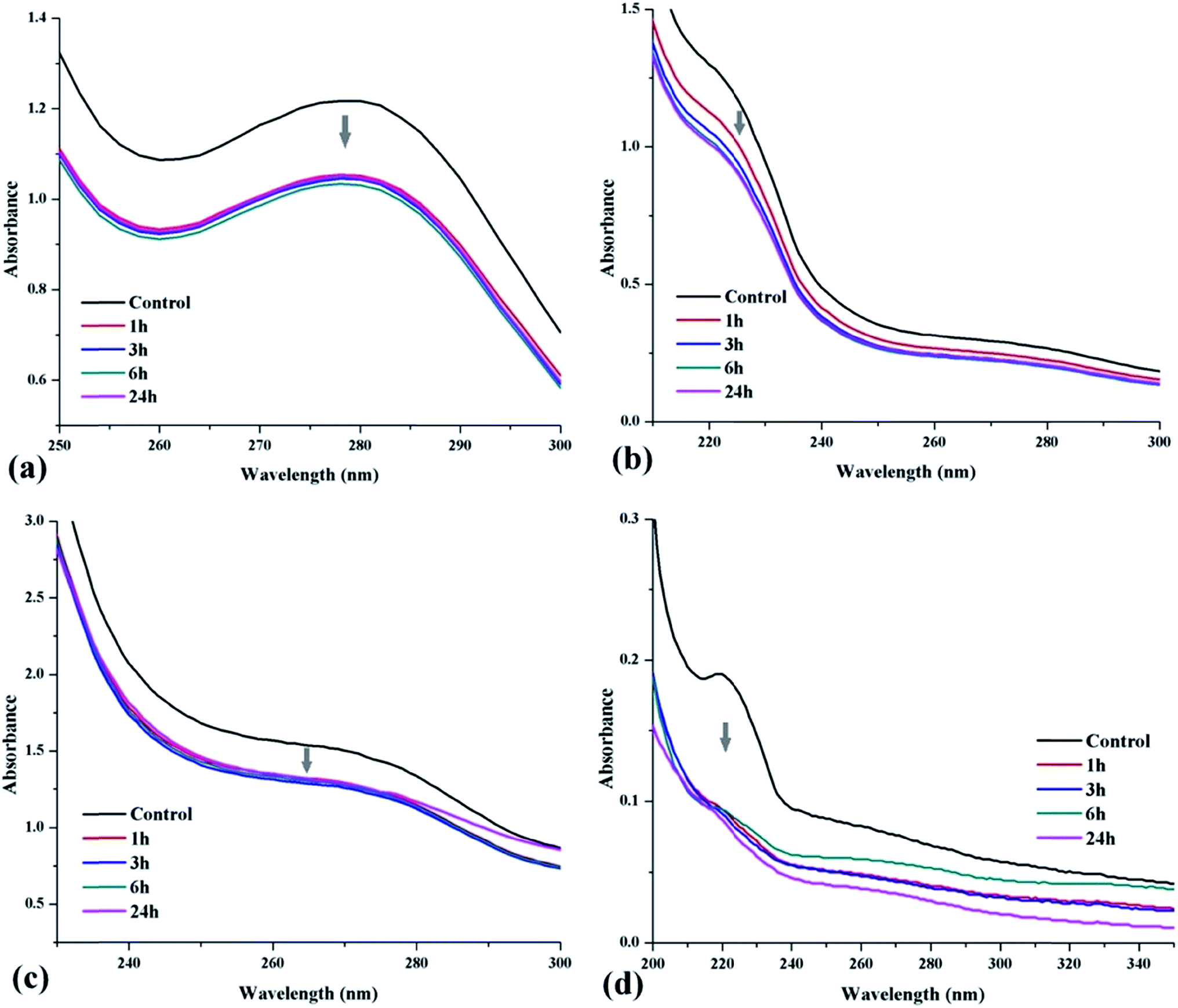 | ||
| Fig. 1 UV-vis absorption spectra of TiO2 dispersions in (a) A. concinna extract, (b) EPS, (c) sludge extract, (d) SDS. | ||
The UV-vis spectra of TiO2 NPs dispersion in A. concinna extract is given in Fig. 1(a). The spectra displayed the decrease in absorbance at 200–300 nm against time during 24 h of incubation. This means that there may be some compounds present in the extract are getting adsorbed on TiO2 NP. Therefore, their concentration decreased in the supernatant and thereby causing decrease in absorbance with passing time. The maximum adsorption took place during initial six hours after that very little, or no further decrease in absorbance was noticed.
In Fig. 1(b), the absorbance pattern of EPS on TiO2 surface is given with respect to time. The decrease in absorbance was observed in the range 200–300 nm. The gradual decrease in absorbance represents the slow adsorption of EPS on NP. The reason may be the higher molecular mass components with steric hindrance (branched protein and polysaccharides) present in EPS.16,21 The UV-vis spectral analysis of TiO2 NP dispersion in sludge extract given in Fig. 1(c) has also shown a considerable decrease in absorbance. The decrease in absorbance noticed within region 225–300 nm. The maximum decrease in absorbance was obtained after first few hours. The UASB sludge contains several dissolved biological and chemical compounds, and that may fall in this range.9,21
For comparative analysis, SDS, an anionic surfactant was also used to disperse the TiO2 NPs. The result of UV-vis absorbance is given in Fig. 1(d). The decrease in absorbance was found within 200–300 nm, and the least absorbance was observed at the end of 24th h. On the basis of difference in initial and final absorbance spectra, it can be said that among all dispersions the Acacia extract showed more adsorption on TiO2 NP surface. The basic reason might be the varied component found in Acacia extract that may lie in an appropriate baseline for the adsorption such as suitable molecular size, functional groups, ionic state, etc. which plays a significant role.
![[double bond, length as m-dash]](https://www.rsc.org/images/entities/char_e001.gif) O stretching vibrations overtone and amines (–NH2, NH) stretching vibrations. A sharp peak at 2361 cm−1 represents the presence of amine groups (
O stretching vibrations overtone and amines (–NH2, NH) stretching vibrations. A sharp peak at 2361 cm−1 represents the presence of amine groups (![[triple bond, length as m-dash]](https://www.rsc.org/images/entities/char_e002.gif) NH+,
NH+, ![[double bond splayed left]](https://www.rsc.org/images/entities/char_e009.gif) NH2+, CNH+). It also suggests the medium stretching vibrations of O–H and N–H bonds. Another major peak obtained at 1599 cm−1 indicate an intense CC stretching vibration conjugated with CO and CC, strong to medium CC stretching vibration from aromatic ring, very strong CN stretching vibration may be from hydrazoketones (1530–1600 cm−1) or anthraquinone type structures that gives very strong CO stretching vibration from 1590 up to 1615 cm−1. The minor peaks near 1118 cm−1 designate the medium range C–H symmetrical deformation vibration and strong C–O stretching vibration from secondary and tertiary alcohol groups. This region shows the presence of carbohydrates. The peak 671 cm−1 corresponds to the aliphatic –COOH group, and amides with medium to broad NH2 deformation vibration. The results clearly illustrate that the above-mentioned chemical groups have been introduced onto the TiO2 NP surface since no such peaks were obtained in control TiO2 spectra. The decrease in TiO2 absorbance peak in the test sample (TiO2 + PE) spectra is due to the NP surface–organic molecule complex formation that may interfere with the absorption properties of TiO2 NP.
NH2+, CNH+). It also suggests the medium stretching vibrations of O–H and N–H bonds. Another major peak obtained at 1599 cm−1 indicate an intense CC stretching vibration conjugated with CO and CC, strong to medium CC stretching vibration from aromatic ring, very strong CN stretching vibration may be from hydrazoketones (1530–1600 cm−1) or anthraquinone type structures that gives very strong CO stretching vibration from 1590 up to 1615 cm−1. The minor peaks near 1118 cm−1 designate the medium range C–H symmetrical deformation vibration and strong C–O stretching vibration from secondary and tertiary alcohol groups. This region shows the presence of carbohydrates. The peak 671 cm−1 corresponds to the aliphatic –COOH group, and amides with medium to broad NH2 deformation vibration. The results clearly illustrate that the above-mentioned chemical groups have been introduced onto the TiO2 NP surface since no such peaks were obtained in control TiO2 spectra. The decrease in TiO2 absorbance peak in the test sample (TiO2 + PE) spectra is due to the NP surface–organic molecule complex formation that may interfere with the absorption properties of TiO2 NP.
 | ||
| Fig. 2 FT-IR spectra of TiO2 NPs surface modified with (a) A. concinna extract (PE), (b) extracellular polymeric substances (EPS), (c) sludge extract (SE), (d) sodium dodecyl sulphate (SDS). | ||
As shown in Fig. 2(b), the spectra of surface modified TiO2 NP show absorption band at 3391 cm−1 which is formed by the stretching vibration of both hydroxyl (carbohydrates) and amino groups (proteins). An strong peak at 2359 cm−1 stand for the presence of groups like NH+, NH2+, CNH+ that usually show absorbance between 2250 and 2700 cm−1. It also suggests the medium stretching vibration in O–H and N–H bonds. Another major peak obtained at 1597 cm−1 suggests strong CC stretching vibration conjugated with CO and CC, strong to medium CC stretching vibration from aromatic ring. The absorption band at 1597 and 1418 cm−1 are related to the stretching vibration of primary amides and RCOO– groups respectively in the presence of proteins. The band at 1122 cm−1 correlated with the C–O stretching vibrations from the carbohydrates and aromatic compounds, various saturated and unsaturated tertiary alcohols. The peak 670 cm−1 corresponds to the aliphatic –COOH group, amides with medium to broad, NH2 deformation vibration. The increase in absorbance of these groups in the test sample (TiO2 + EPS) suggests the presence of mostly proteins on NP surface.
In Fig. 2(c), the region 3407 cm−1 with a high-intensity peak of FT-IR spectrum for treated TiO2 NPs is attributed to the hydroxy and amine groups. An intense peak at 2361 cm−1 stand for the groups like NH+, NH2+, CNH+ that usually exist between 2250 and 2700 cm−1. It also suggests the medium O–H and N–H stretching vibration. Another major peak obtained at 1588 cm−1 suggests strong CC stretching vibration conjugated with CO and CC, strong to medium CC stretching vibration from aromatic ring. The absorption band near 1588 and 1418 cm−1 are related to the stretching vibration of primary amides and RCOO– groups respectively in the presence of proteins. The band at 1116 cm−1 correlated with the C–O stretching vibrations from the carbohydrates, aromatic compounds, and various saturated and unsaturated tertiary alcohols. The peak 673 cm−1 corresponds to the aliphatic –COOH group, amides with medium to broad NH2 deformation vibration. Most of the peaks obtained here resemble from EPS samples suggesting of the origin of EPS from sludge. Moreover, the similar chemical groups have been adsorbed on NPs surface as designated by the presence of concerned peaks in the test sample (TiO2 + SE) spectra.
In Fig. 2(d), the detectable broad transmission band at 3391 cm−1 observed for the surface modified TiO2 NPs are assigned to the hydroxyl group and is concentration dependent. The peak at 2359 cm−1 designates the presence of sulphinic acid with weak O–H stretching vibration. The signal at 1601 cm−1 assigned for acetate salts strong signals and asymmetric –COO− stretching vibration. Other transmission bands at 1421 cm−1 and 1125 cm−1 correspond to R–CH2–S, –CH2 deformation vibration, and R–OC–SR, C–C strong vibration respectively. The region 1125 cm−1 represents sulphinic acid esters (–SO–O–) strong SO stretching vibration. This region also assigned for medium symmetrically SO2 stretching vibration, sulphate ion (SO4−) very strong SO4 stretching vibration. The presence of above mentioned functional groups in all the test sample IR spectra is suggestive of their adsorption on TiO2 NP surface. Since the extracts were rich in various organic and inorganic compounds, the mechanism of TiO2–biomolecule interaction may be of electro-steric type as mentioned elsewhere in the text.
Zeta potential and particle size distribution (PSD) of nanoparticle dispersion
The zeta potential and the thickness of the electrical double layer are the key factors that determine the stability of NP dispersion.22 An increased electrostatic repulsion will result between dispersed NPs on increasing either of them. The thickness of the electrical double layer depends on the ionic strength of the solution and decreases on increasing the ionic strength. The DLVO theory doesn't hold good for very low ionic strength suspensions where the particles remain distantly placed.23 Though, this type of circumstances is rarely found among the samples taken for toxicological studies.Zeta (ζ) potential is a function of the surface charge of dispersed particles. Studies have revealed that the zeta potential of particles depends on several aspects, like the chemical composition of the particle surfaces, the composition of the surrounding solvent, the environmental pH value and ions in the suspension.24 In general, the magnitude of the zeta potential determines the colloidal stability of NP dispersion. In dispersions, the particles having zeta potential near to zero (isoelectric point), are most likely to agglomerate. Particles with high negative or positive values of zeta potential repel each other causing reduced agglomeration. However, the real values required to check re-agglomeration remain controversial, and depend on the solvent, concentration of ions, effective pH, and the chemical entities on the NP surface.25
The Z-average and zeta potential values of TiO2 dispersion in various dispersants are given in Fig. 3(a) and (b). All the dispersions were sonicated, placed on rotary shaker overnight and left standing for the fixed time period, and samples collected periodically from upper layer for the analysis. DLS results are usually expressed in terms of the “Z-average”. It offers a trustworthy assessment of the NPs average size in dispersion. In addition, it is measured without difficulty. Therefore, the Z-average size is considered for presenting particle sizing results by DLS. The initial Z-average value of TiO2 dispersed in PE was found 266.16 ± 6.98 nm that was least among the other dispersions including the dispersion in SDS. However, on comparing the day 15 data, the Z-average of TiO2 NPs in SDS dispersion was found the minimum that is 189.76 nm. The Z-average of TiO2 NP in EPS and SE was comparatively higher than in Acacia extract, even with the higher magnitude of zeta potential. The possible reason could be the presence of higher molecular weight (branched) compounds. Such compounds may attach together the several dispersed NPs present in close vicinity leading to the agglomeration and increase in average size.18 These large size particles eventually settle down due to gravitational effect. The polydispersity index (PDI) obtained during the day 1 analysis was 0.2 to 1.0 for most of the measurements. The higher PDI may be due to the smaller as well as the larger particles that were remained suspended in the dispersion due to the effect of sonication. On the other hand, the day 15 analysis showed lower PDI (0.1–0.3) that was the result of sedimentation of larger particles causing the dispersion more uniform.
 | ||
| Fig. 3 Z-average (a) and zeta potential (b) of TiO2 NP in various dispersants measured on day 1 and day 15. PE = A. concinna extract, EPS = extracellular polymeric substances, SE = sludge extract, SDS = sodium dodecyl sulphate. | ||
The PSD curve of TiO2 NPs in all the dispersions is given in Fig. 4(a)–(d). The PSD of TiO2 NPs in PE (Fig. 4(a)) shows that most of the particle diameter lay in the range 100 to 500 nm on day 1 and day 15 measurement. Similar trend was obtained for TiO2 NPs dispersed in SDS (Fig. 4(d)) where the PSD was 150 to 700 nm for day 1, and 80 to 500 on day 15. The PSD of TiO2 NPs in EPS and SE for day 1 (Fig. 4(b) and (c)) were 175 to 600 nm and 200 to 600 nm respectively. While on day 15, the PSD value remained around 175 to 1000 nm for both the dispersions. The results suggest the better dispersion of TiO2 NPs in PE and SDS as compared to EPS and SE. However, the PSD plots were found monomodal for all dispersions except 15th day PSD of EPS and SE.
 | ||
| Fig. 4 Particle size distribution by intensity (day 1 and day 15) of TiO2 nanoparticles dispersed in (a) plant extract, (b) extracellular polymeric substances, (c) sludge extract, (d) SDS. | ||
Zeta potential values of all dispersions were negative. Such tendency was observed for other nanomaterial suspensions also.26 Moreover, the more negative zeta potential values of TiO2 NP dispersions indicate their substantial stabilities. In case of dispersion in SDS, the higher negative value of zeta potential (−34 to −41 mV) was maintained till the end of day 15. Therefore, the average size of the TiO2 NP was obtained 189.76 nm on day 15. So the TiO2 NPs dispersed in SDS remained stable for a longer period of time. On the other hand, the magnitude of zeta potential in TiO2–biological extract dispersions were below −20 mV that caused them to be unstable comparatively with passing time. The zeta potential distribution (ZPD) of TiO2 NPs in all the dispersions is given in Fig. 5(a)–(d). Exception to the TiO2 NPs dispersed in PE, others showed absolute negative ZPD. Though, the plot obtained were monomodal for all the dispersions.
 | ||
| Fig. 5 Zeta potential distribution (day 1 and day 15) of TiO2 nanoparticles dispersed in (a) plant extract, (b) extracellular polymeric substances, (c) sludge extract, (d) SDS. | ||
The data were analyzed with single factor ANOVA and at level p < 0.05, the Z average and zeta potential among all the dispersions were not significantly different. This result suggests that the average size of TiO2 NPs in all the dispersions were comparable. Though, with passing time, most of the large aggregate settled down in biological dispersions. This behaviour is usual and desirable in order to minimize the toxic effect when the NPs are released in the open environment. However, the chemical surfactants such as SDS cause the NP dispersion to remain stable for an extended period of time.
Dispersion stability analysis
Fig. 6(a) and (b) shows the dispersion state of TiO2 NPs with various dispersants for day 1 and day 15 respectively. In all the dispersions, there was certain stability till the end of day 15. However, in SDS it was found more stable comparatively. On day 15, all the biological dispersions showed sedimentation that can be seen as the increase in transparency through the vials and sediments at the bottom which were the uniform dispersion in day 1 images. This suggests the stability of all the dispersions were comparable during the initial period of analysis, but the SDS kept the TiO2 NPs stable for the longer duration of time. | ||
| Fig. 6 Stability of TiO2 NPs (100 mg L−1) in various dispersants (a) photograph taken on day 1 just after preparation, (b) photograph taken on day 15. | ||
The pericarp of Acacia pod contains a phytochemical called as saponin. Saponin is considered as a non-ionic natural surfactant and is used in cosmetics and pharmaceutical products.20,27 The saponin molecules adsorb on the TiO2 NP surface via hydrophobic interaction and impart a steric stabilization to the dispersion. EPS contains several types of proteins, carbohydrates, humic and fulvic acid that coat the NP surface. An earlier study states that such NP capping can provide them colloidal stability either by electrostatic, steric or electro-steric mechanisms.28 The EPS was used for stabilizing the biosynthesized and chemically synthesized Se NPs via an electro-steric mechanism.29 Kroll et al. also reported the stabilizing ability of EPS for CeO2 and Ag NPs.30 Sludge extract has numerous organic and inorganic compounds dissolve in it that helps to sustain the dispersion stability.31–33 An anionic detergent; SDS coats the NP surface and provides steric stability by increasing the surface charge.34,35
Fig. 7(a)–(d) shows the transmission profiles of TiO2 NPs suspended in various dispersants. The X-axis represents the length of tube in mm while the Y-axis represents the percentage transmission. The transmittance detector received the light which passed through the dispersion at an angle of 180° with respect to the source, while the back scattering detector received the light scattered backwards by the dispersion at an angle of 45°. In this study, the back scattering data was not considered, because at higher concentration the back scattered light may not provide accurate results due to the absorbance of light.36
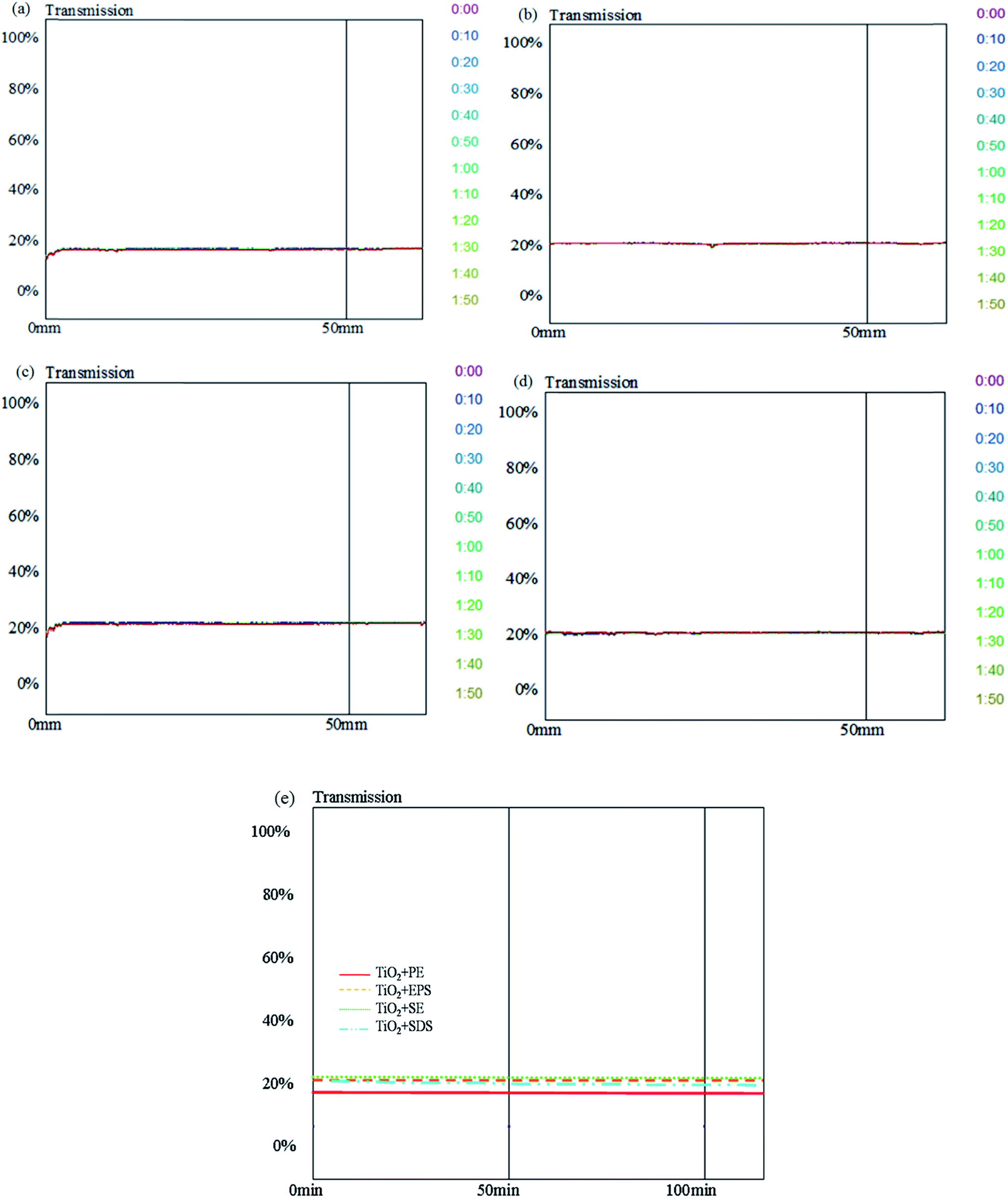 | ||
| Fig. 7 Transmission profile of various dispersions. Dispersion of TiO2 NPs (100 mg L−1) in (a) A. concinna extract, (b) extracellular polymeric substances, (c) sludge extract, (d) SDS, and (e) variation in the mean transmission of various dispersions. These data are represented as a function of time (0:00 to 2:00 h) and of sample height (10 to 80 mm). | ||
The transmitted light flux in % correlated with the density of suspension in the dispersion. The variation of the dispersed NPs between the top and the bottom of the sample vial varies the transmission signal. The faster rise in transmission over the same period of time demonstrates that the samples have been unsteady and have undergone a quicker sedimentation. While, on the other hand, a steady transmission through the length of tube represents good stability. Here the various types of dispersions have shown a similar trend of transmission profile suggesting of adequate stability during the period of observation. The result greatly supports the outcomes of particle size distribution, and zeta potential analysis explained elsewhere in the text. The result also suggests the applicability of biological dispersants for TiO2 NP. A mean transmission profile of various dispersions with respect to time is given in Fig. 7(e). The transmission variation among all the dispersions had the similar behaviour during the period of analysis. The similar slopes in all the dispersions represent similar stability.
Biodegradability of dispersants
Biodegradability is crucial for the NP dispersions when exposed to the open environment. It decides the fate of NP and the level of hazard it may produce after entering into the environment. The Table 3(a) and (b) showing the average COD measured in inoculated dispersions. The dispersions were showing microbial growth within few days of inoculation. Microbial growth was visible only in the dispersions with biological extracts not with the SDS.| (a) | |||
|---|---|---|---|
| Dispersion type | Average COD measured | COD reduction (in %) | |
| Day 1a | Day 15a | ||
| a Mean ± SD (n = 3). | |||
| Acacia extract + TiO2 NP | 8260.44 ± 298.95 | 5530.78 ± 62.49 | 33.04 |
| Sludge extract + TiO2 NP | 159.13 ± 2.23 | 12.72 ± 7.95 | 92.00 |
| EPS + TiO2 NP | 58.2 ± 4.02 | 14.37 ± 1.91 | 75.30 |
| SDS + TiO2 NP | 8800.37 ± 71.63 | 8570.13 ± 210.87 | 02.61 |
| (b) | |||
|---|---|---|---|
| Dispersion type | Average COD measured | COD reduction (in %) | |
| Day 1a | Day 15a | ||
| Acacia extract + TiO2 NP | 7433.97 ± 156.56 | 4670.72 ± 161.46 | 37.17 |
| Sludge extract + TiO2 NP | 448.48 ± 300.03 | 164.59 ± 104.34 | 63.30 |
| EPS + TiO2 NP | 23.51 ± 23.95 | 13.96 ± 5.99 | 40.62 |
| SDS + TiO2 NP | 8457.32 ± 130.84 | 8096.73 ± 83.07 | 04.26 |
In both the aerobic and anaerobic degradation, the maximum COD reduction was found in TiO2 NP dispersed in sludge extract that was 92% and 63% respectively. The COD reduction in EPS dispersion was approximately 75% and 40% in aerobic and anaerobic samples respectively. The higher degree of COD removal in SE and EPS dispersion is due to the suitability of these media for microflora, as the SE and EPS were extracted from sludge itself. Therefore, the microbes readily degraded the SE and EPS. The COD removal for Acacia extract dispersion was approximately 33% and 37% for aerobic and anaerobic samples respectively. The lesser COD removal as compared to SE and EPS may be due to the presence of several antimicrobial compounds in Acacia extract.20
In case of dispersion in a chemical surfactant SDS, the COD removal during the stipulated period was 2% and 4% in aerobic and anaerobic samples respectively. It was the least COD removal among all the dispersants because of the less biodegradability. In such conditions, the NP may remain dispersed for the longer duration, and the surfactant itself would cause considerable damage to the environment. A single factor ANOVA (p < 0.05) was performed to determine the significant difference between the CODs of day 1 and day 15. At level p < 0.05, the result obtained were found significant for both the aerobic and anaerobic degradation.
Conclusions
In present work, the dispersion of TiO2 NPs in various biological originated extracts were studied and compared with that of SDS. All the biological extracts were able to stabilize the TiO2 NP dispersion for a satisfactory period of time and were comparable to chemical surfactant SDS. The aerobic and anaerobic degradation of biological extracts proves their suitability for commercial applications. The biodegradability of such stabilizers ensures the easy removal of NPs from environmental matrices once released after use. The aggregation and settlement of NPs also render them comparatively less toxic to the ecosystem. However, the biological extracts can be further improved in terms of stability. Currently, more research is required to find the applications of biological extracts for commercial products.Compliance with ethical standards
We would like to affirm that the study was carried out at Chemical Engineering Department, Sardar Vallabhbhai National Institute of Technology, Surat, INDIA. Results consist of a part of our ongoing work and have not been published previously and are not under consideration for publication elsewhere. We also affirm the approval by all authors and tacitly or explicitly by the responsible authorities where the work was carried out, and that, if accepted, it will not be published elsewhere in the same form. The authors declare that there is no conflict of interest. No human and/or animal was used as an experimental subject for the present work.Acknowledgements
We thank Chemical Engineering Department, SVNIT, Surat for providing basic infrastructure for work, MHRD for providing fellowship, and Shree Dhanvantary Pharmaceutical Analysis and Research Centre, Surat for analytical facilities.References
- B. V. Derjaguin and L. D. Landau, Acta Physicochim. URSS, 1941, 14, 733 Search PubMed.
- E. J. W. Verwey and J. T. G. Overbeek, Theory of the stability of lyophobic colloids, Elsevier, Amsterdam, 1948 Search PubMed.
- H. Kamiya and M. Iijima, J. Soc. Powder Technol., Jpn., 2009, 46(8), 605 CAS.
- D. B. Warheit, B. R. Laurence, K. L. Reed, D. H. Roach, G. A. M. Reynolds and T. R. Webb, Toxicol. Sci., 2004, 77, 117 CrossRef CAS PubMed.
- A. A. Shvedova, E. R. Kisin, R. Mercer, A. R. Murray, V. J. Johnson and A. I. Potapovich, et al., Am. J. Physiol.: Lung Cell. Mol. Physiol., 2005, 289, 698 CrossRef PubMed.
- B. Nowack and T. D. Bucheli, Environ. Pollut., 2007, 150, 5 CrossRef CAS PubMed.
- I. Bhatt and B. N. Tripathi, Chemosphere, 2011, 82, 308 CrossRef CAS PubMed.
- A. Sarkar, M. Ghosh and P. C. Sil, J. Nanosci. Nanotechnol., 2014, 14, 730 CrossRef CAS PubMed.
- A. K. Mungray and P. Kumar, J. Environ. Manage., 2008, 88, 995 CrossRef CAS PubMed.
- T. M. Sager, D. W. Porter, V. A. Robinson, W. G. Lindsley, D. E. Schwegler-Berry and V. Castranova, Nanotoxicology, 2007, 1(2), 118 CrossRef CAS PubMed.
- Z. Ji, X. Jin, S. George, T. Xia, H. Meng, X. Wang, E. Suarez, H. Zhang, E. M. V. Hoek, H. Godwin, A. E. Nel and J. I. Zink, Environ. Sci. Technol., 2010, 44, 7309 CrossRef CAS PubMed.
- D. Lin, J. Ji, Z. Long, K. Yang and F. Wu, Water Res., 2012, 46, 4477 CrossRef CAS PubMed.
- D. Palomino, C. Yamunake, P. L. Coustumer and S. Stoll, J. Colloid Sci. Biotechnol., 2013, 2(1), 62 CrossRef CAS PubMed.
- A. Maurya, P. Chauhan, A. Mishra and A. K. Pandey, J. Res. Updates Polym. Sci., 2012, 1, 43 CAS.
- C. Chen, Q. Liu, S. Gao, K. Li, H. Xu, Z. Lou, B. Huang and Y. Dai, RSC Adv., 2014, 4, 12098 RSC.
- S. Yang and X. Li, Process Biochem., 2009, 44, 91 CrossRef CAS PubMed.
- F. Rodrigues, D. P. Jaisi and M. Elimelech, Environ. Sci. Technol., 2013, 47, 625 CrossRef PubMed.
- S. H. Othman, S. A. Rashid, T. I. M. Ghazi and N. Abdullah, J. Nanomater., 2012, 718214 Search PubMed.
- W. I. Park, H. S. Kim, S. M. Kwon, Y. H. Hong and H. J. Jin, Carbohydr. Polym., 2009, 77, 457 CrossRef CAS PubMed.
- K. Khanpara, Renuka, V. J. Shukla and C. R. Harisha, J. Curr. Pharm. Res., 2012, 9(1), 6 Search PubMed.
- J. Wingender, T. R. Neu and H. C. Flemming, Microbial extracellular polymeric substances: characterization, structures and function, Springer, Berlin, Heidelberg, 1st edn, 1999 Search PubMed.
- I. D. Morrison and S. Ross, Colloidal dispersions: suspensions, emulsions, and foams. Wiley-Interscience, New York, 2nd edn, 2002 Search PubMed.
- J. Widegren and L. Bergstrom, J. Am. Ceram. Soc., 2002, 85, 523 CrossRef CAS PubMed.
- D. L. Liao, G. S. Wu and B. Q. Liao, Colloids Surf., A, 2009, 348, 270 CrossRef CAS PubMed.
- N. Mandzy, E. Grulke and T. Druffel, Powder Technol., 2005, 160, 121 CrossRef CAS PubMed.
- W. Chen, L. Duan, L. L. Wang and D. Q. Zhu, Environ. Sci. Technol., 2008, 42, 6862 CrossRef CAS.
- C. Schmitt, B. Grassl, G. Lespes, J. Desbrieres, V. Pellerin, S. Reynaud, J. Gigault and V. A. Hackley, Biomacromolecules, 2014, 15(3), 856 CrossRef CAS PubMed.
- A. M. El-Badawy, T. P. Luxton, R. G. Silva, K. G. Scheckel, M. T. Suidan and T. M. Tolaymat, Environ. Sci. Technol., 2010, 44, 1260 CrossRef CAS PubMed.
- R. Jain, R. Hubner, N. Jordan, H. Kramer, S. Weiss, H. Foerstendorf, E. D. van Hullebusch, K. Heim, F. Farges, R. Kacker and P. N. L. Lens, Environ. Sci. Technol., 2015, 49, 1713 CrossRef CAS PubMed.
- A. Kroll, R. Behra, R. Kaegi and L. Sigg, PLoS One, 2014, 9(10), e110709 Search PubMed.
- J. E. Schmidt and B. K. Ahring, Biotechnol. Bioeng., 1996, 49, 229 CrossRef CAS.
- E. Z. Harrison, S. R. Oakes, M. Hysell and A. Hay, Sci. Total Environ., 2006, 367, 481 CrossRef CAS PubMed.
- S. Chelme, P. Fonseca, R. Mercado, N. Alarcon and O. Sanchez, Electron. J. Biotechnol., 2008, 11(3), 1 Search PubMed.
- L. Vaisman, H. D. Wagner and G. Marom, Adv. Colloid Interface Sci., 2006, 128–130, 37 CrossRef CAS PubMed.
- D. L. Liao and B. Q. Liao, J. Photochem. Photobiol., A, 2007, 187, 363 CrossRef CAS PubMed.
- H. S. Kim, W. I. Park, M. Kang and H. J. Jin, J. Phys. Chem. Solids, 2008, 69, 1209 CrossRef CAS PubMed.
| This journal is © The Royal Society of Chemistry 2015 |