DOI:
10.1039/C5RA08639F
(Paper)
RSC Adv., 2015,
5, 47108-47116
4-Nitro-2,1,3-benzoxadiazole derivatives as potential fluorescent sigma receptor probes†
Received
9th May 2015
, Accepted 19th May 2015
First published on 19th May 2015
Abstract
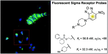
New fluorescent derivatives for σ receptors were designed and synthesized. To achieve this purpose, a 4-nitro-2,1,3-benzoxadiazole fluorescent tag was connected through a piperazine linker to a modified skeleton derived from selected σ receptor agonists or antagonists. Compounds 5g, 7b, 7e and 7g displayed high σ1 affinity and low σ1/σ2 selectivity (Kiσ1 ranging from 31.6 nM to 48.5 nM, Kiσ1/σ2 = 5–18), while compound 5d exhibited high σ2 affinity and selectivity (Kiσ2 = 56.8 nM, Kiσ1 > 5000 nM). Binding affinity studies revealed that compounds 5d, 5g, 7b, 7e and 7g showed no affinity towards several receptors including opioid, dopaminergic, serotonergic, adrenergic, muscarinic, histaminergic, N-methyl-D-aspartate (NMDA), NMDA receptor channel, or dopamine and serotonine transporters. The fluorescent properties, cellular uptake and confocal microscopy studies on 5d suggest a potential use of this probe to further clarify the molecular role of σ2 receptor subtypes in normal and cancer cells.
1. Introduction
A sigma (σ) receptor was initially classified as a subtype of opiate receptors. Subsequent studies revealed that sigma-binding sites are a distinct class of receptors that are located in the central nervous system (CNS), in endocrine tissues, in the liver and kidneys, and in immune system cells.1–4 Early studies using radioligand binding methods and biochemical analyses demonstrated that there are two subtypes of sigma receptors, named σ1 and σ2. They differ from each other in function, pharmacology, and molecular size.5,6
σ1 receptor is involved in the release of neurotransmitters, in particular in the NMDA, serotonergic, dopaminergic and muscarinic neurotransmission, suggesting its potential applications in the treatment of a number of neurological disorders and cognitive disease7 including depression,8 schizophrenia,9 Alzheimer's disease,10–13 frontotemporal lobar degeneration-motor neuron disease14 and drug abuse.15
In recent studies σ1 receptor has been classified as a protein chaperone at the endoplasmatic reticulum (ER)–mitochondrion interface that regulates Ca2+ signaling and cell survival.16
To date, σ2 subtype has not been completely characterized. Although its structure is still unknown, σ2 receives relevant interest related to its overexpression in a wide variety of cancer tissues.17 Therefore, several σ2 ligands are designed for cancer treatment and diagnosis.18–22 Moreover, it was recently proposed that progesterone receptor membrane component 1 (PGMRC1) represents a possible σ2 binding site.23 However, even though PGRMC1 was highly expressed in proliferating tumour cells, its molecular weight (25 kDa)23 is different from the σ2 receptor binding sites observed with [3H]-azido-1,3-di-o-tolylguanidine ([3H]-Az-DTG) photoaffinity labeling (21.5 and/or 18 kDa).6,24
In spite of several studies, there are still major shortcomings related to σ receptors: (i) although σ1 receptor has been cloned and displays a 30% sequence homology with the yeast C8–C7 sterol isomerase,25 it lacks C8–C7 isomerase activity; (ii) moreover, the σ2 receptor has not been cloned, and most of what is known regarding it has been obtained through the use of in vitro receptor binding studies aimed at its pharmacological characterization;26 (iii) finally, the absence of the cloned gene or the purified σ2 receptor protein has also prevented the generation of antibodies to study the subcellular localization of this receptor using the immunohistochemical techniques.27 For these reasons, new fluorescent probes have been synthesised as useful tools to understand and better characterize the biomolecular role of the σ1 and σ2 receptors.28–30
Generally, considering the low molecular weight of selective σ ligands, the introduction of bulky fluorescent probes could provide a loss in receptor affinity. In order to avoid this, new potential σ fluorescent probes, having as typical fluorophore the 4-nitrobenzo-2-oxa-1,3-diazole (NBD), were designed and synthesized.31 This moiety showed reduced steric hindrance and a low impact on the ligand affinity. Moreover, the NBD fluorophore generally showed low emission quantum yield in aqueous solution, but it became highly fluorescent in nonpolar solvents, when it bound to the cell membrane or inserted in a hydrophobic pocket of target protein.28,29
Several N,N′-disubstituted piperazines, like SA-4503 (Fig. 1), and morpholine esters, like PRE-084 (Fig. 1), have been reported as potent and selective σ1 receptor agonist ligands inducing antiamnesic and antidepressive activity,32–34 or able to reduce cocaine-induced toxic effects.15,35–39 Others piperazine (PB28, a σ2 receptors agonist),40,41 piperidine (haloperidol, σ1 antagonist and σ2 agonist),42 and pyrrolidine (AC915, σ1 antagonist)42 derivatives (Fig. 1), showed an antiproliferative activity towards different cancer cell lines.
 |
| Fig. 1 Structures of selected sigma ligands. | |
Thus, starting from the framework of typical sigma ligands, including the above mentioned ones (Fig. 1), we designed and synthesized a new series of piperazine derivatives 5a–g and 7a–g carrying the NBD fluorophore (Schemes 1 and 2), as new potential fluorescent probes for sigma receptors.
 |
| Scheme 1 Synthetic strategy for the preparation of compounds 5a–g. Reagents and conditions: (a) THF, 0 °C, 5 min; (b) SOCl2, CH2Cl2, reflux, 2 h; (c) only for 4a–f, K2CO3/H2O, reflux, 12 h; (d) i-PrOH, reflux, 48 h. | |
 |
| Scheme 2 Synthetic strategy for the preparation of compounds 7a–g. Reagents and conditions: (a) THF, 0 °C, 5 min. | |
2. Results and discussion
2.1. Chemistry
The 4-nitro-2,1,3-benzoxadiazole derivatives (5a–g and 7a–g) were synthesized as outlined in Schemes 1 and 2. Compounds 5a–f were obtained in three steps by reaction of 2-(piperazin-1-yl)ethanol (1) with the commercially available 4-chloro-7-nitrobenzo-2-oxa-1,3-diazole (NBD-Cl). Conversion of alcohol 2 into alkyl chloride 3 with SOCl2, followed by condensation with the potassium salts of the corresponding acids 4a–f (Scheme 1) under refluxing i-PrOH, gave the desired products 5a–f.43,44 On the other hand, 4-(4-chlorophenyl)-4-hydroxypiperidine (4g) reacted directly with 3 to give the desired compound 5g.
Target compounds 7a–g, reported in Scheme 2, were obtained by reaction of suitable N-functionalized piperazines (6a–g) with NBD-Cl according to the literature.45
2.2. Radioligand binding and σ1 and σ2 receptor affinities
The pharmacological results of binding assays for σ receptors were expressed in terms of inhibition constant (Ki) and are reported in Table 1. The ester derivatives 5a and 5e–f displayed irrelevant σ1/σ2 affinity, while compound 5b showed moderate affinity at both σ receptor subtypes. Compound 5c showed negligible σ1 and moderate σ2 affinities, while compound 5d exhibited good σ2 affinity and selectivity (Kiσ2 = 56.8 nM, Kiσ1 > 5000 nM) (Table 1). This result suggests that the replacement of the morpholine ring present in the σ1 selective ligand PRE-084 with the 4-nitro-7-(piperazin-1-yl)-2,1,3-benzoxadiazole moiety, shifted the affinity and selectivity towards the σ2 receptor subtype.
Table 1 Radioligand binding assays
Compound |
Binding constant Ki (nM) ± SEMa |
σ1 |
σ2 |
The value is the mean of three independent experiments, sample in duplicate. Ki has been not obtained and the percentage of radioligand binding inhibition at 10−5 M of tested compound is reported in parentheses. |
5a |
2172 ± 410 |
>10 000 (36%)b |
5b |
355 ± 50 |
278 ± 15 |
5c |
>5000 (23%) |
393 ± 20 |
5d |
>5000 (42%) |
56.8 ± 2 |
5e |
>5000 (24%) |
>10 000 (39%) |
5f |
>5000 (13%) |
>10 000 (28%) |
5g |
41 ± 8 |
211 ± 8 |
7a |
264 ± 40 |
358 ± 15 |
7b |
48.5 ± 9 |
313 ± 25 |
7c |
127 ± 25 |
381 ± 10 |
7d |
944 ± 150 |
>10 000 (44%) |
7e |
32.3 ± 5 |
577 ± 30 |
7f |
526 ± 80 |
4690 ± 150 |
7g |
31.6 ± 3 |
225 ± 25 |
PRE-084 |
33.5 ± 2 |
>10 000 (29%) |
PB 28 |
18.5 ± 2 |
1.0 ± 0.2 |
DTG |
61 ± 5 |
42 ± 4 |
The piperazine derivatives 5g and 7a–g, revealed high or moderate affinity for both σ receptor subtypes. Among these, the greater σ1 affinity is showed by the ligand with a phenethylic substituent 7g (Ki = 31.6 nM). It should be noted that the corresponding benzyl derivative 7f was about 16 fold less potent (Ki = 526 nM). On the other hand, the derivative with cyclohexylmethyl substituent at piperazine moiety showed a reduced σ1 affinity respect to the cyclohexyl substituted one (7c, Ki = 127 nM; 7b, Ki = 48.5 nM). Compounds 5g and 7e, with 4-phenylpiperidine and N-methylpiperidine moiety, respectively, display a σ1 affinity of the same order of 7g (5g, Ki = 41 nM; 7e, Ki = 32.3 nM). However, among these derivatives, compound 7e displays the better selectivity for σ1 receptors. In order to assess the binding profile of compounds 5d, 5g, 7b, 7e and 7g, we investigated their affinities towards different receptors and transporters (Tables S1 and S2†). All tested compounds showed negligible affinity for opioid, dopaminergic (D1, D2, D3, D4.2), serotonergic (5-HT2A, 5-HT2C, 5-HT3, 5-HT4, 5-HT6), adrenergic (α1, α2), muscarinic, histaminergic H1, NMDA, NMDA receptor channel, such as dopamine and serotonine transporters confirming an excellent σ selectivity.
2.3. Permeability experiments
Among the overall synthesized compounds, 5d, 5g, 7b, 7e and 7g, have been tested for their membrane permeability. Apparent permeability (Papp) through a monolayer of Caco-2 cells was assessed in order to define the pharmacokinetic profile.
In this assay, both the fluxes of ligand from basolateral to apical (B → A) and from apical to basolateral (A → B) compartment were determined. The B → A flux describes the passive permeation of tested compound, whereas the A → B flux is representative for active transport P-glycoprotein (P-gp) mediated.
This biological assay permits to establish if a compound is a substrate of P-gp and therefore unable to permeate into CNS or P-gp not transported and consequently brain penetrant. This pharmacokinetic parameter is estimated taking into account both fluxes contributions, (B → A/A → B). Normally, compounds displaying B → A/A → B > 2 are considered P-gp substrates and thus unable to cross BBB, whereas ligands showing B → A/A → B < 2 are defined P-gp not transported and therefore could permeate BBB and meet specific areas of CNS. The five compounds analysed showed a ratio B → A/A → B > 2 (Table 2), thus they can be estimated P-gp substrates.
Table 2 Cell permeability
Comp. |
Papp A → B (nm s−1) ± SEMa |
Papp B → A (nm s−1) ± SEMa |
B → A/A → B |
εb |
λ (nm) |
The value is the mean of three independent determinations, sample in duplicate. ε = molar extinction coefficient measured at 10−5 M at listed λmax for each tested compound. |
5d |
575 ± 78 |
2059 ± 198 |
3.6 |
26 220 |
230 |
5g |
280 ± 21 |
2182 ± 355 |
7.8 |
28 290 |
498 |
7b |
363 ± 57 |
1581 ± 291 |
4.4 |
15 170 |
484 |
7e |
356 ± 18 |
2083 ± 153 |
5.8 |
26 110 |
496 |
7g |
558 ± 30 |
2028 ± 117 |
3.6 |
25 030 |
230 |
In light of these finding, the aforementioned compounds could be useful as diagnostic tools in detecting P-gp mediated chemoresistant tumours in peripheral organs, where a high concentration of σ receptors has been reported.22
2.4. Spectroscopic properties
The absorbance and emission properties of some selected compounds are listed in Table 3. The excitation and emission spectra were registered in organic solvents (EtOH and CHCl3) and in phosphate buffered saline (PBS) at pH 7.4 solutions. The NBD-bearing compounds 5d, 5g, 7b, 7e and 7g displayed absorbance maxima at two different wavelengths (at about 335 and at about 470 nm). After excitation in both these maxima regions, the corresponding emission maximum (λem) was in the range 530–550 nm. Thus, we selected the excitation wavelength (λexc) of 450 nm to avoid autofluorescence phenomena. This was selected in light of potential future studies employing living cells. In the corresponding excitation spectra, the compounds displayed λexc maxima centred in the UVA region (about 330 nm), as expected considering the relative absorbance spectra. All compounds showed an important difference between λexc and λem (Stokes shift). Quantum yields (Φ) were determined in the selected solvents to probe the environment affecting the sensitivity of the final fluorescent ligand. Indeed, the selected fluorophore (NBD) is endowed with environment sensitivity properties, (i.e., low quantum yield in aqueous solution, but high fluorescence in non-polar solvents or when bound to a hydrophobic site). In this view, 5d exhibited about two order lower fluorescence in PBS buffer with respect to that measured in the non-polar organic solvent. For the vast majority of compounds, the highest quantum yields were those recorded in CHCl3: Φ values were generally higher in CHCl3 than in EtOH and several-fold higher than in PBS buffer. The relative polar compound 7b showed a less pronounced increase in Φ values from EtOH to CHCl3 solutions. Molar extinction coefficients (ε) were determined for most of the compounds, with the lowest values often displayed in PBS; low solubility of 5d and 7g in PBS, as well as that of relatively polar 7e in CHCl3, did not allow a reliable evaluation of molar extinction coefficient. On the other hand, solubility limits were sufficient to obtain the emission data.
Table 3 Spectroscopic propertiesa
Comp. |
ΦFL |
Solvent |
λmax1 |
ε1 |
λmax2 |
ε2 |
Fluorescence properties herein reported were evaluated on compounds as free bases, but they were also evaluated on their corresponding hydrochloride salts in EtOH and PBS solutions. nd = not detected. |
5d |
0.142 |
CHCl3 |
323 |
5930 |
454 |
18 476 |
4.47 × 10⁻³ |
EtOH |
332 |
7325 |
472 |
20 808 |
5.93 × 10⁻⁴ |
PBS |
ndb |
nd |
nd |
nd |
5g |
7.46 × 10⁻³ |
CHCl3 |
336 |
8323 |
472 |
29 982 |
2.26 × 10⁻³ |
EtOH |
334 |
8256 |
472 |
24 098 |
1.00 × 10⁻⁴ |
PBS |
351 |
1918 |
486 |
6048 |
7b |
4.05 × 10⁻³ |
CHCl3 |
318 |
5850 |
446 |
19 520 |
3.13 × 10⁻³ |
EtOH |
328 |
6048 |
463 |
17 708 |
1.05 × 10⁻³ |
PBS |
347 |
1412 |
487 |
4925 |
7e |
6.01 × 10⁻³ |
CHCl3 |
nd |
nd |
nd |
nd |
3.24 × 10⁻³ |
EtOH |
335 |
5026 |
471 |
13 188 |
1.34 × 10⁻³ |
PBS |
353 |
7393 |
496 |
30 459 |
7g |
0.100 |
CHCl3 |
336 |
7229 |
472 |
25 599 |
4.05 × 10⁻³ |
EtOH |
334 |
8381 |
473 |
23 670 |
1.63 × 10⁻³ |
PBS |
nd |
nd |
nd |
nd |
2.5. Subcellular localization by confocal microscopy
Both isolated and confluent MCF7 breast cancer cell patches were observed. In both conditions, fluorescence was evenly distributed as punctuated signals throughout the cytoplasm (Fig. 2A and B). At 100 nM, the least dilution tested, the signal was still well distributed, suggesting that compound 5d could be further diluted while keeping its ability to bind to σ2 receptor in vivo (Fig. 3).
 |
| Fig. 2 (A) Microscopic field showing both isolated and confluent MCF-7 cells labelled with compound 5d (300 nM). Arrows point to fluorescence spots at cell periphery. The cells in the red square are further enlarged in (B), which shows orthogonal projections of cells at the top and right sides. | |
 |
| Fig. 3 (A) A clump of MCF-7 cells at the 100 nM dilution. (B) Negative control. | |
The fluorescence signals were sometimes clustered in a paranuclear environment, suggestive of accumulation within membrane-delimited subcellular components, such as ER or lysosomes. This is in agreement with similar observations obtained with living and fixed fluorescent microscopy on the same cells46 as well as on different cell lines, both murine and human.28–30 Moreover, fluorescent aggregates were seen at specific sites in some cell's periphery (Fig. 2A, arrows), suggesting involvement of σ2 receptor with filopodia/lamellipodia system in the course of cell spreading. As expected, nuclei were thoroughly devoid of fluorescent signals, as seen in orthogonal projections of labelled cells (Fig. 2B).
More interestingly, nucleoli were found positive, a finding so far not reported for other σ2-specific ligands. We do not know whether our observation could be due to the high resolution of our pictures or to a different effects of drug uptake by the cells. Vertical sectioning of cells showed no sign of fluorescence within nuclei. On the other hand, being nucleoli, at least partially, membrane-delimited organelles, our observation does not contradict the assumption of co-localization of σ2 receptor with cytomembranes, but further extends the actual knowledge at this regard.
3. Experimental section
3.1. Chemistry
All starting materials were obtained commercially from Aldrich, Inc, and were used without further purification. Flash column chromatography were performed with 60 Å pore size silica gel as the stationary phase (0.040–0.063 mm, 230–400 mesh ASTM, Merck). Melting points were determined in open capillaries on a Büchi 530 apparatus and are uncorrected. Elemental analyses (C, H, N) were performed on a Carlo Erba CHNS-0 EA1106 analyser; the analytical results were within ±0.4% of the theoretical value assuring a purity ≥95%. 1H and 13C NMR spectra were recorded on a Varian Inova 200 MHz using CDCl3 as solvent. The following data were reported: chemical shift (δ) in ppm respect to TMS, multiplicity, and coupling constants in hertz. PBS consisted of sodium phosphate 10 mM at pH 7.4, NaCl 138 mM and KCl 2.7 mM. Chemicals were from Sigma-Aldrich (Milan, Italy) and were used without any further purification.
3.1.1. 2-[4-(7-Nitro-2,1,3-benzoxadiazol-4-yl)-piperazin-1-yl]-ethanol (2). A solution of 4-chloro-7-nitro-2,1,3-benzoxadiazole (NBD-Cl) (2.2 g, 11 mmol) in anhydrous THF (80 mL) was slowly dropped into a solution of 2-piperazin-1-yl-ethanol (1) (5.0 g, 38.4 mmol) in anhydrous THF at 0 °C and the reaction mixture was stirred for 5 min. The solution colour change from orange to red. The solvent was then evaporated in vacuum, the crude was dissolved in the minimum amount of methanol/acetone 1
:
1 and dripped in chloroform (500 mL). The resulting mixture was then washed with water, dried over Na2SO4, the solvent was then evaporated in vacuum, and the residue was purified by flash chromatography (chloroform/ethanol 9
:
1) to give 2 as a red powder (3.0 g, 92% yield). 1H NMR (200 MHz, CDCl3) δ 8.44 (d, 1H, J = 8.8 Hz), 6.32 (d, 1H, J = 9.0 Hz), 4.14 (t, 4H, J = 5.0 Hz), 3.72 (t, 2H, J = 5.4 Hz), 2.8 (t, 2H, J = 5.0 Hz), 2.67 (t, 4H, J = 5.2 Hz). 13C NMR (50 MHz, CDCl3) δ 138.22, 132.04, 125.69, 125.57, 120.08, 102.67, 59.20, 57.95, 52.48, 49.34. Anal. calcd for C12H15N5O4: C, 49.14; H, 5.16; N, 23.88. Found: C, 49.37; H, 5.12; N, 23.79.
3.1.2. 4-[4-(2-Chloroethyl)piperazin-1-yl]-7-nitro-2,1,3-benzoxadiazole (3). SOCl2 (0.2 mL, 309 mg, 2.6 mmol) was added to a solution of compound 2 (200 mg, 0.682 mmol) in CH2Cl2 (10 mL) at 0 °C under magnetic stirring and, after 15 min, the mixture was refluxed for 2 h and successively stirred overnight at room temperature. The solvent was evaporated in vacuum and the crude was washed several times with ether to remove the excess of SOCl2 to give 3 as an orange powder (200 mg, 94% yield). 1H NMR (200 MHz, CDCl3) δ 8.57 (d, 1H, J = 9.0 Hz), 6.22 (d, 1H, J = 9.0 Hz), 4.07 (t, 4H, J = 4.8 Hz), 3.58 (t, 2H, J = 6.6 Hz), 2.78 (t, 2H, J = 6.8 Hz), 2.74 (t, 4H, J = 5.2 Hz). 13C NMR (50 MHz, CDCl3) δ 144.96, 144.58, 135.83, 135.11, 123.02, 102.52, 59.08, 52.42, 49.23, 40.77. Anal. calcd for C12H14ClN5O3: C, 46.24; H, 4.53; N, 22.47. Found: C, 46.08; H, 4.55; N, 22.41.
3.2. Preparation of potassium salt of acids 4a–f
To an aqueous solution of K2CO3 at 120 °C was added an aqueous solution of the corresponding acid 4a–f (ratio 1
:
1) and the reaction mixture was refluxed for 12 h until the solution become clear. The water was evaporated in vacuum and the crude was washed with ether and MeOH several times. The product was stored under argon at room temperature.
3.3. General procedure for the synthesis of compounds 5a–g
An isopropanolic solution of the potassium salt of acid 4a–f or compound 4g (3.2 mmol) was added to an heated solution of 3 (200 mg, 0.642 mmol) in i-PrOH (100 mL). The mixture changed colours from orange to red and was thus refluxed for 48 h. The solvent was evaporated in vacuum and the crude was dissolved in CH2Cl2 (20 mL). The solution was extracted with an 1 M aqueous solution of NaHCO3, dried over Na2SO4, the solvent evaporated under reduced pressure and the residue was purified by flash chromatography using CHCl3 as eluent. The purified compounds 5a–g were converted into the corresponding chlorohydrates by treatment with an anhydrous solution of hydrogen chloride in diethyl ether.
3.3.1. 2-[4-(7-Nitro-2,1,3-benzoxadiazol-4-yl)piperazin-1-yl]ethyl 2-methyl-2-phenylpropanoate (5a). 246 mg, 72% yield, orange solid: mp 214–217 °C. 1H NMR (200 MHz, CDCl3) δ 8.41 (d, 1H, J = 7.4 Hz), 7.41–7.23 (m, 5H), 6.24 (d, 1H, J = 9.0 Hz), 4.24 (t, 2H, J = 5.2 Hz), 3.95 (t, 4H, J = 5.2 Hz), 2.62 (t, 2H, J = 5.2 Hz), 2.54 (t, 4H, J = 5.0 Hz), 1.60 (s, 6H). 13C NMR (50 MHz, CDCl3) δ 170.50, 141.04, 138.22, 132.04, 128.96, 128.06, 127.50, 126.74, 125.57, 120.08, 85.16, 63.50, 59.06, 56.73, 50.34, 45.34, 27.77. Anal. calcd for C22H25N5O5·HCl: C, 55.52; H, 5.51; N, 14.72. Found: C, 55.66; H, 5.52; N, 14.67.
3.3.2. 2-[4-(7-Nitro-2,1,3-benzoxadiazol-4-yl)piperazin-1-yl]ethyl 2-(4-chlorophenyl)-2-methylpropanoate (5b). 320 mg, 80% yield, orange solid: mp 211–215 °C. 1H NMR (200 MHz, CDCl3) δ 8.39 (d, 1H, J = 9.0 Hz), 7.45–7.18 (m, 4H), 6.27 (d, 1H, J = 9.0 Hz), 4.24 (t, 2H, J = 5.4 Hz), 3.99–3.95 (m, 4H), 2.64 (t, 2H, J = 5.4 Hz), 2.60–2.55 (m, 4H), 1.59, (s, 6H). 13C NMR (50 MHz, CDCl3) δ 174.97, 144.78, 144.69, 144.58, 143.02, 136.22, 131.52, 128.29, 127.91, 123.00, 104.83, 59.73, 53.90, 50.49, 46.00, 45.76, 25.98. Anal. calcd for C22H24ClN5O5·HCl: C, 51.77; H, 4.94; N, 13.72. Found: C, 51.56; H, 4.92; N, 13.78.
3.3.3. 2-[4-(7-Nitro-2,1,3-benzoxadiazol-4-yl)piperazin-1-yl]ethyl (3,4-dichlorophenyl)acetate (5c). 294 mg, 79% yield, orange solid: mp 215–218 °C. 1H NMR (200 MHz, CDCl3) δ 8.43 (d, 1H, J = 9.0 Hz), 7.42 (dd, 1H, J = 1.2 J = 6.4 Hz), 7.26 (d, 1H, J = 0.5 Hz), 7.14 (dd, 1H, J = 1.2, 2.4 Hz), 6.29 (d, 1H, J = 9.0 Hz), 4.29 (t, 2H, J = 5.4 Hz), 4.03 (t, 4H, J = 5.0 Hz), 3.62 (s, 2H), 2.70 (m, 6H). 13C NMR (50 MHz, CDCl3) δ 167.29, 138.22, 136.22, 133.29, 132.54, 132.53, 131.26, 129.66, 125.69, 125.57, 120.08, 85.16, 62.49, 59.06, 56.73, 45.34, 39.76 Anal. calcd for C20H19Cl2N5O5·HCl: C, 46.48; H, 3.90; N, 13.55. Found: C, 46.36; H, 3.92; 13.50.
3.3.4. 2-[4-(7-Nitro-2,1,3-benzoxadiazol-4-yl)piperazin-1-yl]ethyl 2-phenylcycloesancarbossilate (5d). 251 mg, 68% yield, orange solid: mp 225–228 °C. 1H NMR (200 MHz, CDCl3) δ 8.38 (d, 1H, J = 7.6 Hz), 7.45–7.18 (m, 5H), 6.23 (d, 1H, J = 9.0 Hz), 4.24 (t, 2H, J = 5.4 Hz), 3.94 (t, 4H, J = 5 Hz), 2.62 (t, 2H, J = 5.0 Hz), 2.53 (t, 4H, J = 4.8 Hz), 1.80–1.25 (m, 10H). 13C NMR (50 MHz, CDCl3) δ 174.91, 145.03, 144.72, 144.70, 143.59, 135.12, 128.36, 126.74, 125.98, 123.20, 102.36, 61.79, 56.11, 52.42, 50.70, 49.32, 34.47, 25.50, 23.52. Anal. calcd for C25H29N5O5·HCl: C, 58.19; H, 5.86; N, 13.57. Found: C, 57.97; H, 5.84; N, 13.57.
3.3.5. 2-[4-(7-Nitro-2,1,3-benzoxadiazol-4-yl)piperazin-1-yl]ethyl naphthalen-1-ylacetate (5e). 294 mg, 82% yield, orange solid: mp 196–202 °C. 1H NMR (200 MHz, CDCl3) δ 8.42 (d, 1H, J = 9.0 Hz), 8.07 (m, 1H), 7.82 (m, 1H), 7.48 (m, 4H), 7.25 (s, 1H), 6.15 (d, 1H, J = 9.0 Hz), 4.25 (t, 2H, J = 5.0 Hz), 4.10 (s, 2H), 3.73 (t, 4H, J = 4.5 Hz), 2.57 (t, 2H, J = 4.8 Hz), 2.36 (t, 4H, J = 4.8 Hz). 13C NMR (50 MHz, CDCl3) δ 174.07, 171.21, 144.73, 138.00, 137.23, 135.06, 133.75, 132.08, 130.54, 128.73, 128.17, 128.06, 126.36, 125.84, 125.53, 123.88, 102.32, 62.19, 56.07, 52.36, 49.09, 39.47. Anal. calcd for C24H23N5O5·HCl: C, 57.89; H, 4.86; N, 14.06. Found: C, 57.64; H, 4.85; N, 14.12.
3.3.6. 2-[4-(7-Nitro-2,1,3-benzoxadiazol-4-yl)piperazin-1-yl]ethyl naphthalen-2-ylacetate (5f). 305 mg, 85% yield, orange solid: mp 210–214 °C. 1H NMR (200 MHz, CDCl3) δ 8.38 (d, 1H, J = 8.0 Hz), 7.79 (m, 3H), 7.45 (m, 3H), 6.03 (d, 1H, J = 9.0 Hz), 5.30 (s, 2H), 4.29 (t, 2H, J = 5.4 Hz), 3.78 (m, 6H), 2.66 (t, 2H, J = 5.2 Hz), 2.52 (t, 4H, J = 5.2 Hz). 13C NMR (50 MHz, CDCl3) δ 171.24, 169.11, 144.71, 141.10, 139.22, 135.02, 133.33, 132.37, 131.50, 128.21, 128.02, 127.64, 127.55, 127.43, 126.31, 125.93, 102.38, 62.23, 56.08, 52.54, 49.06, 41.78. Anal. calcd for C24H23N5O5·HCl: C, 57.89; H, 4.86; N, 14.06. Found: C, 57.75; H, 4.85; N, 14.11.
3.3.7. 4-(4-Chlorophenyl)-1-{2-[4-(7-nitro-2,1,3-benzoxadiazol-4-yl)piperazin-1-yl]ethyl}-piperidin-4-ol (5g). Directly starting from 4g; 233 mg, 75% yield, orange solid: mp 190–194 °C. 1H NMR (200 MHz, CDCl3) δ 8.41 (d, 1H, J = 9.0 Hz), 7.47–7.27 (m, 4H), 6.31 (d, 1H, J = 9.0 Hz), 4.13 (t, 4H, J = 4.2 Hz), 2.86–2.55 (m, 12H), 2.14–1.70 (m, 5H). 13C NMR (50 MHz, CDCl3) δ 149.05, 145.25, 144.72, 136.25, 130.74, 128.30, 127.69, 126.80, 121.00, 103.47, 69.34, 58.77, 55.48, 53.33, 50.09, 49.58, 38.48. Anal. calcd for C23H27ClN6O4·HCl: C, 52.78; H, 5.39; N, 16.06. Found: C, 52.97; H, 5.40; N, 15.99.
3.4. General procedure for the synthesis of compounds 7a–g
A solution of NBD-Cl (0.4136 g, 2.07 mmol) in anhydrous THF was added to a solution of substituted piperazine 6a–g (1.727 mmol) in anhydrous THF at 0 °C and the reaction mixture was stirred for 5 min. The solution colour change from orange to red. After the solvent evaporation the crude was dissolved in CHCl3, extracted with water, dried over Na2SO4 and, after the evaporation of the solvent under reduced pressure the residue was purified by flash chromatography (CHCl3/EtOH 9
:
1).
3.4.1. 4-(4-Butylpiperazin-1-yl)-7-nitro-2,1,3-benzoxadiazole (7a). 468 mg, 83% yield, orange solid: mp 250–255 °C. 1H NMR (200 MHz, CDCl3) δ 8.41 (d, 1H, J = 9.0 Hz), 6.30 (d, 1H, J = 9.0 Hz), 4.13 (t, 4H, J = 5.2 Hz), 2.69 (t, 4H, J = 5.2 Hz), 2.43 (t, 2H, J = 7.8 Hz), 1.60–1.25 (m, 4H), 0.95 (t, 3H, J = 7.2 Hz). 13C NMR (50 MHz, CDCl3) δ 166.41, 145.16, 144.81, 135.15, 129.97, 102.39, 57.69, 52.74, 49.45, 28.87, 20.56, 13.98. Anal. calcd for C14H19N5O3: C, 55.07; H, 6.27; N, 22.94. Found: C, 55.31; H, 6.29; N, 22.87.
3.4.2. 4-(4-Cyclohexylpiperazin-1-yl)-7-nitro-2,1,3-benzoxadiazole (7b). 415 mg, 72% yield, red solid: mp 240–246 °C. 1H NMR (200 MHz, CDCl3) δ 8.35 (d, 1H, J = 9.0 Hz), 6.24 (d, 1H, J = 9.0 Hz), 4.07 (t, 4H, J = 5.0 Hz), 2.78 (t, 4H, J = 5.0 Hz), 1.84–1.03 (m, 10H). 13C NMR (50 MHz, CDCl3) δ 145.14, 144.81, 144.76, 135.21, 102.24, 96.99, 63.32, 49.96, 48.68, 28.86, 26.12, 25.66. Anal. calcd for C16H21N5O3: C, 57.99; H, 6.39; N, 21.13. Found: C, 57.76; H, 6.37; N, 21.18.
3.4.3. 4-[4-(Cyclohexylmethyl)piperazin-1-yl]-7-nitro-2,1,3-benzoxadiazole (7c). 412 mg, 77% yield, orange solid: mp 230–237 °C. 1H NMR (200 MHz, CDCl3) δ 8.42 (d, 1H, J = 9.0 Hz), 6.29 (d, 1H, J = 9.0 Hz), 4.12 (t, 4H, J = 5.0 Hz), 2.65 (t, 4H, J = 5.0 Hz), 2.21 (d, 2H, J = 7.2 Hz), 1.83–0.82 (m, 11H). 13C NMR (50 MHz, CDCl3) δ 145.18, 144.79, 135.80, 135.21, 123.13, 102.30, 65.09, 53.12, 49.48, 34.94, 31.69, 26.68, 26.02. Anal. calcd for C17H23N5O3: C, 59.12; H, 6.71; N, 20.28. Found: C, 58.93; H, 6.72; N, 20.53.
3.4.4. 4-Nitro-7-[4-(tetrahydrofuran-2-ylmethyl)piperazin-1-yl]-2,1,3-benzoxadiazole (7d). 490 mg, 81% yield, orange solid: mp 223–227 °C. 1H NMR (200 MHz, CDCl3) δ 8.42 (d, 1H, J = 8.8 Hz), 6.30 (d, 1H, J = 9.2 Hz), 4.16–3.73 (m, 7H), 2.80 (t, 4H, J = 5.2 Hz), 2.58–2.47 (m, 2H), 2.11–1.47 (m, 4H). 13C NMR (50 MHz, CDCl3) δ 171.41, 145.14, 144.78, 137.20, 135.18, 102.37, 76.47, 68.31, 62.81, 53.23, 49.35, 30.18, 25.36. Anal. calcd for C15H19N5O4: C, 54.05; H, 5.75; N, 21.01. Found: C, 53.81; H, 5.74; N, 21.11.
3.4.5. 4-[4-(1-Methylpiperidin-4-yl)piperazin-1-yl]-7-nitro-2,1,3-benzoxadiazole (7e). 400 mg, 70% yield, orange solid: mp 280–284 °C. 1H NMR (200 MHz, CDCl3) δ 8.42 (d, 1H, J = 9.0 Hz), 6.30 (d, 1H, J = 9.0 Hz), 4.12 (t, 4H, J = 4.8 Hz), 2.98–2.79 (m, 7H), 2.30 (s, 3H), 2.05–1.60 (m, 6H). 13C NMR (50 MHz, CDCl3) δ 175.00, 162.20, 149.70, 145.98, 135.41, 102.56, 61.49, 55.39, 50.03, 49.01, 46.28, 28.29. Anal. calcd for C16H22N6O3: C, 55.48; H, 6.40; N, 24.26. Found: C, 55.75; H, 6.42; N, 24.21.
3.4.6. 4-(4-Benzylpiperazin-1-yl)-7-nitro-2,1,3-benzoxadiazole (7f). 430 mg, 73% yield, red solid: mp 176–180 °C. 1H NMR (200 MHz, CDCl3) δ 8.41 (d, 1H, J = 8.8 Hz), 7.36–7.23 (m, 5H), 6.28 (d, 1H, J = 9.2 Hz), 4.12 (t, 4H, J = 5.0 Hz), 3.61 (s, 2H), 2.71 (t, 4H, J = 5.0 Hz). 13C NMR (50 MHz, CDCl3) δ 145.00, 144.12, 137.15, 130.84, 128.10, 127.91, 125.69, 125.57, 120.08, 102.08, 62.70, 59.20, 46.69. Anal. calcd for C17H17N5O3: C, 60.17; H, 5.05; N, 20.64. Found: C, 59.94; H, 5.03; N, 20.69.
3.4.7. 4-Nitro-7-[4-(2-phenethyl)piperazin-1-yl]-2,1,3-benzoxadiazole (7g). 395 mg, 71% yield, red solid: mp 193–197 °C. 1H NMR (200 MHz, CDCl3) δ 8.42 (d, 1H, J = 9.0 Hz), 7.35–7.22 (m, 5H), 6.31 (d, 1H, J = 9.0 Hz), 4.15 (t, 4H, J = 5.0 Hz), 2.91–2.68 (m, 8H). 13C NMR (50 MHz, CDCl3) δ 145.22, 144.01, 136.80, 129.07, 128.63, 126.05, 125.69, 125.57, 120.08, 102.08, 59.05, 58.20, 45.69, 34.16. Anal. calcd for C18H19N5O3: C, 61.18; H, 5.42; N, 19.82. Found: C, 61.43; H, 5.44; N, 19.78.
3.5. Cell permeability experiments
3.5.1. Preparation of Caco-2 monolayer. This procedure has been previously reported.47 Briefly, Caco-2 cells were harvested with trypsin–EDTA and seeded onto MultiScreen Caco-2 assay system at a density of 10
000 cells per well. The culture medium was replaced every 48 h for the first 6 days and every 24 h thereafter, and after 21 days in culture, the Caco-2 monolayer was utilized for the permeability experiments. The transepitelial electrical resistance (TEER) of the monolayers was measured daily before and after the experiment using a epithelial volt–ohm meter (Millicell®-ERS; Millipore, Billerica, MA). Generally, TEER values obtained are greater than 1000× for a 21 day culture.
3.5.2. Drug transport experiment. Papp A → B and Papp B → A of drugs were measured at 120 min and at various drugs concentrations (1–100 μM).48 drugs were dissolved in Hank's balanced salt solution (HBSS, pH 7.4) and sterile filtered. After 21 days of cell growth, the medium was removed from filter wells and from the receiver plate. The filter wells were filled with 75 μL of fresh HBSS buffer and the receiver plate with 250 μL per well of the same buffer. This procedure was repeated twice, and the plates were incubated at 37 °C for 30 min. After incubation time, the HBSS buffer was removed and drug solutions added to the filter well (75 μL). HBSS without the drug was added to the receiver plate (250 μL). The plates were incubated at 37 °C for 120 min. After incubation time, samples were removed from the apical (filter well) and basolateral (receiver plate) side of the monolayer and then were stored in a freezer (−20 °C) for pending analysis.The concentration of compounds was analysed using UV-Vis spectroscopy.
The Papp, in units of nm s−1, was calculated using the following equation:
where
VA is the volume (in mL) in the acceptor well; area is the surface area of the membrane (0.11 cm
2 of the well); time is the total transport time in seconds (7200 s); [Drug]
acceptor is the concentration of the drug measured by UV spectroscopy; [Drug]
initial is the initial drug concentration (1 × 10
−4 M) in the apical or basolateral wells.
3.6. Absorption and emission spectroscopy
Emission spectra of compounds were determined in EtOH, CHCl3 and in PBS solutions. In all experiments, the excitation and the emission bandpass was set at 5 nm (S/R mode; response time 0.5 s). The emission spectra were obtained from about 300 to 700 nm, with excitation set at the appropriate excitation wavelength. Fluorescence quantum yields were calculated with respect to quinine sulphate in 0.5 M H2SO4 as a standard (ΦFL = 0.546).49 Solutions of both the sample and the reference were prepared from original solutions diluted with the appropriate solvent so that absorbance was below 0.2 at the same excitation wavelength (356 nm). Fluorescence measurements were carried out for each solution with the same instrument parameters, and the fluorescence spectra were corrected for instrumental response before integration. The quantum yield for each sample was calculated according the following equation:
ΦS = ΦQ(AQ/AS)(FS/FQ)(ηS/ηQ)2 |
where Φ is the emission quantum yield, A is the absorbance at the excitation wavelength, F is the area under the corrected emission curve, η is the refractive index of the solvent (1.34, 1.36 and 1.44 for 0.5 M H2SO4, EtOH and CHCl3, respectively) for the sample (S) and the standard (Q). Absorption spectra were recorded with a Beckman DU 650 spectrophotometer, and fluorescence spectra were obtained with a Yvon Jobin Spex Fluorolog-2 (model F-111) spectrofluorimeter. Molar extinction coefficients (ε) of soluble sample dissolved in the various solvents were determined. ε values were determined by fitting the Beer's law: A = εCl, where (A) is the absorbance at the λexc; (C) is the molar concentration of the sample, and (l) was the optical path length (l = 1 cm) of standard quartz cuvettes. Triplicates of measurements were always made.
3.7. Radioligand binding assays
σ1 and σ2 receptor binding experiments were carried out with [3H]-1,3-di-o-tolylguanidine ([3H]-DTG) (30 Ci mmol−1) and (+)-[3H]-pentazocine (34 Ci mmol−1). These were purchased from Perkin-Elmer Life Sciences (Zavantem, Belgium). [3H]-DTG and unlabeled DTG were purchased from Tocris Cookson Ltd., UK. (+)-Pentazocine was obtained from Sigma-Aldrich (Milan, Italy). Male Dunkin-Hartley guinea pigs and Wistar Hannover rats (250–300 g) were from Harlan, Italy. The specific radioligands and tissue sources were respectively:
(a) σ1 receptor, (+)-[3H]-pentazocine, (2S,6R,11R)-6,11-dimethyl-3-(3-methylbut-2-en-1-yl)-1,2,3,4,5,6-hexahydro-2,6-methano-3-benzazocin-8-ol, guinea pig brain membranes without cerebellum; nonspecific binding was determined in the presence of 1 μM (+)-pentazocine.
(b) σ2 receptor, [3H]-DTG in the presence of 1 μM (+)-pentazocine to mask σ1 receptors, rat liver membranes. Nonspecific binding was determined in the presence of 10 μM DTG.
Concentrations required to inhibit 50% of radioligand specific binding (IC50) were determined by using six-nine different concentrations of the drug studied in two or three experiments with samples in duplicate. Scatchard parameters (Kd and Bmax) and apparent inhibition constants (Ki) values were determined by nonlinear curve fitting by using the Prism, version 3.0, GraphPad software.
Binding affinities towards the target receptors and transporters listed in Table S2† were achieved as previously reported.50 Briefly, different rat brain regions were dissected to assess the following binding assays; rat striatum for D1 and D2 receptors and DAT; rat olfactory tubercle for D3 receptors; rat cortex for muscarinic, 5-HT2A, 5-HT3, α1, α2, H1, and NMDA receptors and SERT; guinea pig cortex for 5-HT4 receptors. 5-HT2C binding assays were carried out on pig brains (frontal cerebral cortex) obtained from a local slaughterhouse and promptly placed on ice until use. Binding experiments on 5-HT6 receptors were performed using a rat-cloned serotonin receptor subtype 6 in HEK-293 cells, as reported by Boess et al.51
Tissue preparation steps and binding assays were performed according to Gobbi et al.52 and Mennini et al.53 After incubation the samples were filtered through Whatman GF/B or GF/C glass fiber filters using a Brandel apparatus (model M-48R) or Millipore filter apparatus. The filters were washed with ice-cold buffer (3 × 4 mL). Radioactivity was counted in 4 mL of “Ultima Gold MV” or Filter Count Cocktail (Packard) in a DSA 1409 (Wallac) or 1414 Winspectral Perkin-Elmer Wallac liquid scintillation counter. For inhibition experiments, the drugs were added to the binding mixture at seven to nine different concentrations. Total opioid receptor binding assays were performed using [3H]-naloxone (55.5 Ci mM−1; Kd = 6.6 ± 0.7 nM; n = 3), and rat brain membranes were prepared as previously reported.54 Nonspecific binding was assessed in the presence of 10 μM of unlabeled naloxone. Inhibition constants (Ki values) were calculated using the “Allfit” program or the EBDA/LIGAND program purchased from Elsevier/Biosoft.
3.8. Confocal microscopy
3.8.1. Cell culture and analysis. MCF7 cells were grown in Dulbecco's MEM supplemented with 10% fetal calf serum, 2 mM glutamine, 100 units per mL penicillin, and 100 μg mL−1 streptomycin, in a humidified incubator at 37 °C in 5% CO2. About 3000 cells were seeded in each well of a 16-wells glass Chamberslide (Lab-Tek, Thermo Scientific Nunc). After 24 hours, the medium was replaced with fresh complete medium containing serial dilutions of 5d from a 20 mM stock solution in DMSO, added in duplicate to the wells from 1 mM to 100 nM. Some wells were kept aside for negative control. The incubation time was limited to one hours in order to keep cells alive, since it is well known σ2 receptor agonists may lead to cell death by apoptosis.30 Medium was then withdrawn, wells washed twice with prewarmed PBS and fixed with 4% formaldehyde in PBS for 2 hour, then washed in PBS two more times. Before microscopic examination, the well strips, with their silicone gasket, were teared from the glass slide; fixed cells were again PBS washed, briefly air dried and mounted in DAPI-containing antifade (Vectashield Mounting Medium, Vector Labs, Peterborough, UK). Several fields, including both isolated and confluent cells, were observed in a Zeiss LSM 700 confocal microscope at 40× and 60× magnifications; optical sections (0.25 μm) were taken when appropriate.
4. Conclusions
A new series of 4-nitro-2,1,3-benzoxadiazole derivatives, as potential fluorescent σ receptor ligands, were synthesized. Binding results displayed that compounds 5d, 7e and 7g showed high sigma receptor affinity as well as a different selectivity feature. Cell permeability evaluation led to address these compounds as useful fluorescent in vitro probes for detecting peripheral chemoresistant tumors where sigma receptors are overexpressed.
Indeed, the fluorescent properties of σ2 selective ligand 5d and preliminary confocal microscopy analysis on MCF7 breast cancer cells, provide evidence that this new ligand represent a useful probe to characterize the function and molecular mechanisms in which this enigmatic receptors are involved.
Acknowledgements
A free academic license to OpenEye Scientific Software and ChemAxon for their suites of programs is gratefully acknowledged.
Notes and references
- T. P. Su, J. Pharmacol. Exp. Ther., 1982, 223, 284–290 CAS.
- D. B. Vaupel, Eur. J. Pharmacol., 1983, 92, 269–274 CrossRef CAS.
- G. A. Young and N. Khazan, Neuropharmacology, 1984, 23, 1161–1165 CrossRef CAS.
- I. P. Berzetei-Gurske and L. Toll, Eur. J. Pharmacol., 1992, 212, 283–286 CrossRef CAS.
- Y. Itzhak and I. Stein, Life Sci., 1990, 47, 1073–1081 CrossRef CAS.
- S. B. Hellewell and W. D. Bowen, Brain Res., 1990, 527, 244–253 CrossRef CAS.
- V. Antonini, O. Prezzavento, M. Coradazzi, A. Marrazzo, S. Ronsisvalle, E. Arena and G. Leanza, J. Neurochem., 2009, 109, 744–754 CrossRef CAS PubMed.
- E. J. Cobos, J. M. Entrena, F. R. Nieto, C. M. Cendan and E. Del Pozo, Curr. Neuropharmacol., 2008, 6, 344–366 CrossRef CAS PubMed.
- G. Skuza and Z. Rogoz, Pharmacol. Rep., 2006, 58, 626–635 CAS.
- V. Antonini, A. Marrazzo, G. Kleiner, M. Coradazzi, S. Ronsisvalle, O. Prezzavento, G. Ronsisvalle and G. Leanza, J. Alzheimer's Dis., 2011, 24, 569–586 CAS.
- K. L. Jansen, R. L. Faull, P. Storey and R. A. Leslie, Brain Res., 1993, 623, 299–302 CrossRef CAS.
- M. Mishina, K. Ishiwata, K. Ishii, S. Kitamura, Y. Kimura, K. Kawamura, K. Oda, T. Sasaki, O. Sakayori, M. Hamamoto, S. Kobayashi and Y. Katayama, Acta Neurol. Scand., 2005, 112, 103–107 CrossRef CAS PubMed.
- M. Mishina, M. Ohyama, K. Ishii, S. Kitamura, Y. Kimura, K. Oda, K. Kawamura, T. Sasaki, S. Kobayashi, Y. Katayama and K. Ishiwata, Ann. Nucl. Med., 2008, 22, 151–156 CrossRef CAS PubMed.
- A. A. Luty, J. B. Kwok, C. Dobson-Stone, C. T. Loy, K. G. Coupland, H. Karlstrom, T. Sobow, J. Tchorzewska, A. Maruszak, M. Barcikowska, P. K. Panegyres, C. Zekanowski, W. S. Brooks, K. L. Williams, I. P. Blair, K. A. Mather, P. S. Sachdev, G. M. Halliday and P. R. Schofield, Ann. Neurol., 2010, 68, 639–649 CrossRef CAS PubMed.
- R. R. Matsumoto, F. H. Potelleret, A. Mack, B. Pouw, Y. Zhang and W. D. Bowen, Pharmacol., Biochem. Behav., 2004, 77, 775–781 CrossRef CAS PubMed.
- T. P. Su, T. Hayashi, T. Maurice, S. Buch and A. E. Ruoho, Trends Pharmacol. Sci., 2010, 31, 557–566 CrossRef CAS PubMed.
- W. T. Bem, G. E. Thomas, J. Y. Mamone, S. M. Homan, B. K. Levy, F. E. Johnson and C. J. Coscia, Cancer Res., 1991, 51, 6558–6562 CAS.
- K. W. Crawford and W. D. Bowen, Cancer Res., 2002, 62, 313–322 CAS.
- H. Kashiwagi, J. E. McDunn, P. O. Simon, Jr, P. S. Goedegebuure, J. Xu, L. Jones, K. Chang, F. Johnston, K. Trinkaus, R. S. Hotchkiss, R. H. Mach and W. G. Hawkins, Mol. Cancer, 2007, 6, 48 CrossRef PubMed.
- V. Megalizzi, M. Le Mercier and C. Decaestecker, Med. Res. Rev., 2012, 32, 410–427 CrossRef CAS PubMed.
- M. S. Ostenfeld, N. Fehrenbacher, M. Hoyer-Hansen, C. Thomsen, T. Farkas and M. Jaattela, Cancer Res., 2005, 65, 8975–8983 CrossRef CAS PubMed.
- A. van Waarde, A. A. Rybczynska, N. Ramakrishnan, K. Ishiwata, P. H. Elsinga and R. A. Dierckx, Curr. Pharm. Des., 2010, 16, 3519–3537 CrossRef CAS.
- J. Xu, C. Zeng, W. Chu, F. Pan, J. M. Rothfuss, F. Zhang, Z. Tu, D. Zhou, D. Zeng, S. Vangveravong, F. Johnston, D. Spitzer, K. C. Chang, R. S. Hotchkiss, W. G. Hawkins, K. T. Wheeler and R. H. Mach, Nat. Commun., 2011, 2, 380 CrossRef PubMed.
- S. B. Hellewell, A. Bruce, G. Feinstein, J. Orringer, W. Williams and W. D. Bowen, Eur. J. Pharmacol., 1994, 268, 9–18 CrossRef CAS.
- M. Hanner, F. F. Moebius, A. Flandorfer, H. G. Knaus, J. Striessnig, E. Kempner and H. Glossmann, Proc. Natl. Acad. Sci. U. S. A., 1996, 93, 8072–8077 CrossRef CAS.
- T. L. Collier, R. N. Waterhouse and M. Kassiou, Curr. Pharm. Des., 2007, 13, 51–72 CrossRef CAS.
- G. Alonso, V. L. Phan, I. Guillemain, M. Saunier, A. Legrand, M. Anoal and T. Maurice, Neuroscience, 2000, 97, 155–170 CrossRef CAS.
- C. Abate, J. R. Hornick, D. Spitzer, W. G. Hawkins, M. Niso, R. Perrone and F. Berardi, J. Med. Chem., 2011, 54, 5858–5867 CrossRef CAS PubMed.
- C. Zeng, S. Vangveravong, L. A. Jones, K. Hyrc, K. C. Chang, J. Xu, J. M. Rothfuss, M. P. Goldberg, R. S. Hotchkiss and R. H. Mach, Mol. Imaging, 2011, 10, 420–433 CAS.
- C. Zeng, S. Vangveravong, J. Xu, K. C. Chang, R. S. Hotchkiss, K. T. Wheeler, D. Shen, Z. P. Zhuang, H. F. Kung and R. H. Mach, Cancer Res., 2007, 67, 6708–6716 CrossRef CAS PubMed.
- A. A. Elbashir, F. E. O. Suliman and H. Y. Aboul-Enein, Appl. Spectrosc. Rev., 2011, 46, 222–241 CrossRef PubMed.
- K. Matsuno, M. Nakazawa, K. Okamoto, Y. Kawashima and S. Mita, Eur. J. Pharmacol., 1996, 306, 271–279 CrossRef CAS.
- J. R. Lever, J. L. Gustafson, R. Xu, R. L. Allmon and S. Z. Lever, Synapse, 2006, 59, 350–358 CrossRef CAS PubMed.
- G. Skuza and Z. Rogoz, Pharmacol. Rep., 2009, 61, 1179–1183 CrossRef CAS.
- Y. Zhang, W. Williams, C. Torrence-Campbell, W. D. Bowen and K. C. Rice, J. Med. Chem., 1998, 41, 4950–4957 CrossRef CAS PubMed.
- B. R. de Costa, X. S. He, J. T. Linders, C. Dominguez, Z. Q. Gu, W. Williams and W. D. Bowen, J. Med. Chem., 1993, 36, 2311–2320 CrossRef CAS.
- S. Younes, Y. Labssita, G. Baziard-Mouysset, M. Payard, M. Rettori, P. Renard, B. Pfeiffer and D. Caignard, Eur. J. Med. Chem., 2000, 35, 107–121 CrossRef CAS.
- S. Bedurftig and B. Wunsch, Bioorg. Med. Chem., 2004, 12, 3299–3311 CAS.
- M. Weigl and B. Wunsch, Eur. J. Med. Chem., 2007, 42, 1247–1262 CrossRef CAS PubMed.
- F. Berardi, C. Abate, S. Ferorelli, V. Uricchio, N. A. Colabufo, M. Niso and R. Perrone, J. Med. Chem., 2009, 52, 7817–7828 CrossRef CAS PubMed.
- M. Yarim, M. Koksal, D. Schepmann and B. Wunsch, Chem. Biol. Drug Des., 2011, 78, 869–875 CAS.
- A. A. Rybczynska, R. A. Dierckx, K. Ishiwata, P. H. Elsinga and A. van Waarde, J. Nucl. Med., 2008, 49, 2049–2056 CrossRef PubMed.
- H. Nate, K. Matsuki, A. Tsunashima, H. Ohtsuka, Y. Sekine, K. Oda, Y. Honma, A. Ishida, H. Nakai, H. Wada, M. Takeda, H. Yabana, Y. Hino and T. Nagao, Chem. Pharm. Bull., 1987, 35, 2394–2411 CrossRef CAS.
- J. Wolinski and D. Plachta, Acta Pol. Pharm., 1975, 32, 303–306 CAS.
- R. Nudelman, O. Ardon, Y. Hadar, Y. Chen, J. Libman and A. Shanzer, J. Med. Chem., 1998, 41, 1671–1678 CrossRef CAS PubMed.
- C. Abate, M. Niso, R. Marottoli, C. Riganti, D. Ghigo, S. Ferorelli, G. Ossato, R. Perrone, E. Lacivita, D. C. Lamb and F. Berardi, J. Med. Chem., 2014, 57, 3314–3323 CrossRef CAS PubMed.
- N. A. Colabufo, F. Berardi, R. Perrone, S. Rapposelli, M. Digiacomo and A. Balsamo, J. Med. Chem., 2006, 49, 6607–6613 CrossRef CAS PubMed.
- J. Rautio, J. E. Humphreys, L. O. Webster, A. Balakrishnan, J. P. Keogh, J. R. Kunta, C. J. Serabjit-Singh and J. W. Polli, Drug Metab. Dispos., 2006, 34, 786–792 CrossRef CAS PubMed.
- W. H. Melhuish, J. Phys. Chem., 1961, 65, 229–235 CrossRef CAS.
- A. Marrazzo, E. J. Cobos, C. Parenti, G. Arico, G. Marrazzo, S. Ronsisvalle, L. Pasquinucci, O. Prezzavento, N. A. Colabufo, M. Contino, L. G. Gonzalez, G. M. Scoto and G. Ronsisvalle, J. Med. Chem., 2011, 54, 3669–3673 CrossRef CAS PubMed.
- F. G. Boess, F. J. Monsma, Jr, C. Carolo, V. Meyer, A. Rudler, C. Zwingelstein and A. J. Sleight, Neuropharmacology, 1997, 36, 713–720 CrossRef CAS.
- M. Gobbi, D. Crespi, M. C. Foddi, C. Fracasso, L. Mancini, L. Parotti and T. Mennini, Naunyn-Schmiedeberg's Arch. Pharmacol., 1997, 356, 22–28 CrossRef CAS.
- T. Mennini, P. Bernasconi and M. G. Fiori, Invest. Radiol., 1993, 28, 821–827 CrossRef CAS PubMed.
- G. Ronsisvalle, A. Marrazzo, O. Prezzavento, A. Cagnotto, T. Mennini, C. Parenti and G. M. Scoto, Pure Appl. Chem., 2001, 73, 1499–1509 CrossRef CAS.
Footnotes |
† Electronic supplementary information (ESI) available: Radioligand binding profile of compounds 5d, 5g, 7b, 7e and 7g against different receptors and transporters, and tabulation of radioligand binding assay conditions; NMR spectra. See DOI: 10.1039/c5ra08639f |
‡ These authors contributed equally. |
|
This journal is © The Royal Society of Chemistry 2015 |
Click here to see how this site uses Cookies. View our privacy policy here.