Mahesh Akula,
Yadagiri Thigulla,
Amit Nag and
Anupam Bhattacharya*
Department of Chemistry, Birla Institute of Technology and Science-Pilani(Hyderabad Campus), Hyderabad-500078, India. E-mail: anupam@hyderabad.bits-pilani.ac.in; Fax: +91-40-66303998; Tel: +91-40-66303522
First published on 25th June 2015
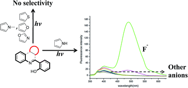
Selective detection of fluoride by 2-(3H-pyrrolo[2,3-c]quinolin-4-yl)phenol is reported using a turn-on fluorescence method. The presence of a pyrrole ring along with substituted o-phenol is essential as their absence results in either loss of selectivity or decrease in fluorescence intensity.
Several approaches are currently used for detection of fluoride ion, notably colorimetric and fluorescence sensing, 19F NMR analysis and electrode method.6–8 Although, 19F NMR analysis and electrode methods are well established, they suffer from several drawbacks. Detection of micromolar levels of fluoride is a key problem associated with NMR technique; whereas fragile instrumentation is the shortcoming for electrochemical methods. All these concerns are effectively addressed by colorimetric and fluorescence detection techniques, which allow sub-ppm level detection and the possibility of intracellular monitoring. Fluorescent chemosensors are generally categorized into two types; fluorescence enhancers (turn-on sensors) and fluorescent quenchers (turn-off sensors). Literature reports clearly indicate that turn-on sensors are more sensitive, as fluorescence enhancement can be more easily monitored than fluorescence quenching.9 Hydrogen bonding or Lewis acid–base interactions are the main driving force behind these techniques.10 In case of hydrogen bonding led detection, fluoride either acts as a contributor or a disrupter. Most of the fluoride detection systems in literature are based on nitrogen containing heterocycles, where sp2 hybridized nitrogen forms hydrogen bonding.11
In this work, we have demonstrated utilization of a 4-substituted pyrrolo[2,3-c]quinoline ligand for detection of fluoride in micromolar level (S32, ESI†). Sensors bearing pyrrole and Schiff base groups are well known in literature as fluoride sensors.12 The ligand used herein is based on a hybrid pyrrole-Schiff base model. The main rational was to incorporate the important structural and functional features of the aforementioned sensors so as to increase its sensitivity.
Attempted development of 2-(3H-pyrrolo[2,3-c]quinolin-4-yl)phenol[PQP] as a fluoride sensor was centred on availability of two possible binding sites on the molecule (Scheme 1). Interestingly, the molecule also shows structural similarity to known salicaldimine based Schiff base receptors13 (Scheme 1). Presence of fused pyrrole ring was expected to increase the overall conjugation thereby aiding in the enhancement of fluorescence signal.
Synthesis and characterization of PQP has been recently reported from our group.14 Our initial attempt was to look at steady state fluorescence behaviour of PQP in presence of F− and other anions such as Cl−, Br−, I−, AcO−, H2PO4− and HSO4−, in THF. Preliminary study showed promising F− detection capability of ligand. When we added 3.32 equivalent of F− to the ligand solution in THF, it underwent a 90 nm red-shift with approximately 250 fold enhancement. However, interference by acetate anion was also noticed, while other anions did not bring about any change (S25, ESI†). Similar fluorescence behaviour of acetate and fluoride is well documented in literature and mostly this problem is solved using silyl protecting groups.15 We adopted the same approach here; hydroxyl group in PQP was protected with TBDPS group.15 The modified PQP ligand thus obtained, was fully characterized before embarking on further F− detection studies. The results obtained show clearly selective sensing behaviour of PQP towards F− (Fig. 1).
![]() | ||
Fig. 1 Anion selectivity of TBDPS-PQP in THF. Inset: Fluorescent response (λex = 340 nm) of TBDPS-PQP in THF to various anions (3.32 equivalents), measured at 490 nm; 1: only TBDPS-PQP. |
Competitive fluorescence experiments (S33, ESI†) were then performed with other anions to further establish the selectivity of F− anions. It showed that PQP as well as TBDPS-PQP were highly selective towards F− even in presence of anions like Cl−, Br− and I−. As expected, presence of AcO− along with F− results in significant enhancement of fluorescence signal. In addition, detection limits were also calculated (S32, ESI†): 19 × 10−6 M and 16.5 × 10⁻⁶ M for PQP and TBDPS-PQP, respectively.
Our initial success with PQP prompted us to completely replace fused pyrrole ring and explore fluoride sensing potential of the resultant ligand. For this purpose 2-(quinolin-2-yl)phenol, 2-(pyridin-2-yl)phenol and 2-(4-phenylquinolin-2-yl)phenol [ligands 2, 3 and 4; Table 1] were synthesized.16 The ligands did not show any selectivity towards F− sensing when their steady state fluorescence behaviour was monitored in presence of various anions.
Ligand | Structure | Anion selectivitya (red shift with intensity enhancement) |
---|---|---|
a Ligands showing selective F− sensing also show AcO− sensing, which can be easily removed by using TBDPS protection of hydroxyl group.b No red shift and no AcO− interference. | ||
1 | ![]() |
F− selective |
2 | ![]() |
No selectivity |
3 | ![]() |
No selectivity |
4 | ![]() |
No selectivity |
5 | ![]() |
No selectivity |
6 | ![]() |
No selectivity |
7 | ![]() |
No selectivity |
8 | ![]() |
No selectivity |
9 | ![]() |
No selectivity |
10 | ![]() |
No selectivity |
11 | ![]() |
F− selectiveb |
12 | ![]() |
F− selective |
13 | ![]() |
F− selective |
14 | ![]() |
F− selective |
Further fused thiophene, furan and 1,3-oxazole based analogues of PQP [ligands 5, 6 and 7; Table 1] were also synthesized. None of the compounds displayed any selective fluoride sensing capability. Thus the above studies clearly indicated to us criticality of pyrrole ring for selective fluoride detection.
Subsequent studies were focussed on understanding the stoichiometry and nature of fluoride interaction with PQP. To probe the intermolecular interaction between F− anion and PQP, F− anions were added to DMSO-d6 solution of PQP and changes in corresponding 1H NMR spectrum were monitored (Fig. 2). Simultaneous disappearance of OH and NH peaks on titration with F− as tetra butyl ammonium salt was noticed, which indicates deprotonation of both phenol and pyrrole hydrogen. Also downfield shift of C-2 protons (adjacent to pyrrole nitrogen), further demonstrates interaction between pyrrole NH and F−. Hence it can be argued that the red shift in fluorescence maxima seen on F− addition is due to dianion formation. Dianion formation was also established by the Job's plot (S31, ESI†), which showed 2:
1 ratio of F− to PQP.
In order to further understand the dianion formation we carried out minor changes on PQP structure. The compounds synthesized with this focus were ligands 8, 9, 10 and 11 (Table 1). In cases of ligands 7 and 8, pyrrole NH and hydroxyl were blocked, respectively. Main outcome of this study was lack of any F− detection by either ligand 8 or 9. In ligand 10 and 11 hydroxyl position was adjusted to meta and para position, respectively. While ligand 10 failed to show selective F− sensing, ligand 11 displayed selective F− detection with fluorescence enhancement. Interestingly, fluorescence enhancement seen for ligand 11 was without any red shift and no interference from AcO− was observed. The observations from studies conducted on ligands 8–11 help us to conclude two important aspects of F− sensing. Firstly, dianion formation is essential and also continuous delocalization of electron cloud around the entire ligand is necessary for F− detection. In case of ligand 10 where there is a dianion formation but disruption of electron delocalization, no selectivity was observed.
After demonstrating suitability of PQP as fluoride sensor attention was focused on compounds bearing similar structure as PQP. Main motivation behind this effort was to explore flexibility in PQP structure while still retaining the fluoride sensing ability. As structural variations were already carried out on quinoline half of the molecule to understand the binding and mechanism of F− sensing, only few structural variations on PQP were planned on phenol half of the molecule. This involved replacement of o-hydroxyphenyl with α-hydroxynaphthyl, β-hydroxynaphthyl and 7-hydroxycoumarin systems [ligands 12, 13 and 14; Table 1]. The ligands were prepared by using the same synthetic protocol as was used for preparation of PQP. On complete characterization, these ligands were screened for fluorescence behaviour in presence of the previously mentioned anions (S28–30, ESI†). In case of α-hydroxynaphthyl analogue of PQP, the fluorescence intensity was similar but on changing to β-hydroxynaphthyl and 7-hydroxycoumarin analogues, decrease in intensity was noticed. Main rational behind the above observation could be loss of planarity in case of ligand 13 and 14, which makes delocalization of electrons ineffective, thereby decreasing the fluorescence intensity.
Footnote |
† Electronic supplementary information (ESI) available. See DOI: 10.1039/c5ra08626d |
This journal is © The Royal Society of Chemistry 2015 |