DOI:
10.1039/C5RA08274A
(Paper)
RSC Adv., 2015,
5, 71190-71202
Albumin stabilized silver nanoparticles–clotrimazole β-cyclodextrin hybrid nanocomposite for enriched anti-fungal activity in normal and drug resistant Candida cells
Received
5th May 2015
, Accepted 12th August 2015
First published on 14th August 2015
Abstract
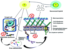
Nanotechnology unlocked distinctive platforms to move inside a hybrid therapeutic zone. Currently, nano-metal technology is the targeted field with exceptional advantages. Exceptionally small size and dominance of the surface properties such as high surface charge has raised a great deal of interest. This work is designed to exploit an interesting mechanistic feature i.e. multiple therapeutic targets possessed by metal nanoparticles. In this study we selected silver nanoparticles (AgNPs), which possess well documented anti-fungal activity and a standard antifungal molecule i.e. “clotrimazole”. A hybrid of AgNPs and clotrimazole was aimed to tackle clotrimazole resistance. Clotrimazole was firstly included into a β-cyclodextrin cavity to render it water soluble; subsequently the drug loaded dextrin moiety is functionalized on the surface of bovine serum albumin (BSA) stabilized silver nanoparticles using 1-ethyl-3-(3-dimethylaminopropyl)carbodiimide/N-hydroxysulfosuccinimide (EDC/NHS) chemistry. BSA stabilisation was essential to shield the physical interaction of AgNPs with the clotrimazole β-cyclodextrin complex that is otherwise experienced. Spectral and morphological characterization of the complex assures the synthesis of a hybrid metal complex. A cellular toxicity assay was performed to determine the toxic nature of the hybrid. This hybrid was then evaluated for its fungicidal activity on normal and clotrimazole resistant Candida cells. The toxicity and efficacy outcomes revealed a potent profile with a handy therapeutic window. Mechanistic explanations for this hybrid nature were supported by aggravated apoptotic cell percentage and reactive oxygen species production in both resistant and non-resistant cells. Cell cycle arrest studies further revealed G2/M phase cell cycle arrest, directing towards compromised cell membrane and DNA synthesis process equivalently in clotrimazole resistant cells.
1. Introduction
Drug resistance is a relative inattentiveness of a pathogenic and non-pathogenic species towards an active molecule.1 Presently drug resistance is classified depending on the mode and conduct of exposed specie. Primary resistance (in organisms never exposed in that host to the drug of interest), secondary resistance (defined as acquired resistance, only after exposure), intrinsic resistance (resistance of all or almost all isolates of one species) and clinical resistance (therapy failure or relapse of infection, which is not evident during in vitro study).2–4
Azoles (or azole derivatives) are among one of the category of antifungal drugs that inhibit the 14α lanosterol demethylase, a key enzyme in ergosterol (a most prevalent sterol in the fungal plasma membrane) biosynthesis. Some reports also suggest that they may also target fungal plasma membrane lipids and may upset methylsterol biosynthesis by interacting with an associated 3-ketosteroid reductase.5 Mechanism of azole resistance can be easily understood as it resembles just another antimicrobial resistance process viz. adaptation of the target enzyme, altered drug efflux and influx, mutation of genes encoding for target enzymes. Development of active efflux pumps with the up regulation of CDR (with almost all azoles) and MDR (two different gene families of transporters encoded in Candida species; CDR genes of the ATP binding cassette super family, and the MDR genes of the major facilitators class) has been demonstrated in azole-resistance.6 Binding of azoles to the enzymatic site gets halted with alterations in ERG11 (a gene encoding for lanosterol C14α-demethylase).7 Studies also revealed some Candida species have elevated intracellular concentration of ERG11p which leads to reduced azole susceptibility. Mutation of the ERG3 gene prevents the formation of 14α-methyl-3,6-diol from 14α-methylfecosterol after the azole exposure, which is toxic product primarily responsible for the growth arrest.8,9 Co-existing multiple resistance mechanisms can be operative in any given fungal strain with additive effects.4 Consequently the problem of antifungal resistance in immunocompromised and high-risk patients is becoming a major concern.
Metal nanoparticles exhibit numerous fascinating properties, and it is just a matter of time until more of these assets will be exploited.10,11 Medicine and biology, diagnostic, biomedical, engineering, catalysis, pigments, high density storage, obscurant smokes, metallurgy, nanotechnology are the areas under focus.12,13 More specifically AgNPs have also been revealed to possess potential inhibitory activity against fungal strains.14,15 AgNPs may exert an antifungal activity by disrupting the structure of the cell membrane and inhibiting the normal budding process due to the destruction of the membrane integrity.16 Studies revealed that 80% inhibitory concentration (IC-80) on ATCC strains of T. mentagrophytes and C. albicans ranges from 1 to 7 μg ml−1.17 Many other reports also described the MICs of AgNPs from 0.4 to 3.3 μg ml−1 against C. albicans, C. glabrata adhered cells and biofilm and at 10 μg ml−1 against Trichophyton rubrum.18 The properties of nanoparticles can be altered by changing their size, surface properties and concentration especially when at the nanometer scale.15
Inspiring from the fact that co-existing multiple resistance mechanisms may result in additive effects, we propose a multiple targeting approach to weaken or eliminate the kinetics as well as possibility of drug resistance.19,20 Entirely different targets, if approached at the same time, may synergise the overall effect and raise the probability to disorganise the resistance machinery. With such a theme we developed a hybrid macromolecular system employing two mechanistically different antifungal moieties with common target. Clotrimazole; an imidazole derivative with a broad spectrum of antimycotic activity, inhibiting the biosynthesis of the sterol ergosterol and rendering the membrane leaky with apparent disruption of the membrane bound enzyme systems.21 On the on other hand Ag-NPs possess excellent physico-chemical properties, especially surface charge. Direct membrane disruption achieved by virtue of its surface properties and sustained release of Ag+ ions in local environment establish its potential in this field.14 Reports are already available highlighting its antibacterial spectrum to antibiotic resistant organisms.
According to our hypothesis, simultaneous exposure of AgNPs and clotrimazole may synergise the possible inhibition; moreover concurrent exposure may weaken the resistance machinery to work against the loaded drug of the macromolecule. In this research work we have synthesised an engineered macromolecule with nanosilver in its core and the branches of clotrimazole containing nanopockets on its surface. Silver nanoparticles (∼40 nm) were initially stabilized by bovine serum albumin (BSA) and the engineered silver surface is then functionalised with clotrimazole-β-cyclodextrin (CbC) complex using ethyl(dimethylaminopropyl) carbodiimide (EDC)/N-hydroxysuccinimide (NHS) coupling. Direct functionalization of the clotrimazole molecule alone, as well its dextrin complex was also tried, but a potential interaction was experienced. Thus, non-specific adsorption of BSA on AgNP surface by the virtue of electrostatic attraction was achieved. Fig. 1 describes the cartoon representing this macromolecular strategy.
 |
| | Fig. 1 Macromolecular strategy using nanometal hybrid. Multiple targets of this hybrid nanocomposite, where AgNPs can cause direct damage to the fungal cell membrane, interruption of DNA replication and ATP production and generation of reactive oxygen species. Indifferently clotrimazole released from this complex provide fungal cell inhibition by inhibiting the ergosterol biosynthesis, leading to compromised fungal plasma membrane. Entirely different targets can be approached at a single time with this hybrid nanometal composite, which may improve the overall effect and the resistance development process may get disorganised. | |
2. Material and methods
2.1 Synthesis of BSA stabilized silver nanoparticles (BSAAGNPs) and estimation of hard BSA corona
Sodium borohydride (NaBH4) reduction method was used to synthesise AgNPs with slight modifications. Briefly, 10 ml of (1.0) mM AgNO3 was reduced with 30 ml of (2.0) mM NaBH4 under controlled temperature and stirring conditions.22
19.5 ml lemon yellow coloured AgNPs nanosuspension (100 μg ml−1) was then incubated with 0.5 ml of 0.5 M BSA solution in double distilled water. The reaction was allowed to proceed for 2 h at ambient temperature and the resultant bioconjugate (BSAAGNPs) was centrifuged for 1 h at 13
000 rpm.23
Synthesised AgNPs and BSAAGNPs were then characterized for particle size, polydispersity index (PDI), zeta potential using (Zeta sizer, DLS 4C Beckman Coulter, Japan), UV-VIS spectroscopy using (UV-1700 spectrophotometer, Shimadzu, Japan), FTIR Spectrophotometer (Nicolet-380, Thermo, USA), X-ray powder diffractometry using (Bruker AXS D8 Advance Diffractometer), and morphology using transmission electron microscope (TEM – Hitachi H-7500, USA) operating at 100 kV.
Flocculation experiments were performed to assure this surface conjugation.24 BSAAGNPs were exposed to saline phosphate buffer (pH 7.4 and 155 mM NaCl) in 1
:
1 volume ratio and precipitation was checked.
Hard BSA corona was estimated as the quantity of BSA forming the tight cloud around the nanometal surface. The supernatant left after centrifugation step was subjected to the quantification of unconjugated BSA. Bicinchoninic acid (BCA) assay was performed for the quantitative estimation of free protein at 562 nm on UV-Vis spectrophotometer and using eqn (1) hard BSA corona was quantified.
| | |
BSAhard corona = BSAtotal − BSAsupernatant
| (1) |
2.2 Synthesis of clotrimazole-β-cyclodextrin (CβC) inclusion complex
Inclusion complex was prepared by solvent evaporation encapsulation method with 1
:
1 molar ratio of clotrimazole and β-CD (following methodology as detailed in our previous work).25 β-CD was dissolved in 20 ml deionized water and clotrimazole solution in 2 ml methanol was added to β-CD solution under stirring at 800 rpm. Stirring was continued for 12 h with open cap. Highly water soluble clotrimazole-β-cyclodextrin complex then was separated from the supernatant after centrifugation at 1500 rpm and then recovered by lyophilizer (Alpha 1-2 LD plus, Martin Christ, Germany). Inclusion complex was characterized by FTIR Spectrophotometer (Nicolet-380, Thermo, USA), differential scanning calorimetry (DSC-Mettler Toledo DSC 822e), scanning electron microscopy (JSM-840 SEM, Jeol, Japan). Clotrimazole loading inside β-CD cavity was assessed by methanol based extraction method.
2.3 Synthesis of clotrimazole-β-cyclodextrin–BSAAgNPs (CβC–BSAAgNPs) hybrid nanocomposite
CβC–BSAAgNPs nanocomposite was synthesised by using EDC based crosslinking chemistry.26 Firstly the linker was attached to the surface exposed carboxylic groups of CβC and then the crosslinking was done with exposed amino groups of hard BSA corona on the nanometal surface, to achieve a stable nanobioconjugate. The synthetic scheme for the preparation of CβC–BSAAgNPs nanobioconjugate is displayed in Fig. 2. 20 mg of CβC was dissolved in 20 ml PBS buffer solution (pH-6). Then, the solution was added into a centrifuge tube in the presence of 60 mg EDC and 20 mg NHS. After the tube was rotated constantly for 90 min, 5 mg BSAAgNPs was added into the tube and again the tube was rotated overnight. Afterwards the CβC–BSAAgNPs nanobioconjugate was purified by dialysis to remove the uncoupled CβC. Dialysis was done using the dialysis bag with 1000 molecular weight cutoff against PBS buffer (pH-7.2). Finally the hybrid was recovered by centrifugation at 2000 rpm for 10 min and was characterized for particle size, polydispersity index (PDI), morphology using transmission electron microscope (TEM-Hitachi H-7500, USA) operating at 100 kv, zeta potential using (Zeta sizer, DLS 4C Beckman Coulter, Japan), FTIR spectroscopy (Nicolet-380, Thermo, USA), UV-VIS spectroscopy using (UV-1700 spectrophotometer, Shimadzu, Japan) and X-ray powder diffractometry using (Bruker AXS D8 advance diffractometer).
 |
| | Fig. 2 Synthesis scheme of albumin stabilized silver nanoparticles–clotrimazole-β-cyclodextrin (CβC–BSAAgNPs) hybrid nanocomposite. | |
CβC–BSAAgNPs was further evaluated for clotrimazole and Ag content per mg of the complex. For clotrimazole content 1 mg of CβC–BSAAgNPs was digested in 1 ml methanol for 6 h. After digestion the clotrimazole content was determine by HPLC (Waters-2707, C-18 column and 2998 PDA detector) with mobile phase consisting of water 0.1% triethylamine (pH-3.2 adjusted by using ortho-phosphoric acid): methanol in ratio of (25
:
75 v/v%) with a flow rate of 1.0 ml min−1 at a detection wavelength of 215 nm. Ag content present in per mg complex was observed by atomic absorption spectroscopy (AAS, Shimadzu AA-7000 Atomic Absorption Spectrophotometer).
2.4 In vitro release studies
Release of both clotrimazole and Ag from CβC–BSAAgNPs was evaluated using dialysis bags (containing 5 mg CβC–BSAAgNPs) in 100 ml phosphate buffer saline (PBS 7.4), at 37 °C at 100 rpm in a thermostatically shaking incubator (LSB-1005RE, Daihan Labtech. Co. Ltd Korea). 0.5 ml of sample was taken from the buffer solution after time intervals 0, 1, 2, 4, 8, 16, 32, 64 h. Post-sampling, the volume was swapped with fresh medium to maintain the sink conditions. Quantification of clotrimazole was done by HPLC (as per the above mentioned method), whereas quantification of the released Ag was done by atomic absorption spectroscopy (AAS, Shimadzu AA-7000 Atomic Absorption Spectrophotometer).
2.5 Cell cytotoxicity study (MTS assay)
The (3-(4,5-dimethylthiazol-2-yl)-5-(3-carboxymethoxyphenyl)-2-(4-sulfophenyl)-2H-tetrazolium) dye reduction assay in the presence of phenazinemethosulfate (PMS), produces a formazan product that has an absorbance maximum at 490–500 nm in phosphate-buffered saline.27 The MTS assay was performed to determine the cytotoxic potential of CβC, AgNPs, BSAAgNPs and CβC–BSAAgNPs on different cell cultures. Cell viability assay was performed on Hela (Cervical cells), HEL (Human embryonic lung), VERO, and MDCK (Madin Darby canine kidney cell) cultures.28 Minimum cytotoxic concentration (MCC) i.e. the concentration required to cause a microscopically detectable alteration of normal cell morphology and cell cytotoxicity 50% (CC50) i.e. the concentration causing 50% cell death were examined from this study.29
2.6 Antifungal susceptibility testing
2.6.1 Test organisms and test medium. C. albicans (Microbial Type Culture Collection “MTCC-183”) and C. tropicalis (MTCC-184) strains were used in this study. The strains were inoculated onto Sabouraud dextrose agar plates and incubated at 35 °C for 24 h. They were subcultured on the same medium for a further 24 h at 35 °C. The fungal inoculay were prepared by diluting the overnight culture with 0.9% NaCl to 1 × 10⁶ CFU ml−1. The suspensions were then further diluted in test medium i.e. RPMI-1640 (with L-glutamine, without sodium bicarbonate) supplemented with 0.165 M morpholinepropanesulfonic acid (MOPS) to a final count ranging 0.5 × 10³ CFU ml−1. The experiments are done as per the recommendation made by NCCLS standards.30
2.6.2 Minimum inhibitory concentration (MIC) and inhibitory concentration 50% (IC50). The antifungal effectiveness was determined against the final cell concentration of 106 CFU ml−1 for both C. albicans and C. tropicalis cells. Fungal inoculas (100 μl) were added to each well of a sterile, 96-well flat bottomed microtiter plate. Arithmetic concentrations (in 100 μl volume) of clotrimazole, CβC, AgNPs, BSAAgNPs and CβC–BSAAgNPs (ranging from 2–60 μg ml−1) were added systematically in the well. Each concentration was tested in triplicate for each fungal strain. 300 μl of deionized sterile water were added on the exterior perimeter wells of the microplates to minimize the evaporation of the culture medium of the test wells during the incubation. For each strain two wells containing fungal suspension with no drug (growth control) and two wells containing only media (background control) were included in this plate. Optical density (OD) was measured for 24 h at 35 °C using a microplate reader (Bio-Rad) at 405 nm and were automatically recorded every 3 h for each well. Turbidimetric growth curves were obtained depending on the changes in the OD of fungal growth for each drug concentration and the drug-free growth control. MIC and IC50 values were calculated for each treatment using the same plots; the growth percentage for each drug concentration was calculated using eqn (2). OD measurement were reported only after the subtraction of background ODs (ODs of fungal cells free wells). Death kinetic curves were also plotted, by challenging the fungal cells by estimated IC50 values of clotrimazole, CβC, AgNPs, BSAAgNPs and CβC–BSAAgNPs using similar OD measurement spectral method. Slopes observed in these graphs were related to the potency of the treatment.| | |
Growth% = [(OD405 of wells containing the test/OD405 of the drug free well) × 100]
| (2) |
2.6.3 Development of clotrimazole resistance and antifungal susceptibility testing. Cell suspension of both the Candida species (0.5 × 10⁶ CFU ml−1) was exposed to their respective clotrimazole IC10 (concentration inhibiting 10% fungal cells). After 24 h of exposure both the fungal cell suspensions were individually subcultured on sabouraud dextrose agar plates at 35 °C for 24 h. After that passaged fungal cells were recovered by diluting the overnight culture with 0.9% NaCl. This experiment was repeated sequentially by exposing passaged cells with clotrimazole IC20, IC30, IC40 and IC50. Clotrimazole resistant fungal cells obtained after the gradient exposure of clotrimazole to its pre-calculated IC50 were again tested for their modified MIC, IC50 and death kinetics against clotrimazole, CβC, AgNPs, BSAAgNPs and CβC–BSAAgNPs using microdilution assay (as done previously).
2.7 Estimation of early apoptosis and reactive oxygen species (ROS) production
2.7.1 Annexin V–propidium iodide double staining apoptotic changes. The early stages of apoptotic phenomenon in the treated fungal cells can be detected with fluorescein isothiocyanate (FITC)–Annexin V staining (to study the expression of phosphatidylserine on apoptotic fungal cells) and propidium iodide (to study the compromised membrane permeability of treated cells) based double-staining method. Non-resistant and resistant fungal cells were digested for 1 h at 28 °C in a potassium phosphate buffer (pH 6.0) containing 20 mg ml−1 lysing enzyme and 1 M sorbitol. Protoplasts of non-resistant and resistant strains were stained with FITC-labelled Annexin V and PI using the FITC–Annexin V apoptosis detection kit (Sigma Aldrich, USA) according to the staining kit protocol. Protoplasts were incubated with MIC concentrations of clotrimazole, CβC, AgNPs, BSAAgNPs and CβC–BSAAgNPs and incubated for 20 min in an Annexin-binding buffer containing 5 μL FITC–Annexin V per ml and PI. Protoplasts were then examined by a flow cytometer (Becton Dickinson “BD” Accuri C6).
2.7.2 Reactive oxygen species (ROS) production. With the similar treatment protocol (as followed for apoptosis detection), treated fungal cells were incubated for 10 minutes with 5 mg ml−1 dihydrorhodamine 123 (DHR-123). DHR-123 is oxidized by intracellular ROS to the fluorescent chromophore rhodamine 123. Samples were quantitatively analysed for the generated ROS species by flow cytometry (BD Accuri C6).
2.8 Cell cycle arrest after CβC–BSAAgNPs treatment
Non-resistant and resistant fungal cells (in their log-phased with concentration around 1 × 10⁸ cells), cultured in a yeast extract-peptone-dextrose (YPD) medium, were harvested and treated with IC50 concentration of CβC–BSAAgNPs. After 6 h of incubation, the cells were washed with PBS and fixed with 70% ethanol overnight at 4 °C. All the cells were treated with 200 μg ml−1 of RNase A and the mixture was left to react for 2 h at 37 °C. For DNA staining, 50 μg ml−1 of propidium iodide (PI) were added and incubated for 1 h at 4 °C in the dark. Flow cytometric analysis was performed by (BD Accuri C6). The values represent the average of the measurements conducted in triplicate of three independent assays.
2.9 Statistical analysis
All the observations were based on 3 independent experiments. Data were expressed as mean ± SEM or percentage. The results were analyzed by 1-way analysis of variance (ANOVA), and chi-square test, as applicable.
3. Results and discussion
3.1 Surface biocongucation of AgNP with BSA (BSAAgNPs)
AgNPs were successfully synthesized with an average size of 37.5 ± 1.3 nm and a narrow PDI value of 0.254 ± 0.37. Incubation with BSA leads to the surface coating of AgNPs resulting in increased average size to 54.2 ± 2.8 nm and PDI value of 0.291 ± 0.19. Zeta potential value of −2.04 ± 0.01 mV indicated the inherent colloidal instability of synthesised AgNPs nanoparticles but BSA stabilization leads to a drastic change in surface potential to −18.6 ± 0.07 mV. The altered value of surface potential provides a strong conformation of improved colloidal stability of BSAAgNPs. Characteristic absorption maxima at 416 nm and a solo transverse peak indicated the nano-size and isotropic nature of the AgNPs. After BSA stabilization SPR observed a shift to 432 nm with compromised intensity, this significant bathochromic and hypochromic shift resulted due to protein lead quenching of silver colloid's SPR. Comparison of FTIR spectra's revealed no significant shift in the AgNPs peaks after stabilization, and the presence of the characteristic peaks of BSA in the BSAAgNPs spectra suggested the BSA corona formation on the hydrophobic AgNPs surface by virtue of physical interactions. X-ray diffraction (XRD) patterns of AgNPs showed sharp and intense peaks corresponding to the crystalline nature of metallic nanoparticles. Surface stabilized AgNPs showed no alteration in crystalline XRD pattern, additionally a broad amorphous BSA peak was observed at shorter 2θ range. Transmission electron microscope (TEM) revealed that synthesised AgNPs were well dispersed showing a confined distribution with particles ranging from 20–50 nm (25–40 nm size dominating the population). Comparatively, TEM analysis of BSAAgNPs revealed an obvious increase in particle size where the dominating nanoparticle population lies between 45–70 nm. Fig. 3 represents the characterization report of synthesised AgNPs and surface bioconjucated BSAAgNPs.
 |
| | Fig. 3 Characterization parameters of AgNPs and BSA stabilized AgNPs. (a) UV-Visible spectral comparison, (b) zeta potential (AgNPs), (c) zeta potential (BSAAgNPs), (d) XRD spectral comparison, (e) TEM (AgNPs), (f) TEM (BSAAgNPs). | |
Harsh conditions of pH, ionic strength, temperature as well as physical incompatibility with drug molecules lead to the destabilization of the nanosilver colloids. Establishing the protein corona, probably because of ionic or electrostatic and hydrophobic interactions provides a physical shield preventing the direct interaction with the silver core.24 Flocculation experiment confirmed no precipitation of BSAAgNPs in harsh conditions presented by saline phosphate buffer, presenting a straightforward protective effect of protein cloud on metal colloid stability. Further these proteins results in the development of two kinds of protein clouds around the nanometal surface viz. hard corona (essential to prevent colloid destabilization) and second is weak/soft/loose corona (hanging or accompanied protein molecules which for a secondary layer and are not necessary for stabilization process).31 BCA assay performed for the quantitative estimation of hard protein corona revealed the supernatant BSA concentration of 41 mM ml−1. Estimated free BSA amount in the supernatant was around 0.083 M and thus BSA amount in hard protein corona was calculated around 0.417 M. Studies had also shown that the corona BSA protein retains its secondary structure and the helicity to a quite good extent on the nanoparticles surface. Also some studies explored the variables for this BSA functionalization and came out with a significant decrease in the BSA quantity immobilized on the AgNPs surface with the increase in AgNPs size. Its explanation was related to the relation of surface curvature (inversely proportional to the size) of the nanoparticles and the consequent energetics involved in protein adsorption and crowding. It was also suggested that the interaction of BSA and AgNPs is majorly governed by increased changes in protein conformation and to lesser extent hydrophobic interactions.
3.2 Synthesis of clotrimazole-β-cyclodextrin (CβC) inclusion complex
CβC inclusion complex have dull white and fluffy appearance, pointing toward its amorphous nature. Aqueous saturation solubility of the complex at ambient conditions was found to be around 27.5 ± 2.1 mg ml−1 and the clotrimazole loading in the complex was obtained to be 84.5 ± 4.7 μg mg−1. IR (KBr) cm−1 spectra revealed the major existence of β-CD specific peaks with a slight shift to higher/lower wave numbers while very few characteristic peaks of clotrimazole (at shorter wavelength) were observed. This indicated a successful inclusion of clotrimazole in the hydrophobic β-CD cavities. DSC thermograms of CβC complexes showed a substantial reduced intensity and broad thermodynamic peak with a shift to lower temperature (142° to 82 °C) in comparison to sharp melting point of clotrimazole and β-CD. These endotherms indicate the substantial inclusion of clotrimazole in the (β-CD) cavities. Scanning electron microscopy clearly justifies the above observation of having a highly amorphous nature of clotrimazole inclusion complex. Fig. 4 displays the FT-IR, DSC and SEM characterisation of the inclusion complex. Further, drug loading study using methanol extraction method showed that lyophilized CβC complex have a significant clotrimazole loading of 68.4 ± 3.2 μg per mg of complex.
 |
| | Fig. 4 FT-IR, DSC and SEM image of clotrimazole-β-cyclodextrin (CβC) inclusion complex. | |
3.3 Synthesis of clotrimazole-β-cyclodextrin–BSAAgNPs (CβC–BSAAgNPs) hybrid nanocomposite
CβC–BSAAgNPs nanocomposite was synthesised as per the above mentioned procedure. Conjugation of CβC to BSA coating of AgNPs was obtained by carbodiimide coupling. Surface exposed carboxylic group of CβC reacted with EDC to form unstable reactive ester and further addition of NHS resulted in the formation of semi-stable amine-reactive NHS-ester. Afterwards, addition of BSAAgNPs containing amino group on the exposed protein corona of AgNPs, leads to the formation of CβC–BSAAgNPs conjugate by a stable amide bond.
Zeta size reports revealed an increased hydrodynamic size (68.5 ± 1.9 nm) with uniform distribution pattern (PDI-0.274 ± 0.13) due to the surface conjugation. To confirm this surface attachment of CβC on the BSAAgNPs surface, changes in surface potential values were observed. Considerable change in zeta potential to −22.0 ± 0.3 mV revealed the successful surface conjugation of CβC on the surface of BSAAgNPs. TEM images revealed the increased particle size, but nanoparticles possessed compromised surface smoothness. The CβC–BSAAgNPs conjugates was further investigated by infrared spectroscopy where FT-IR spectra showed the characteristic peaks at 1627 cm−1 and 1506 cm−1 as a result of newly formed amide bond. UV-Visible spectroscopy further revelled no sharp peak, rather a wide hump corresponding to the AgNP's surface plasmon resonance was observed in the range of 425–450 nm. X-ray diffraction (XRD) patterns of synthesised conjugates suggest the presence of both BSAAgNPs and cyclodextrin peaks but with compromised intensity. Characterization parameters synthesised CβC–BSAAgNPs nanobioconjugate are mentioned in Fig. 5. Further quantitative analysis revealed that per mg of CβC–BSAAgNPs complex is loaded with of 572.6 ± 4.7 μg of silver content and 83.4 ± 1.9 μg of clotrimazole.
 |
| | Fig. 5 Characterization parameters of AgNPs and BSA stabilized AgNPs. (a) UV-Visible spectral comparison, (b) zeta potential (AgNPs), (c) zeta potential (BSAAgNPs), (d) XRD spectral comparison, (e) TEM (AgNPs), (f) TEM (BSAAgNPs). | |
3.4 In vitro release studies
In vitro release showed a controlled release profile for both clotrimazole and Ag from the synthesised CβC–BSAAgNPs nanobiocomposite. Clotrimazole release followed a first order pattern initial for 16 h, afterwards pseudo-zero order behaviour was observed. This can be attributed to the initial ease of clotrimazole release from the edges of the cyclodextrin cavities, and in later part it become comparatively difficult to elute from the centre of hydrophobic cavity. On the other hand a sustained release pattern of silver (Ag) ions was analysed from the atomic absorption spectroscopy. AgNPs dissolution can be better understood as the ionisation of Ag(0) to Ag(+) and literature showed several relationships between the Ag(+) release and AgNPs size.32 Possible reasons for this kind of Ag release could be the multiple mechanistic aspects responsible for its release from the nanosilver core of the conjugate system. Among several mechanisms provided for the ionic release of silver ions, results revealed an initial and dominant desorption of the chemisorbed silver ions from the BSAAgNPs surface.33 Moreover the presence of clotrimazole in the release environment further played its part (quite understood from the silver and clotrimazole interaction mentioned in the introductory part). Oxidative dissolution could be another simultaneous reason providing an instant release of silver ions. After 64 h, cumulative release observed for clotrimazole was 91.4 ± 3.2% while for Ag ions it was 39.8 ± 4.6%. Fig. 6 describes the release pattern followed by clotrimazole and Ag ions from the synthesised CβC–BSAAgNPs nanobiocomposite.
 |
| | Fig. 6 In vitro release pattern of clotrimazole and Ag from the synthesised CβC–BSAAgNPs nanobiocomposite. | |
In order to completely understand the kinetics of clotrimazole and ionic silver release different kinetic models were applied including zero order, first order and Higuchi. On the basis of regression coefficient (R2) from these curves it was concluded that release pattern of clotrimazole fits first order profile more closely as discussed above. In the case of Ag ions the release had a better fit in Higuchi model, which can be explained as the continuous eroding of silver ions from the nanosilver matrix of AgNPs.
3.5 Cell viability assay
AgNPs showed their inherent toxicity with comparatively lower MCC and CC50 values than, BSAAgNPs, CβC and CβC–BSAAgNPs as mentioned in Table 1. CβC showed negligible toxicity describing their highly safe nature which can be attributed to the highly safe nature of exposed cyclodextrins. BSAAgNPs showed slight safe profile than naked AgNPs and that can be explained as a result of masking of the highly reactive silver colloid surface. A coating of biological amphoteric molecule probably had decreased the adverse effect of silver colloids on challenged cell membrane. Also BSA conjugation resulted in an increased molecular weight of the nanosystem resulting in the compromised cell membrane permeation, which further limited the intracellular cell death mechanisms. Finally the CβC–BSAAgNPs toxicity profile clearly indicated the additive safety effects of surface linked CβC moieties, providing this nanoconjugate a much safer profile than AgNPs and BSAAgNPs.
Table 1 Cytotoxic concentration, as determined by measuring the cell viability with the colorimetric formazan-based MTS assay
| Cell line/sample |
HeLa |
HEL |
VERO |
MDCK |
| MCC μg ml−1 |
CC50 μg ml−1 |
MCC μg ml−1 |
CC50 μg ml−1 |
MCC μg ml−1 |
CC50 μg ml−1 |
MCC μg ml−1 |
CC50 μg ml−1 |
| AgNPs |
6.3 ± 0.7 |
431.7 ± 4.1 |
4.9 ± 1.2 |
396.2 ± 5.7 |
5.5 ± 0.3 |
403.5 ± 2.5 |
6.8 ± 0.8 |
465.2 ± 7.5 |
| BSAAgNPs |
11.2 ± 1.5 |
620.8 ± 12.5 |
16.9 ± 2.2 |
528.2 ± 2.3 |
8.5 ± 1.2 |
603.5 ± 5.8 |
13.4 ± 1.7 |
703.8 ± 4.6 |
| CβC |
>100 |
>1000 |
>100 |
>1000 |
>100 |
>1000 |
>100 |
>1000 |
| CβC–BSAAgNPs |
21.2 ± 0.8 |
770.5 ± 2.5 |
18.2 ± 1.6 |
714.3 ± 6.4 |
14.7 ± 0.9 |
843.4 ± 1.6 |
24.2 ± 2.0 |
782.4 ± 5.2 |
3.6 Antifungal susceptibility testing
Serial dilution antifungal assays revealed the hybrid nature of synthesised nanobioconjugate “CβC–BSAAgNPs”. MIC and CC50 of all the challenged treatments in both the resistant and non-resistant strains are provided in Table 2. It is very clear from this experiment that plain clotrimazole was displaying more than three times MIC and CC50 values in both the resistant C albicans and C tropicalis cells. Following these results, CβC also showed an elevated MIC and CC50 values in these clotrimazole resistant strains, but this increase is not of same potential as seen with plain clotrimazole. The only reason responsible for this can be explained by the fact of controlled clotrimazole release from the dextrin cavity which is already mentioned in the CβC complex in vitro release evaluation part.
Table 2 Antifungal susceptibility testing, estimation of MIC and IC50 of all the challenged treatments in both the resistant and non-resistant strains
| Challenge |
C. albicans |
C. tropicalis |
C. albicans (resistant cells) |
C. tropicalis (resistant cells) |
| MIC |
IC50 |
MIC |
IC50 |
MIC |
IC50 |
MIC |
IC50 |
| Clotrimazole |
6.3 ± 0.2 |
88.3 ± 2.1 |
7.1 ± 0.4 |
112.5 ± 1.4 |
21.3 ± 0.2 |
275 ± 3.5 |
19.3 ± 1.8 |
364.0 ± 0.7 |
| AgNPs |
24.6 ± 1.7 |
186.7 ± 4.6 |
21.3 ± 0.9 |
177.4 ± 2.3 |
27.2 ± 0.8 |
198.2 ± 1.7 |
26.1 ± 1.6 |
184.7 ± 1.2 |
| CβC |
31.8 ± 0.9 |
456.5 ± 5.3 |
37.5 ± 1.4 |
589.2 ± 3.5 |
45.3 ± 1.7 |
690.4 ± 2.6 |
42.4 ± 0.6 |
658.7 ± 4.5 |
| BSAAgNPs |
27.9 ± 2.1 |
218.3 ± 2.5 |
26.3 ± 1.7 |
191.4 ± 0.3 |
29.9 ± 1.3 |
218.2 ± 3.7 |
29.2 ± 0.4 |
203.5 ± 2.7 |
| CβC–BSAAgNPs |
4.1 ± 0.3 |
64.8 ± 0.4 |
3.9 ± 0.5 |
71.4 ± 2.6 |
5.3 ± 0.1 |
93.7 ± 2.5 |
5.0 ± 0.3 |
109.8 ± 3.8 |
Further the potential antifungal activity of AgNPs and BSAAgNPs can be explained by their inherent nature to disrupt fungal cell membrane, produce ROS species and potential to cause intracellular damage. There was a noticeable difference between the in vitro antifungal profile of naked and BSA stabilized AgNPs. Interestingly BSAAgNPs showed slightly higher MIC and IC50 values, which can be understood by the fact that protein corona on the surface of AgNPs controls the direct surface interaction of metal colloid and fungal cell membrane. Whereas nanobioconjugate “CβC–BSAAgNPs” displayed a potent antifungal profile on both the resistant and non-resistant cells. With the MIC value ∼4 μg ml−1, which was significantly lower than its individual components, a potentiation was observed. Hybrid nature of the nanobioconjugate can be explained by the multi-targets attained to inhibit the fungal cell growth as explained in hypothesis proposed in Fig. 1. There are reports explaining the potentiated activity of dextrin functionalized AgNPs, which also justifies this potentiation. Moreover the nanoconjugate provide a higher localized antifungal concentration at the around or inside the fungal cell which leads to increased destruction. Inhibition kinetic study as depicted in Fig. 7 clearly explains the potential of CβC–BSAAgNPs to provide a hybrid antifungal therapeutics. In both the non-resistant and resistant fungal strains the death inhibition kinetic pattern after CβC–BSAAgNPs exposure was having maximum negative slope.
 |
| | Fig. 7 Inhibition kinetics after the exposure of IC50 concentrations of clotrimazole, CβC, AgNPs, BSAAgNPs and CβC–BSAAgNPs against both non-resistant and clotrimazole resistant fungal cells. | |
It is important to discuss the mechanism behind the potential of CβC–BSAAgNPs against clotrimazole resistant strains. Well it is very clear that boosted effectiveness was observed with the conjugate of cyclodextrin entrapped clotrimazole (CβC) and BSAAgNPs. Much improved response from the CβC part (within CβC–BSAAgNPs) in case of clotrimazole resistant strains is the major question. This can be explained by the fact that presence of stabilized colloidal silver would definitely worked against the molecular fungal cell operations responsible for this azole resistance.
3.7 Apoptotic changes (Annexin V–propidium iodide double staining)
Study revealed the mechanistic comparison in the fungal cell inhibition after treatment with clotrimazole, CβC, AgNPs, BSAAgNPs and CβC–BSAAgNPs (Fig. 8). Clotrimazole treatment showed a sharp reduction in gated apoptotic cells in both the resistant cells, but this difference is not observed with CβC treatment. AgNPs and BSAAgNPs both showed a comparatively high percentage of early apoptotic cells and it was very clear that no such discrimination is seen between resistant and non-resistant cells. Early apoptosis observed with the treatment of synthesised nanobiocomposite was uppermost and again the effect was unanimous in both resistant and non-resistant cells. Generation and accumulation of intracellular ROS is the possible reason behind the significantly higher apoptotic induction observed with nanosilver and AgNP based nanobiocomposite.34,35
 |
| | Fig. 8 Apoptotic effect quantified by Annexin V–propidium iodide double staining procedure. | |
3.8 ROS production
Production of ROS species due to the metabolism of cellular oxygen has been associated in the regulation of the apoptosis in fungal cells. Apoptosis study results made a strong mark for the involvement of the ROS species and this study confirms it on quantitative basis (as shown in Fig. 9). Minor level of ROS generation was observed in clotrimazole and CβC treated non-resistant cells, and these levels further fall down (comparative to control) in resistant cells. Treatment with naked AgNPs and BSAAgNPs showed significantly higher level of ROS accumulation with similar quantitative values in both resistant and non-resistant cells. ROS accumulation in CβC–BSAAgNPs treated cells is marginally higher when compared to AgNPs and BSAAgNPs treated cells. Complying with the earlier antifungal and apoptotic effects results, it is very clear that AgNPs, BSAAgNPs and CβC–BSAAgNPs did not discriminated the clotrimazole resistance cells. ROS accumulation could be one of the major cell death mechanism involved in fungal cell inhibition, but these results again cleared the effectiveness of synthesised nanobiocomposite and the simultaneous failure of fungal resistance machinery to work against the inhibition process of CβC–BSAAgNPs.
 |
| | Fig. 9 Quantitative ROS generation estimated by dihydrorhodamine 123 (DHR-123) positive fungal cells. | |
3.9 Cell cycle arrest after CβC–BSAAgNPs treatment
Major percentage of treated cells was associated with G2/M phase cell cycle arrest in all the treated fungal strains. As shown in Fig. 10, there was a significant elevation in the cell percentage detected in G2/M phase, reflecting that treated cells were not able to enter the normal mitosis phase. Conversely a significant decrease in cells percentage observed in G1 phase revealed a certain incapability of cellular machinery to enter into DNA synthesis and replication phase. So a general possibility of compromised fungal cell membrane and simultaneous disturbance of normal DNA synthesis process can be drafted due to the additive inhibitory effect of nanosilver and available clotrimazole moiety from CβC–BSAAgNPs.
 |
| | Fig. 10 Effect of CβC–BSAAgNPs on the cell cycle process of non-resistant and clotrimazole resistant fungal cells treated with its IC50 concentration. | |
4. Conclusion
Synthesised hybrid nanocomposite CβC–BSAAgNPs was successfully synthesised using EDC/NHS chemistry, and characterization reports confirmed it. In vitro release studies revealed a controlled and pseudo-first order release pattern of clotrimazole moiety and Ag ions. Cell cytotoxicity assay of different cell lines provided a comparatively safe profile when compared to naked and BSAAgNPS. On challenging this biomacromolecule with stabilized silver core and surface attached pockets of clotrimazole against Candida cells (both normal and clotrimazole resistant) a much improved and possible additive effect was seen. Mechanistic evaluation further revealed that early apoptosis due to frequent ROS generation in case of CβC–BSAAgNPs treatment could be the possible reason. Undifferentiated inhibition in both clotrimazole resistant and normal fungal cells was solely reliant on the multiple inhibitory mechanisms possessed by nanosilver and the controlled release of the clotrimazole from the dextrin pockets at the target site. Cell cycle analysis further confirmed the potential of the nanobioconjugate to interrupt with the normal cellular process such as mitosis and DNA replication. The work aimed at designing a nanosized biomacromolecule with improved antifungal activity on even resistant cells. The scope of this strategy can be well exploited where limited choice of therapeutics and the problem of resistance are overlapped. Moreover, use of a nanometal core/base provides a dual benefit of both inherent therapeutic potential and nanobase to develop a functionalized hybrid biomacromolecule.
Acknowledgements
Author acknowledges Department of Biotechnology (DBT) India, Punjab Technical University, Jalandhar, Punjab State Council of Science and Technology (PSCST) India, Department of Virology, Haffkine Institute for Training Research and Testing, Parel, Mumbai, India for providing the supportive facility and financial base for this research. Author also acknowledges the insightful discussions provided by Dr Jitender Bhariwal (Department of Medicinal Chemistry, ISF-CP, Moga, Punjab, India) and technical support from Prof. R. Snoeck and Prof. G. Andrei (Laboratory of Virology and Chemotherapy, Department of Microbiology and Immunology, Rega Institute, Belgium).
References
- D. Mazel and J. Davies, Antibiotic resistance in microbes, Cell. Mol. Life Sci., 1999, 56(9–10), 742–754 CrossRef CAS.
- C. Walsh, Antibiotics: actions, origins, resistance, American Society for Microbiology (ASM), 2003 Search PubMed.
- M. M. Gottesman, Mechanisms of cancer drug resistance, Annu. Rev. Med., 2002, 53(1), 615–627 CrossRef CAS PubMed.
- J. Davies and D. Davies, Origins and evolution of antibiotic resistance, Microbiol. Mol. Biol. Rev., 2010, 74(3), 417–433 CrossRef CAS PubMed.
- A. Lupetti, R. Danesi, M. Campa, M. Del Tacca and S. Kelly, Molecular basis of resistance to azole antifungals, Trends Mol. Med., 2002, 8(2), 76–81 CrossRef CAS.
- J. F. Ryley, R. G. Wilson and K. J. Barrett-Bee, Azole resistance in Candida albicans, Sabouraudia, 1984, 22(1), 53–63 CrossRef CAS PubMed.
- M. M. Canuto and F. G. Rodero, Antifungal drug resistance to azoles and polyenes, Lancet Infect. Dis., 2002, 2(9), 550–563 CrossRef CAS.
- E. M. Johnson, D. W. Warnock, J. Luker, S. R. Porter and C. Scully, Emergence of azole drug resistance in Candida species from HIV-infected patients receiving prolonged fluconazole therapy for oral candidosis, J. Antimicrob. Chemother., 1995, 35(1), 103–114 CrossRef CAS PubMed.
- T. C. White, Increased mRNA levels of ERG16, CDR, and MDR1 correlate with increases in azole resistance in Candida albicans isolates from a patient infected with human immunodeficiency virus, Antimicrob. Agents Chemother., 1997, 41(7), 1482–1487 CAS.
- R. A. Masitas, I. V. Khachian, B. L. Bill and F. P. Zamborini, Effect of Surface Charge and Electrode Material on the Size-Dependent Oxidation of Surface-Attached Metal Nanoparticles, Langmuir, 2014, 30(43), 13075–13084 CrossRef CAS PubMed.
- F. Viñes, J. R. Gomes and F. Illas, Understanding the reactivity of metallic nanoparticles: beyond the extended surface model for catalysis, Chem. Soc. Rev., 2014, 43(14), 4922–4939 RSC.
- J. Jain, S. Arora, J. M. Rajwade, P. Omray, S. Khandelwal and K. M. Paknikar, Silver nanoparticles in therapeutics: development of an antimicrobial gel formulation for topical use, Mol. Pharm., 2009, 6(5), 1388–1401 CrossRef CAS PubMed.
- D. L. Fedlheim and C. A. Foss, Metal nanoparticles: synthesis, characterization and applications, CRC Press, 2001 Search PubMed.
- A. Panáček, M. Kolář, R. Večeřová, R. Prucek, J. Soukupová and V. Kryštof, et al. Antifungal activity of silver nanoparticles against Candida spp., Biomaterials, 2009, 30(31), 6333–6340 CrossRef PubMed.
- P. K. Jain, X. Huang, I. H. El-Sayed and M. A. El-Sayed, Noble metals on the nanoscale: optical and photothermal properties and some applications in imaging, sensing, biology and medicine, Acc. Chem. Res., 2008, 41(12), 1578–1586 CrossRef CAS PubMed.
- K.-J. Kim, W. S. Sung, B. K. Suh, S.-K. Moon, J.-S. Choi and J. G. Kim, et al. Antifungal activity and mode of action of silver nano-particles on Candida albicans, BioMetals, 2009, 22(2), 235–242 CrossRef CAS PubMed.
- D. Monteiro, L. Gorup, S. Silva, M. Negri, E. de Camargo and R. Oliveira, et al. Silver colloidal nanoparticles: antifungal effect against adhered cells and biofilms of Candida albicans and Candida glabrata, Biofouling, 2011, 27(7), 711–719 CrossRef CAS PubMed.
- J. Sardi, L. Scorzoni, T. Bernardi, A. Fusco-Almeida and M. M. Giannini, Candida species: current epidemiology, pathogenicity, biofilm formation, natural antifungal products and new therapeutic options, J. Med. Microbiol., 2013, 62(1), 10–24 CrossRef CAS PubMed.
- J. Loeffler and D. A. Stevens, Antifungal drug resistance, Clin. Infect. Dis., 2003, 36(suppl. 1), S31–S41 CrossRef CAS PubMed.
- R. Pelletier, J. Peter, C. Antin, C. Gonzalez, L. Wood and T. J. Walsh, Emergence of resistance of Candida albicans to clotrimazole in human immunodeficiency virus-infected children: in vitro and clinical correlations, J. Clin. Microbiol., 2000, 38(4), 1563–1568 CAS.
- F. C. Odds, A. J. Brown and N. A. Gow, Antifungal agents: mechanisms of action, Trends Microbiol., 2003, 11(6), 272–279 CrossRef CAS.
- K. M. A. El-Nour, A. Eftaiha, A. Al-Warthan and R. A. Ammar, Synthesis and applications of silver nanoparticles, Arabian J. Chem., 2010, 3(3), 135–140 CrossRef PubMed.
- P. Murawala, S. Phadnis, R. Bhonde and B. Prasad, In situ synthesis of water dispersible bovine serum albumin capped gold and silver nanoparticles and their cytocompatibility studies, Colloids Surf., B, 2009, 73(2), 224–228 CrossRef CAS PubMed.
- S. Dominguez-Medina, J. Blankenburg, J. Olson, C. F. Landes and S. Link, Adsorption of a protein monolayer via hydrophobic interactions prevents nanoparticle aggregation under harsh environmental conditions, ACS Sustainable Chem. Eng., 2013, 1(7), 833–842 CAS.
- C. Gaurav, R. Goutam, K. N. Rohan, K. T. Sweta, C. S. Abhay and G. K. Amit, (Copper–curcumin) β-cyclodextrin vaginal gel: delivering a novel metal–herbal approach for the development of topical contraception prophylaxis, Eur. J. Pharm. Sci., 2014, 65, 183–191 CrossRef CAS PubMed.
- M. J. Fischer, Amine coupling through EDC/NHS: a practical approach, Surface plasmon resonance, Springer, 2010, pp. 55–73 Search PubMed.
- G. Malich, B. Markovic and C. Winder, The sensitivity and specificity of the MTS tetrazolium assay for detecting the in vitro cytotoxicity of 20 chemicals using human cell lines, Toxicology, 1997, 124(3), 179–192 CrossRef CAS.
- C. Caviglia, K. Zor, L. Montini, V. Tilli, S. Canepa and F. Melander, et al. Impedimetric toxicity assay in microfluidics using free and liposome-encapsulated anticancer drugs, Anal. Chem., 2015, 87(4), 2204–2212 CrossRef CAS PubMed.
- F. Dal Pozzo, G. Andrei, D. Daelemans, M. Winkler, J. Piette and E. de Clercq, et al. Fluorescence-based antiviral assay for the evaluation of compounds against vaccinia virus, varicella zoster virus and human cytomegalovirus, J. Virol. Methods, 2008, 151(1), 66–73 CrossRef CAS PubMed.
- A. L. Castilho, K. R. Caleffi-Ferracioli, P. H. Canezin, V. L. D. Siqueira, R. B. de Lima Scodro and R. F. Cardoso, Detection of drug susceptibility in rapidly growing mycobacteria by resazurin broth microdilution assay, J. Microbiol. Methods, 2015, 111, 119–121 CrossRef CAS PubMed.
- M. N. Martin, A. J. Allen, R. I. MacCuspie and V. A. Hackley, Dissolution, Agglomerate Morphology, and Stability Limits of Protein-Coated Silver Nanoparticles, Langmuir, 2014, 30(38), 11442–11452 CrossRef CAS PubMed.
- S. Kittler, C. Greulich, J. Diendorf, M. Koller and M. Epple, Toxicity of silver nanoparticles increases during storage because of slow dissolution under release of silver ions, Chem. Mater., 2010, 22(16), 4548–4554 CrossRef CAS.
- T. M. Benn and P. Westerhoff, Nanoparticle silver released into water from commercially available sock fabrics, Environ. Sci. Technol., 2008, 42(11), 4133–4139 CrossRef CAS.
- S. J. Dixon and B. R. Stockwell, The role of iron and reactive oxygen species in cell death, Nat. Chem. Biol., 2014, 10(1), 9–17 CrossRef CAS PubMed.
- S. V. Avery, Oxidative Stress and Cell Function, Systems Biology of Free Radicals and Antioxidants, 2014, pp. 89–112 Search PubMed.
|
| This journal is © The Royal Society of Chemistry 2015 |
Click here to see how this site uses Cookies. View our privacy policy here.