Meena Kumaria,
Muriel Billambozb,
Estelle Leonardb,
Christophe Len*bc,
Christoph Böttchere,
Ashok K. Prasada,
Rainer Haagd and
Sunil K. Sharma*a
aDepartment of Chemistry, University of Delhi, Delhi-110007, India. E-mail: sksharma@chemistry.du.ac.in; Fax: +91-11-27666950
bSorbonne Universités, Université de Technologie de Compiègne, Ecole Supérieure de Chimie Organique et Minérale, Centre de Recherche Royallieu, CS 60319, F-60203 Compiègne, France. E-mail: christophe.len@utc.fr
cDepartment of Chemistry, University of Hull, Hull, HU6 7RX, England, UK
dInstitut für Chemie und Biochemie, Freie Universität Berlin, Takustrasse 3, 14195 Berlin, Germany
eForschungszentrum für Elektronenmikroskopie, Institut für Chemie und Biochemie, Freie Universität Berlin, Fabeckstraße 36a, 14195 Berlin, Germany
First published on 22nd May 2015
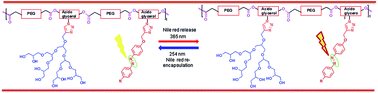
Photoresponsive polymeric amphiphiles were developed by first synthesizing the polyester chain via Novozym 435 catalyzed step growth, condensation polymerization of poly[ethylene glycol bis(carboxymethyl) ether]diethylester and 2-azidopropan-1,3-diol followed by grafting with 4′-butyl-4-propargyloxy(azobenzene) and [G2.0] polyglycerol dendron by using a ‘Click chemistry’ approach. The resulting polymers were observed to form supramolecular micellar aggregates in aqueous solution. The critical aggregation concentration (CAC) was determined via fluorescence measurements and using ‘Nile red’ as a probe. The nano-structures formed in the aqueous solution were characterized by dynamic light scattering (DLS) and cryo-TEM measurements. The encapsulation potential of polymeric amphiphiles for Nile red and curcumin as well as their release via trans–cis photoisomerization of the embedded azobenzene moiety was studied, by absorbance and fluorescence spectroscopy techniques. The developed polymeric micellar systems behave as efficient photoresponsive smart nanocarriers.
n: 1000 g mol−1) and azido glycerol via transesterification reaction under solventless conditions, developed by our group.22–27 The azido group of the base co-polymer was used for further functionalization using ‘grafting to’ approach via copper(I) catalyzed Huisgen-1,3-dipolar cycloaddition reaction,28 with 4′-butyl-4-propargyloxy(azobenzene) (2) and propargylated [G2.0] polyglycerol dendron (3), synthesized separately. The resulting polymeric systems were fully characterized and observed to self-assemble in the aqueous medium above the critical aggregation concentration (CAC), forming supramolecular architectures that closely resemble the micelles/micellar aggregates. The particle size and the CAC in the aqueous solution were determined by using the dynamic light scattering (DLS) supplemented by cryo-TEM and fluorescence measurements using ‘Nile red’ as a probe, respectively. The trans–cis photoisomerization of azo group in both the polymers was studied through absorbance spectroscopy in the aqueous solution, above the CAC values by irradiating at 365 nm. The kinetics of isomerization was compared by calculating their rate constants. The encapsulation potential of these polymers was evaluated for Nile red and curcumin but quantified for former only. The effect of trans–cis geometrical isomerization of azobenzene moiety was also evaluated on the release behavior of encapsulated guest. Nile red being a solvatochromic dye,29,30 is a most frequently used probe for studying the photo-controlled release using fluorescence spectroscopy. Trans–cis isomerization is mainly caused by the irradiation with UV light (approx. 350–400 nm)5,31–38 while cis–trans isomerization could occur either thermally by keeping the samples in dark31,33,34 or by irradiating them using visible light32,36–38 or with UV light (approx. 250–300 nm).9,39 The trans–cis isomerization mainly leads to an increase in dipole moment with the subsequent reduction in fluorescence intensity of Nile red, while the reverse occurs for the cis–trans isomerization. This is the most commonly used strategy for azopolymeric systems to study their release potential. The majority of the systems has reported the decrease in the fluorescence intensity of Nile red on trans to cis isomerization, however the literature suggests a lot of variation in the regain of fluorescence intensity due to re-encapsulation of the dye on subjecting the system to cis–trans isomerization either thermally or photochemically. Yu's group has synthesized azobenzene functionalized block co-polymers, responsive to both temperature and light which showed a complete regain of fluorescence intensity of Nile red in 20 s up to 9 cycles of trans–cis–trans reverse isomerization.36 Oriol et al. have synthesized amphiphilic linear-dendritic block copolymers (LSBCs) by linking poly(ethylene glycol) on one end and 4-isobutyloxyazobenzene (AZO)/hydrophobic chain (C18) on the other end of fourth generation, 2,2-di(hydroxymethyl)propionic acid (bis-MPA) based dendron, with varying composition of hydrophobic content i.e., PEG-b-d(isoAzo/C18) – 25/75, 75/25 and 50/50.33 They have reported that low content of azo moiety accelerates the rate of geometrical isomerization without any release of encapsulated Nile red while the release was triggered by increasing the azo content. A complete regain in the fluorescence intensity of Nile red on cis–trans isomerization keeping the UV irradiated solutions in dark for 24 h was observed for PEG-b-d(isoAzo/C18) – 25/75 while a partial regaining in fluorescence intensity leading to net release of encapsulated material was reported for PEG-b-d(isoAzo/C18) – 75/25 and 50/50 systems.33 Also, they have reported an another miktoarm copolymeric system having azobenzene moiety that showed complete regain in the fluorescence intensity of Nile red.34 However few other groups have emphasized the stirring effect along with the conditions required for cis–trans isomerization (photochemical or thermal restoration in dark) for regaining the fluorescence intensity of Nile red e.g. Das et al. have developed azobenzene based co-polymers forming spherical aggregates which showed only 50% regain in the fluorescence intensity of Nile red on photochemical cis–trans isomerization while it was completely restored by keeping the UV-irradiated solution in dark for 3 days with constant stirring.37 Xu's group has explained the partial regain in fluorescence intensity of Nile red for azobenzene functionalized polymeric systems, on trans–cis–trans reverse isomerization, on the basis of higher rate of micelle formation compared to drug loading.38 Lafleur's et al. has created non-phospholipid photosensitive liposomes which showed switchable photo-controlled
release of sulforhodamine B (SRB), on applying photo-cycles of alternate UV (350 nm) and visible light (450 nm).5 They have shown that release from SRB encapsulated liposomes could be initiated on irradiating with 350 nm light for 120 or 150 s and continues further without prolonged irradiation and could be stopped when desired by irradiating at 450 nm for 180 s thereby achieving the precisely controlled release.5 The systems which regain the fluorescence intensity of encapsulated dye on trans–cis–trans isomerization are reported to find applications in sensor technologies and catalytic systems,36 while the systems having partial regain indicate photoresponsive release and could find potential applications as carriers for drug delivery.5,33–35,38 Herein, we have studied the release profile of Nile red on trans–cis–trans photoisomerization as a function of photo-cycles using fluorescence spectroscopy for our recently developed azobenzene functionalized polymeric systems.
w,
n,
z and PDI (polydispersity index) of polymers were determined using an Agilent GPC system equipped with Agilent 1100 pump, refractive index detector, and PLgel columns. THF was used as an eluent at a flow rate of 1.0 mL min−1 and molecular weights were calibrated using polystyrene standards. CAC of the polymers was determined by fluorescence method using ‘Nile red’ as a model dye.40 A stock solution of Nile red was made by dissolving 0.3 mg of Nile red in 1 mL of ethanol to form 9.4 × 10⁻⁴ M solution. The Nile red solution (10 μL) was taken into empty vials and the ethanol was evaporated completely. The polymer solutions of different concentrations (0.25 mM to 0.12 mM) were also prepared up to 2 mL using 1× PBS buffer. These polymer solutions were allowed to stir for 1 h and then transferred to the vials having Nile red in the same sequence and allowed to stir overnight. The final concentration of Nile red was kept at 4.7 × 10⁻⁶ M. All the solutions were filtered using 0.45 μm polytetrafluoroethylene (PTFE) filter to remove non-encapsulated dye. The fluorescence spectra was recorded for the filtered clear solutions and the fluorescence data (λ: 636 nm) obtained was plotted with the log[polymer concentration] for different samples to obtain the CAC values. Malvern Zetasizer Nano ZS analyzer integrated with 4 mW He–Ne laser, λ: 633 nm based on back scattering detection (scattering angle θ = 173°) using an avalanche photodiode detector was used for characterizing the nanostructures (micelles/micellar aggregates) formed in the aqueous solution. The Zetasizer is equipped with a thermostated sample chamber. The samples were prepared in Milli-Q water at a concentration of 10 mg mL−1, well above their CACs, by vigorously stirring for 20 h. The samples were filtered using 0.45 μm PTFE filter and equilibrated for 1 h at room temperature before the measurements. The size of the nano-particles formed from polymer 4a in the aqueous solution was also evaluated by cryo-TEM image using a Tecnai F20 transmission electron microscope (FEI company, Oregon, USA) operated at 160 kV accelerating voltage. For cryo preparation, droplets of the sample solution were applied to 1 μm hole diameter perforated carbon film, covered with 200 mesh grids (R1/4 batch of the Quantifoil Micro Tools GmbH, Jena, Germany), which had been hydrophilized before use. The supernatant fluid was removed with a filter paper until an ultra thin layer of the sample solution spanned the holes of the carbon film. The samples were immediately vitrified by propelling the grids into liquid ethane at its freezing point (90 K) with a guillotine-like plunging device. The vitrified samples were then transferred to the microscope using a Gatan (Gatan, Inc., California) cryoholder and stage (model 626). The samples were kept at a temperature of 94 K. Imaging was performed using the low-dose protocol of the microscope at a calibrated primary magnification of 62
000× with the defocus set to 1.8 μm. Data were recorded by an Eagle 4k CCD-camera set to binning factor 2. The Nile red and curcumin encapsulation was studied by using UV and fluorescence spectra measurements. The curcumin was used in excess for encapsulation but quantification could not be done because of overlap of its absorbance spectra with that of polymers. The encapsulation of curcumin was done by following the solid dispersion method.41 The Nile red encapsulation was studied quantitatively along with its release triggered by photo irradiation with the help of UV and fluorescence spectroscopy. The Nile red encapsulation was done by following the film method.42 For that, 0.12 mg of Nile red was dissolved in a minimum amount of anhydrous THF and the solvent was allowed to evaporate so that a uniform layer of Nile red was obtained at the bottom of the vial. Then, 1 mL of aqueous polymer solution in 1× PBS buffer (having a concentration of 1 mg mL−1) was added to the vial having the thin film of Nile red. The final concentration of dye in the sample for encapsulation becomes 0.4 mM which is then stirred for 20 h. The non-encapsulated fraction of Nile red was removed by filtering it (twice), slowly through 0.45 μm pore size PTFE filter. The Nile red encapsulated samples were lyophilized and re-dissolved in anhydrous methanol for quantification. The absorbance (200–800 nm) spectra were recorded using Cary-300 series UV-vis spectrophotometer from Agilent Technologies in a thermostated UV cell (1 cm). Fluorescence emission spectra (450–800 nm) were recorded from Cary Eclipse fluorescence spectrophotometer using a variable slit system from 575–800 nm for Nile red, and 450 to 700 nm for curcumin. For recording fluorescence emission spectra, the excitation was done at 550 nm for Nile red, and at 420 nm for curcumin with excitation and emission slit set at 5 nm. Microsoft Excel® and Origin 6 softwares were used for data analysis.
![]() | ||
| Scheme 1 Synthesis of azobenzene functionalized polymer and its dendronized analog: (i) Novozym 435, 80 °C, vacuum, 60 h; (ii) [Cu(PPh3)3]Br, DIPEA, DCM/DMF. | ||
In a microwave vial were introduced 4′-butyl-4-azophenol (1.016 g, 4 mmol), KOH (448 mg, 8 mmol), water (10 mL) and propargyl bromide (0.90 mL, 8 mmol). The vial was cap-sealed and the mixture was heated at 70 °C for 15 min under the microwave. Then, after the extraction with ethyl acetate (3 × 20 mL) and evaporation of the solvent, the crude mixture was purified on flash chromatography on silica gel cartridge (40 g) with cyclohexane/ethyl acetate (100/0 to 70/30) to afford pure 4′-butyl-4-propargyloxy(azobenzene) (768 mg) in 66% yield. IR (neat): 3447.84, 3274.80, 2928.14, 2857.83, 2129.53, 1599.82, 1585.44, 1497.76, 1458.05, 1376.34, 1297.81, 1233.05, 1144.96, 1107.03, 1017.14, 840.78, 707.12, 677.65, 530.26 cm−1; 1H NMR (400 MHz, CDCl3, δ): 7.92 (d, J = 9.16 Hz, 2H, H-2′ & H-6′), 7.82 (d, J = 8.52 Hz, 2H, H-2 & H-6), 7.32 (d, J = 8.56 Hz, 2H, H-3′ & H-5′), 7.10 (d, J = 9.16 Hz, 2H, H-3 & H-5), 4.78 (d, J = 2.44 Hz, 2H, H-1′′), 2.70 (t, J = 7.96 Hz, 2H, H-1′′′), 2.57 (t, J = 2.44 Hz, 1H, H-3′′), 1.68 (q, J = 7.96 Hz, 2H, H-2′′′), 1.43 (m, J = 7.32 Hz, 2H, H-3′′′), 0.91 (t, J = 7.32 Hz, 3H, H-4′′′); 13C NMR (100.5 MHz, CDCl3, δ): 159.5, 150.9, 147.6, 145.9, 129.0, 124.4, 122.5, 115.1, 78.0, 75.9, 55.9, 35.5, 33.4, 22.3, 13.9; HRMS [M + H]+: calculated 293.1654, found 293.1653.
n (NMR analysis) = 9404 g mol−1; GPC (THF, 1 mL min−1):
w = 6510 g mol−1,
n = 4795 g mol−1, PDI = 1.4.
:
1 mixture of dichloromethane and dimethylformamide, as a viscous oil in 85% yield. IR (neat): 3433.74, 2921.04, 2872.40, 1752.23, 1599.30, 1460.09, 1350.38, 1297.61, 1249.37, 1105.52, 951.52, 846.54, 719.79, 563.63 cm−1; 1H NMR (400 MHz, CDCl3, δ): 8.04–7.88 (m, 4H, H-1′, H-2′′′ & H-6′′′), 7.77 (d, J = 7.96 Hz, 3H, H-2′′ & H-6′′), 7.30 (d, J = 7.92 Hz, 3H, H-3′′′ & H-5′′′), 7.10 (d, J = 8.56 Hz, 3H, H-3′′ & H-5′′), 5.28–5.13 (m, 4H, H-2′a & H-2a), 4.94–4.59 (m, 8H, H-1a & H-3a), 4.21–4.11 (m, 9H, H-α, H-α′ & H-2′b), 3.80–3.45 (m, 192H, –(OCH2CH2)n∼20), 2.69 (t, J = 7.92 Hz, 3H, H-1′′′′), 1.67 (q, J = 7.96 Hz, 3H, H-2′′′′), 1.42 (m, J = 7.32 Hz, 3H, H-3′′′′), 1.29 (t, J = 7.32 Hz, CH3 of ethyl end group), 0.95 (t, J = 7.32 Hz, H-4′′′′); 13C NMR (100.5 MHz, CDCl3, δ): 169.8, 169.4, 169.1 (–COO–), 159.8, 159.6, 150.2, 146.7, 146.6, 145.3, 145.3, 128.4, 123.9, 122.7, 121.9, 114.3, 70.3, 70.2, 69.8, 67.7, 67.7, 67.7, 62.7, 62.5, 61.9, 61.4, 60.8, 60.6, 57.8, 51.8, 51.1, 48.8, 34.9, 32.8, 31.2, 29.0, 28.7, 22.0, 21.6, 13.5, 13.3;
n (NMR analysis) = 10
612 g mol−1; GPC (THF, 1 mL min−1):
w = 5960 g mol−1,
n = 4312 g mol−1, PDI = 1.4.
n and
w values of 10
583 and 13
579 g mol−1, respectively, were obtained from GPC with polydispersity of 1.3 by using THF as a solvent and pullulan as standard (Fig. S9†). The degree of polymerization was found to be approximately 9 by both NMR and GPC results. The lower polydispersity (1.3 from GPC) of the co-polymer 1 compared to that given by Carother's equation (PDI = 1.89) in the Novozym 435 catalyzed step-growth polymerization could be explained by the unique chain length or mass selectivity of the lipase.27,45–48 The polymerization reaction carried out in bulk conditions also contribute to lower polydispersity as it increases in solvent-based polymerization reactions due to a greater rate of interchain transesterification reactions.46 The constitution of 4′-butyl-4-propargyloxy(azobenzene) (2) was confirmed by both 1H and 13C NMR spectra (Fig. S13†). In 1H NMR, the protons of the phenyl rings for trans and cis isomers of azobenzene appear separately with the protons of the cis isomer being slightly upfield compared to the protons of the former isomer (Fig. S13†). The integration of the peaks for the two isomers in 1H NMR suggests the trans
:
cis ratio of 19
:
1 for 4′-butyl-4-propargyloxy(azobenzene) (2). The base polymer 1 was then functionalized with 4′-butyl-4-propargyloxy(azobenzene) (2) and propargylated [G2.0] PG dendron (3) to synthesize functionalized polymeric amphiphiles. The functionalization of the base co-polymer ‘under Click regime’ was monitored by the disappearance of azide peak at around 2109 cm−1 in the IR spectra and the product was confirmed by the observance of aromatic protons for the azobenzene and triazole moieties in the 1H NMR spectrum (Fig. S14 and S15†). The 13C NMR spectrum also supported the formation of desired product by the observance of additional peaks in the aromatic region corresponding to 4′-butyl-4-propargyloxy(azobenzene) (2) and triazolyl carbons (Fig. S14 and S15†). The triazole ring formation also resulted in the characteristic downfield shift for H-2 of glycerol from δ 3.89–3.98 ppm to δ 5.12–5.28 ppm and H-1 and H-3 of glycerol from δ 4.00–4.40 ppm to δ 4.59–4.94 ppm (Fig. S14 and S15†). The 1H, 13C NMR spectra of 4′-butyl-4-propargyloxy(azobenzene) (2), both the polymeric amphiphiles (4a and 4b) and 2D HETCOR NMR spectra (Fig. S16) of 4b are given in ESI.†
![]() | ||
| Fig. 1 (A) Plot of fluorescence intensity vs. log[4a]; (B) plot of fluorescence intensity vs. log[4b] for calculating the critical aggregation concentration. | ||
The CAC of non-dendronized polymer 4a, was found to be 1.07 × 10⁻⁵ M while that of its dendronized analog 4b increases to 1.66 × 10⁻⁵ M. A similar behavior was also observed in our earlier reported polymeric systems having hydrophobic alkyl chain and polyglycerol based dendrons.26
![]() | ||
| Fig. 3 Photoisomerization of polymer 4a in aqueous solution above critical aggregation concentration, (A) irradiation at 365 nm (trans–cis); (B) irradiation at 254 nm (cis–trans). | ||
![]() | ||
| Fig. 4 Photoisomerization of polymer 4b in aqueous solution above critical aggregation concentration, (A) irradiation at 365 nm (trans–cis); (B) irradiation at 254 nm (cis–trans). | ||
In the UV absorption spectrum of polymer 4b above its CAC, the delocalized π–π* transitions in trans form get slightly blue-shifted to 337 nm (Fig. 4). This blue-shift could be explained owing to the more non-polar environment when inserted in the hydrophobic core,5,31,49 of the resulting micellar aggregates because of [G2.0] PG dendrons on the outer surface which acts as a shield to the azobenzene moieties in the core from the aqueous environment. Photoisomerization of an aqueous solution of azobenzene functionalized polymers 4a and 4b was carried both above and below their CAC to study the effect of trans–cis isomerization on the micellar aggregates, using a 6 W mercury lamp. The trans isomers could be isomerized to cis form on irradiating π–π* transitions with 365 nm light while the reverse cis–trans isomerization could be achieved either by allowing the cis isomer dominated samples to stay in darkness (thermal isomerization),7 owing to higher stability of trans isomers over cis or irradiating the n–π* transitions with visible light (400–500 nm),5 or π–π* transitions with UV light (250–270 nm),9 of the cis azobenzene. The trans–cis isomerization was studied by irradiating with the light of 365 nm as the molar absorptivity of the trans isomer is much higher than the later one,5 at this wavelength. Trans to cis isomerization led to a decrease in the intensity of the absorbance band at 351 and 337 nm for polymers 4a and 4b respectively, and an increase in the intensity of absorbance bands at 263 and 440 nm until a photostationary state is reached. The trans–cis photoisomerization above CAC occurs at a faster rate in polymer 4a (Fig. 3A) as its photostationary state is reached in approximately 60 seconds, while it takes 300 seconds for polymer 4b (Fig. 4A). The rapid of trans–cis photoisomerization of polymer 4a as compared to 4b may be due to more steric hindrance and embedding of azobenzene moieties,5 within the core of micelles in 4b, which is also supported by the observance of blue shift of 14 nm for its π–π* absorption maxima as compared to 4a. A reverse isomerization i.e., cis–trans was observed on irradiating the polymer solutions at 254 nm,9 this is accompanied by the enhancement in intensity of the absorbance band at 351 and 337 nm for polymer 4a and 4b respectively and decrease in the intensity of absorbance bands at 263 and 440 nm. After irradiation of the cis isomer at 254 nm above the CAC, approximately 95% of initial absorbance for π–π* transitions of azobenzene in trans form was recovered, indicated the efficient reversible isomerization in 300 and 600 seconds for polymers 4a and 4b respectively (Fig. 3B and 4B). A thermal isomerization of the cis isomer was also observed on keeping in darkness for 24 h or more. The rate of cis–trans isomerization was found to be comparable for both polymers. The photoisomerization rates were also compared in terms of their rate constants (ESI†).31,32 The rate of photoisomerization increases significantly below CAC (ESI†). This can be explained by the fact that the azobenzene moieties get embedded in the interior core of the micellar structure above their CAC which limits their mobility and also lowers the free volume availability, both of these factors are reported to be crucial for efficient isomerization.5,50 A similar observation has been made by Lo et al.,32 while studying the azobenzene moiety based micelles in organic solvent.
![]() | ||
| Fig. 5 (A) UV absorbance spectra of Nile red encapsulated samples in methanol; (B) fluorescence spectra of Nile red encapsulated samples in methanol. | ||
The dendronized polymer 4b exhibits better transport potential as compared to the non-dendronized analog (4a) with encapsulation efficiency of 1.87 and 1.45%, respectively. The transport efficiency of dendronized polymer 4b was found to be 7 and 3 times to the transport efficiency of corresponding dendronized polymers having C12 and C14 alkyl chain and [G2.0] PG dendron respectively.26 The higher encapsulation potential of azobenzene functionalized dendronized polymers compared to alkyl chain (C12/C14) functionalized polymers could be explained due to additional π–π interactions between the azobenzene moieties in the core of micellar aggregates and the guest molecule (Nile red).
This decrease in the fluorescence intensity could be explained partially by the release of dye from the core of micellar structures into the aqueous solution due to changes in the size and shape of internal core and partially by the increase in the polarity of the core resulting from the conversion of trans isomer into cis.33,34 However, on irradiating the polymer, predominant with the cis form, with 254 nm light, the fluorescence intensity of Nile red was observed to increase partially suggesting that dye is now available in the more hydrophobic microenvironment because of a decrease in the dipole moment of internal core as a result of reconversion of cis isomers into trans (Fig. 6B and 7B).33,34,36 However, a complete regain of the fluorescence intensity of Nile red could not be observed and this may be because some of dye molecules might have been released from the internal cavity due to change in its size and shape into the aqueous medium and thus could not be pushed back completely into the hydrophobic core. The rate of re-encapsulation of released Nile red from the aqueous solution into the micellar core is slow as compared to the rate of cis–trans isomerization process.37 Thus, this reduced fluorescence intensity on trans–cis–trans reverse isomerization marks the net release of Nile red molecules form micellar structures. The %release of Nile red was calculated by measuring the fluorescence intensity before and after trans–cis–trans reverse isomerization, and found to be approximately 50 ± 5 and 29 ± 5% for polymers 4a and 4b respectively on completion of 1st photocycle. The relatively higher release of dye by polymer 4a as compared to 4b could be explained due to more rapid and efficient trans–cis isomerization in case of polymer 4a above the CAC compared to 4b due to the absence of bulkier [G2.0] PG dendrons corona on the outer surface of micellar structures in the case of polymer 4a. The method and data used for the calculation of %release using fluorescence measurements is given in ESI.† The effect of the geometrical isomerization of azomoiety incorporated in the polymeric system was also studied by applying the photo-cycling process for non-dendronized polymer 4a. The isomerization of trans isomers into cis and then its re-conversion back to trans, thus constitute one photo-cycle. The further repetition of photocycles without allowing any thermal restoration does not lead to any further release of Nile red (Fig. 8, S4 and S6†). However, on keeping the Nile red encapsulated samples for thermal restoration in dark after performing one or more photocycles, led to a net increase in the intensity of Nile red (Fig. 8, 5A and 7A), this could be attributed to the partial re-encapsulation of dye from the aqueous environment into the micellar core. Thus, performing the photocycles by allowing a thermal restoration in dark for more than 2 h, (Fig. 8, S5 and S7†) led to a net release of Nile red, of the same magnitude (40–50%) as observed in 1st photo-cycle (Fig. S3†). The %cumulative release of Nile red as a function of photoisomerization cycles with different thermal restoration time intervals is given in Fig. 8, which is calculated from the fluorescence intensity of Nile red as in Fig. S3–S7,† for polymer 4a.
Footnote |
| † Electronic supplementary information (ESI) available. See DOI: 10.1039/c5ra08047a |
| This journal is © The Royal Society of Chemistry 2015 |