DOI:
10.1039/C5RA07613G
(Paper)
RSC Adv., 2015,
5, 54352-54363
A reversible turn-off fluorescence probe (HNAPP) for Zn(II) ion and inorganic phosphate ions (H2P and HP) at physiological pH†
Received
26th April 2015
, Accepted 5th June 2015
First published on 8th June 2015
Abstract
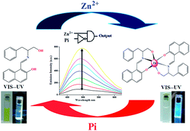
A simple and low-cost new Schiff base compound (HNAPP) of 2-hydroxynaphthaldehyde and 2-amino-3-phenyl-1-propanol was synthesized and characterized by 1H NMR and mass spectroscopic technique. The optimized structure of the probe in different polarity solvents and the potential energy scan reveal that the probe is stable as an enol tautomer in polar solvent, while in less polar solvent the keto tautomer is more prominent. In dichloromethane, the keto tautomeric form of the probe undergoes an excited state intramolecular proton transfer, ESIPT, into a stable enol structure that reveals unusual relaxation routes after electronic excitation. The results, accompanied by TDDFT calculations, are used to diagram the relaxation routes of an excited HNAPP molecule. The probe exhibited highly selective and sensitive turn-on fluorescence emission towards Zn2+ over other common metal ions in a physiological pH window with a 2
:
1 binding mode. The association constant, Kassoc, was observed at 5.2 × 10⁴ M−1. The fluorescence decay constant (s) values were determined from time-resolved fluorescence study. The addition of inorganic phosphate ions quenched the fluorescence intensity of the complex at different pH values, enabling the receptor HNAPP to act as a reversible chemosensor and a pH modulator. On the basis of these observations, I developed a unique molecular system capable of performing logic functions such as INHIBIT by simply varying the level of various ionic inputs in a systematic manner.
Introduction
The development of chemosensors that have the capability to selectively recognize and sense metal ions is one of the most challenging fields in chemistry.1 Amongst the available detection methods, chemosensors based on ion-induced fluorescence changes have become predominantly attractive in terms of sensitivity, selectivity, response time, simplicity, high degree of specificity and low detection limit.2 Zinc is the second-most abundant transition metal in the human body after iron and plays important roles in various pathological processes.3 So far, the search for reagents that can efficiently act as fluorescence probes for Zn(II) at physiological pH has been an active area of research.4
The Zn(II) ion is spectroscopically as well as magnetically silent due to its 3d10 electronic configuration. However, it can modulate ligand luminescence properties by means of chelation-enhanced fluorescence (CHEF).5 The generated CHEF effect is regulated by photoinduced electron transfer (PET) mechanism. In this context, the Schiff base complexes of Zn(II) ion are particularly interesting due to their potential photochromic applications.6
In addition, the development of chemosensors that can discriminate Zn2+ from Cd2+ is still a challenge7 because cadmium and zinc are in the same group of the periodic table and have similar properties, which usually cause similar spectral changes after interacting with chemosensors. In this sense, the design and synthesis of fluorescent selective Zn2+ chemosensors are of great interest. Despite some commercial fluorescent probes for Zn2+,8 the design of facile, easy to synthesize, nontoxic Zn2+-selective probes is still a challenging task, and there is a need to design and synthesize such chemosensors that are small molecules and highly sensitive for real-time detection in biological systems at physiological pH. My synthesized probe, HNAPP, selectively detects Zn2+ ions over the presence of other cations, especially Cd2+, at physiological pH window.
Phosphates are common target anions, as they play significant roles in many biological processes such as cellular ATP hydrolysis, DNA and RNA polymerization, and many enzymatic reactions.9 Recently, the detection of inorganic phosphate ions, such as monohydrogen phosphate (HP) and dihydrogen phosphate (H2P) has become an important issue for cancer research and for rheumatological disorders that arise due to the accumulation of crystals of calcium pyrophosphate dihydrate in the connective tissues.10 Thus, the specific recognition and sensing of inorganic phosphate under physiological conditions is of immense significance.
In this report, I show that the aminopropanol–naphthalene based ligand HNAPP is highly fluorescent when forming a complex with Zn2+. This [Zn–NAPP2] complex has been utilized as a receptor for both HP and H2P in mixed aqueous medium by a metal displacement approach, which results in the quenching of fluorescence. Some aspects of these unanticipated findings were streamlined by DFT and TDDFT calculations.
Experimental section
Materials
The transition metal salts used in this investigation are as follows: Zn(ClO4)2·6H2O, Cr(ClO4)3·6H2O, Cu(ClO4)2·6H2O, Mn(ClO4)2·6H2O, Ni(ClO4)2·6H2O, Co(ClO4)2·6H2O and Cd(ClO4)2·6H2O. The metal salts were procured locally and used as received. Perchlorate salts were preferred because of the low coordinating ability of the anionic counterpart. Tetrabutylammonium (TBA) salts of the respective anions ([A] = F−, Cl−, Br−, I−, OAc−, H2PO4− (H2P), HPO42− (HP), P2O74− (PPi) and CN−; “A” stands for anion) and sodium salts of ATP, ADP and AMP were used as received from Sigma-Aldrich, U.S.A. MES (2-(N-morpholino)ethanesulfonic acid, HEPES (4-(2-hydroxyethyl)piperazine-1-ethanesulfonic acid), TRIS (tris(hydroxymethyl) aminomethane), potassium chloride and hydrochloric acid were purchased from Cyno-chem, India. All other solvents and reagents, such as dichloromethane (DCM), methanol (MeOH), acetonitrile (CH3CN) and dimethylformamide (DMF), were of spectroscopic grade (Spectrochem, India) and used after proper distillation. The solvent was found free from impurities and appeared transparent in the spectral region of interest. The purity was also verified by recording the emission spectra in the studied spectral region. CDCl3 for NMR experiments was used as received from Sigma-Aldrich, U.S.A.
Caution! Perchlorate salts are highly explosive and should be handled with care and in small amounts.
Physical measurements
UV-vis spectra were recorded on a PerkinElmer LAMBDA 25 spectrophotometer. Electrospray ionization mass spectrometry (ESI-MS) measurements were done on a Micromass Qtof YA 263 mass spectrometer. The molar conductivity was determined using Systronics Conductivity Meter 304 in acetonitrile solution at room temperature. Elemental analyses (C, H, N) were performed on a PerkinElmer 2400 series II analyzer. The emission data were collected on a PerkinElmer LS 55 fluorescence spectrometer. All pH measurements were made with a pH-10C digital pH meter. Quantum yields of the ligand and complex were determined in freeze–pump–thaw-degassed solutions of the ligand and complex by a relative method using quinine sulfate in the same solvent as the standard11 and calculated using eqn (1),12a where Φr and Φstd are the quantum yields of unknown and standard samples [Φstd = 0.54 (at 298 K) in methanol at λex = 350 nm]; Ar and Astd (<0.1) are the solution absorbance at the excitation wavelength (λex); Ir and Istd are the integrated emission intensities; and ηr and ηstd are the refractive indices of the solvent. |  | (1) |
The fluorescence decay data were collected on a Hamamatsu MCP photomultiplier (R3809) and analyzed by using IBH DAS6 software. The observed decays of the complex fitted well with a bi-exponential function as in eqn (2) and (3), where τ1 and τ2 are the fluorescence lifetime, and α is the pre-exponential factor. For the fits, reduced χ2 values are around one, and the distribution of weighted residuals was random among the data channels. τf is the mean fluorescence lifetime (meanings of the symbols are as usual).12b All absorption and emission spectral measurements were performed with proper background corrections and with freshly prepared solutions only.
| I(t) = [α1 exp(−t/τ1) − α2 exp(−t/τ2) | (2) |
Computational details
I also calculated and analyzed the singlet ground state natural transition orbitals (NTOs) derived from TDDFT results and compared them with the ground state molecular orbitals (MOs) obtained from DFT calculations. The computational modeling of the NMR parameter is also of abiding interest, and such DFT calculations have emerged as a promising approach for the prediction of nuclear shielding and coupling constants of NMR active nuclei.13 Thus, i computed the proton NMR chemical shifts and also the 1H–1H spin–spin coupling constant using the gauge-independent atomic orbital (GIAO)-DFT method, which was aimed at providing the definitive characterization of the probe. The geometric structure of the ligand (HNAPP) in the singlet ground (S0) and excited states (S1) were optimized by the DFT14 and time-dependent DFT (TDDFT)15 method with B3LYP exchange correlation functional16 approach associated with the conductor-like polarizable continuum model (CPCM).17 The geometry of the probe was fully optimized in different polarity solvents without any symmetry constraints. The vibrational frequency calculation was also performed for ligands to ensure that the optimized geometries represent the local minima and that there are only positive eigenvalues. On the basis of the optimized ground (S0) geometry of the ligand, the absorption properties in DCM media were calculated by TDDFT approach. To calculate the stability of the keto tautomeric form over the enol form for the ligand in ground S0 state, I performed the potential energy scan according to the “distinguished coordinate approach,”18i.e., by specifying a reaction coordinate (in the present case, it is the coordinate for translocation of the proton from Ndonor to Oacceptor, i.e., elongation of the Ndonor−H bond axis) along which energy change is observed. For the ground S1 state, all of the other degrees of freedom are relaxed without imposing any symmetry constraints. For H atoms, I used the 6–31(g) basis set and for C, N and O atoms, the 6–31 + g as basis set for the optimization of the ground state geometries. The calculated electronic density plots for frontier molecular orbitals were prepared by using the GaussView 5.0 software. All the calculations were performed with the Gaussian 09W software package.19 GaussSum 2.1 program20 was used to calculate the molecular orbital contributions from groups or atoms.
In addition, the 1H NMR properties of the HNAPP were calculated with the magnetic field perturbation method using the GIAO algorithm21 with the NMR = spin–spin keyword incorporated in the Gaussian 09W program. The relative chemical shift of a given nucleus X in the molecule was defined as δcalcX [ppm] = σrefX – σcalcX, where TMS was used as a reference molecule optimized at the same level of theory.22 In order to account for the solvent effect, I used the integral equation–formalism polarizable continuum model (IEFPCM) method.23a,b
Synthesis of probe (HNAPP)
To a methanolic solution (20 mL) of 2-hydroxynaphthaldehyde (345 mg, 2 mmol), 2-amino-3-phenyl-1-propanol (305 mg, 2 mmol) was refluxed in water bath for 2 hours. After cooling to room temperature, the solvent was removed under reduced pressure. The crude mass was then subjected to column chromatography on a silica gel column (60–120 mesh). A light yellow band was eluted using 5% ethyl acetate in hexane solution. A yellow solid was obtained after removal of solvent under reduced pressure to afford the desired ligand. Yield: 433 mg (71%). Elemental anal. calcd. for C20H19NO2: C, 78.66; H, 6.27; N, 4.59; found: C, 78.30; H, 6.25; N, 4.82. 1H NMR {300 MHz, CD3Cl, δ (ppm), J (Hz)}: 14.20–14.15 (ArOH, bs), 8.41 (N
CH2, s), 7.52 (1H, d, J = 8.3 Hz), 7.32–7.20 (8H, m, ArH), 7.11–7.06 (1H, m, ArH), 6.68 (1H, d, J = 9.2 Hz), 4.63–4.61 (AlOH, bs), 3.93–3.90 (PHCH2, m), 3.76–3.70 (CH2OH, m), 3.06–2.88 (1H, m). ESI-MS (CH3CN): m/z calcd. 306.1416, found: 306.1296 (100%) (HNAPP + H)+.
Synthesis of complex
[Zn-NAPP2], 1. An aqueous solution of Zn(ClO4)·6H2O (0.037 g, 0.1 mM) was added to a methanolic solution of HNAPP (0.061 g, 0.2 mM), and the reaction mixture was warmed in a water bath with care. A dilute methanolic solution of Et3N (0.040 g, 0.40 mM) was then added to the reaction mixture to maintain at a pH of 7–8, then refluxed for 2 hours in air and allowed to cool to room temperature. The reaction mixture was then filtered, and the volume of solvent was reduced via rotary evaporator to obtain a colorless residue. The residue was filtered and washed by methanol and then dried in vacuum. The solution was kept for slow evaporation, which yielded a colorless crude product in good amount. Yield: 47 mg (70%); elemental anal. calcd. for C40H34N2O4Zn: C, 71.48; H, 5.10; N, 4.17; found: C, 71.34; H, 5.17; N, 4.15. 1H NMR {300 MHz, CD3CN, δ (ppm), J (Hz)}: 8.69 (N
CH2, s), 7.72 (1H, d, J = 6 Hz ArH), 7.46–7.13 (9H, m, ArH), 6.69 (1H, d, J = 10 Hz ArH), 3.65 (2H, PHCH2), 3.47 (2H, CH2OH) and 3.06–2.88 (1H, m). ESI-MS (CH3CN): m/z calcd. 673.2045, found: 673.2388 (100%) (1 + 3H)+. Molar conductance, ΛM: (CH3CN) 265 Ω−1 cm2 mol−1.
Results and discussion
Synthesis
The tridentate N,O,O ligand (HNAPP) was made to react with Zn(ClO4)2·6H2O in a ratio of 2
:
1 in methanol at room temperature in air to produce complexes of composition [Zn–NAPP2] 1 in excellent yield. NAPP is the deprotonated form of the ligand. A schematic representation for the synthesis of ligand and complex is given in Scheme 1.
 |
| Scheme 1 Synthesis of receptor and complex. | |
Conductance measurement and mass spectra
The molar conductivity of the complex was determined in acetonitrile solution at room temperature. The value of the molar conductivity was 265 S cm2 mol−1, which corresponded to a 2
:
1 type electrolyte. The ligand and complex were diluted with acetonitrile for mass spectrometry. Mass spectral analysis in the positive ion mode showed a major peak at m/z (%) = 306.1296 (100), which is assigned to the monocationic protonated ligand [HNAPP + H]+. For the complex, a major spectra appeared at m/z (%) = 673.2388 (100), allocated to the bischelate monomeric form of [Zn–NAPP2+3H]+. The mass spectra are given in ESI Fig. S1 and S2†.
NMR spectra
The ligand and complex are diamagnetic and display well resolved 1H NMR spectra in CD3Cl solution. HNAPP shows two distinguishable broad peaks at 14.20 and 4.6 ppm (Fig. 1). The peak that appeared at the deshielding region is assigned to phenolic proton, whereas the shielding region peak accounts for alcoholic proton. Both peaks completely disappear during complex formation (ESI Fig. S3†). The singlet sharp peak near 8.41 ppm is from the azomethine proton of the ligand going to the downfield region at around 8.69 ppm during complexation. The aromatic proton spans are 7.11–7.06 ppm and 7.46–7.13 ppm for the ligand and complex, respectively. It was observed that due to complex formation, almost all the aromatic protons suffer in the deshielding regions, and the larger splitting of the aromatic span is attributed to symmetry lost during complexation. The methylene protons appeared at 3.92 ppm and 3.73 ppm for the ligand. The correlation between the experimental and calculated 1H NMR chemical shift of HNAPP is shown in the inset of Fig. 1 as a representative case.
 |
| Fig. 1 The 1H NMR spectra of HNAPP with the spectral nature of both aromatic and aliphatic regions. Inset: linear correlation between the experimental and calculated 1H NMR chemical shifts of HANPP in aliphatic and aromatic regions. | |
Ligand photophysical study
The UV-vis absorption spectrum of the probe shows two well resolved peaks at around 400 and 300 nm in solvents of different polarity at room temperature. The peak around 400 nm is split into two distinguishable humps. As shown in Fig. 2, HNAPP shows a low-energy band at approximately 400 nm, which is attributed to the n–π* electronic transition, whereas the prominent high-energy band at around 300 is due to the π–π* electronic transition. Li et al. showed a ground-state keto–enol tautomerisation by varying the percentage of water in ethanolic medium.23c Herein, I also established a similar type of ground state equilibrium by using different polarity solvents. The natures of electronic transitions are well established from TDDFT studies on the ground-state optimized structure (S0) of keto and enol forms of the ligand. The theoretical UV-vis spectra of enol and keto forms are given in ESI (Fig. S4 and S5†). The spectra obtained from the keto form shows a peak at 315 nm with higher oscillating strength, while for the enol form the peak appears at 405 nm (Table S1†). The absorbance intensity of low-energy band transitions corresponding to electronic transition n–π* of the enol form and the high-energy band corresponding to electronic transition π–π* of the keto form of HNAPP showed a dependence on the nature of solvent polarity. The dynamic keto–enol equilibrium preferred the enol tautomeric form in the more polar solvent, whereas the keto form is predominant in the less polar solvent. The selective O–H, C–O, N–H and N–C bond distance of the ground-state optimized structure (S0) of the probe in different polarity solvents clearly indicates that polarity of the solvent controlled the equilibrium constant of keto–enol tautomerization (Table 1). The ground-state optimized structure in DCM, MeOH and DMF are given in ESI Fig. S6†.
 |
| Fig. 2 The absorbance spectra of HNAPP (1 × 10⁻⁵ M) in different solvents at room temperature. | |
Table 1 Optimized structural parameter of HNAPP in different polarity solvents
Solvent |
H–O (Å) |
C–O (Å) |
N–H (Å) |
N–C (Å) |
DCM |
1.96 |
1.27 |
1.03 |
1.33 |
MeOH |
1.06 |
1.36 |
1.60 |
1.29 |
DMF |
0.96 |
1.37 |
1.80 |
1.29 |
The absorption energies associated with oscillator strengths, the main configurations and their assignments calculated using TDDFT method using the S0 geometry for HNAPP are discussed here, and the related data are given in Table 2. In the case of ground state (S0), the electron density at HOMO and HOMO−1 are delocalized over the naphthalene and phenyl moieties, respectively, while in the case of LUMO it originates from the contribution of both naphthalene (61%) and azomethine (35%) moieties. The calculated absorption bands located at 418 and 328 nm are in good agreement with the experimental result. The lower-energy band can be assigned to the S0 → S1 with distribution of electron density of non-bonded oxygen to the antibonding orbital of the azomethine moiety (n–π*), whereas the higher-energy band arises due to S0 → S3, transitions, with allocation of electron density of the π orbital of the phenyl ring to the π* orbital of naphthalene and azomethine moieties. Frontier molecular orbitals involved in the UV-vis absorption for HNAPP are given in Fig. 3.
Table 2 Selected parameters for the vertical excitation (UV-vis absorptions) and the emission of HNAPP; electronic excitation energies (eV) and oscillator strengths (f); configurations of the low-lying excited states of HNAPP; calculation of the S0–S1 energy gaps based on optimized ground-state geometries (UV-vis absorption) and the optimized excited-state geometries (fluorescence). (DCM used as solvent)
Process |
Electronic transitions |
Composition |
Excitation energy |
Oscillator strength (f) |
CI |
λ
exp (nm) |
Absorption |
S0 → S1 |
HOMO → LUMO |
2.9848 eV (418 nm) |
0.6669 |
0.70493 |
400 |
S0 → S3 |
HOMO−1 → LUMO |
3.7698 eV (328 nm) |
0.3798 |
0.64327 |
300 |
HOMO−3 → LUMO |
0.25444 |
Emission |
S1 → S0 |
HOMO → LUMO |
2.4653 eV (503 nm) |
0.1381 |
0.69282 |
492 |
 |
| Fig. 3 Frontier molecular orbitals involved in the UV-vis absorption and emission of HNAPP. CT stands for conformation transformation. Excitation and radiative decay process are marked as solid lines, and the non-radiative processes are marked by dotted lines. | |
The fluorescence spectral measurement for the receptor in the absence of Zn2+ ions was carried out in DCM at room temperature (ESI Fig. S6†). Free HNAPP upon excitation at 400 nm has shown an emission band at 492 nm with a Stokes shift of 92 nm and quantum yields (ΦF) around 0.051. The ground S0 state of HNAPP in DCM is the keto form (Fig. S4c†), whereas the S1 state is the enol form (Fig. S4d†).
It was clear from the S1 state optimized structure in DCM and the fluorescence excitation spectrum that there were two routes of creation of the excited enol tautomer: tautomerization via the excited keto form (the ESIPT route) and direct excitation of the keto tautomer. In order to study the emission property, the potential energy scan of HNAPP was performed, which reveals that the two tautomeric forms exist at ground state, in which the enol form is more stable than keto tautomeric form over the amount of energy (ΔG) 0.1338 eV mol−1. The transition energy required (ΔG#) for this keto–enol tautomerization is 0.1679 eV mol−1 (Fig. 4a).
 |
| Fig. 4 (a) Potential energy curves for HNAPP calculated at DFT/B3LYP level. (b) Spectrophotometric titrations of HNAPP (10 μM) with various equivalents of Zn2+ at room temperature ([Zn2+] = (0–7 × 10⁻⁶ M). Insets: the corresponding titration profiles confirm the 2 : 1 (HNAPP : Zn2+) binding stoichiometry (the absorbance values take the corresponding wavelength at 250 nm). | |
The energy gap between the S0 and S1 state, calculated with the optimized S1 state geometry, is the fluorescence emission wavelength. This geometry relaxation upon photo excitation imparts remarkable effect on the energy level of the molecular orbitals. In the case of HNAPP, the LUMO is stabilized by 0.75 eV at the S1 state geometry compared to that at S0 state geometry, while the HOMO is destabilized by 0.36 eV for S1 state geometry compared to that at S0 state geometry. As a result, the energy difference between the HOMO and LUMO is greatly decreased at the S1 state compared to that at the S0 state, and this geometry relaxation is the main origin of the large Stokes shift. The fluorescence wavelength was calculated as 503 nm (in DCM), which is in very good agreement with the experimental value of 492 nm (Fig. 3).
UV-vis titration of HL with Zn(II)
Fig. 4b shows a representative UV-vis titration curve of HNAPP with various concentrations of Zn2+ ions. The probe showed two humps at around 400 nm. It has been observed that the absorbance intensity of both humps decreases with increasing concentration of Zn2+ ion with some blue shift (∼10 nm). A similar trend is also observed at 320 nm, whereas the reversed trend appears at the shorter wavelength of 250 nm, and the yellow color of the ligand finally becomes colorless, which can be observed by the naked eye. The whole process has been passing through four distinguishable isosbestic points at 240, 292, 348 and 384 nm, respectively. It is to be noted that there is no change in absorption intensity at 250, 320 and 400 nm after the addition of an excess of ∼1.0 equivalent of Zn2+ ion with respect to ∼2.0 equivalent of HNAPP. Jobs plot of the maximum absorption intensity shows the [M]/([M] + [L]) value is 0.321, which indicates 2
:
1 complex formation of HNAPP with Zn2+ (inset of Fig. 4b).
Fluorogenic Zn(II) sensing
To determine the practical applications, the fluorescence response behavior of the probe was examined upon treatment with various metal ions in 10 mM HEPES aqueous buffer–CH3OH (3
:
2, v/v). Fig. 5 shows the bar diagram of fluorescence intensity of HNAPP in the presence of different metal ions. Only Zn2+ resulted in a pronounced fluorescence enhancement, whereas other transition metal ions including Cu2+, Ni2+, Co2+, Mn2+, Cd2+ and Cr3+ did not induce fluorescence, as there is a probability of an electron and energy transfer between the metal ion and probe. When the experiment was carried out with ubiquitous intracellular metal ions such as K+, Na+ and Ca2+, which exist at very high concentrations inside the cell, no significant fluorescence was observed, even at concentrations that were 10-fold higher than Zn2+ ion concentration (Fig. 5, blue bar). Metal-ion selectivity was also examined to probe if HNAPP could be used as a selective probe for Zn2+ in the presence of other competitive cations found in biological systems. Emission spectra were measured for a 2
:
1 mixture of HNAPP and Zn2+ in the presence of other metal ions. The prominent fluorescence enhancement observed upon mixing HNAPP and Zn2+ remained unchanged even in the presence of a 10-fold excess of metal ions such as K+, Na+, and Ca2+ (Fig. 5, purple bar).
 |
| Fig. 5 Metal-ion sensitivity of HNAPP in the presence of different metals. Blue bars represent the fluorescence sensitivity of HNAPP (2 × 10⁻⁵ M) to various metal ions. Purple bars represent the fluorescence response measured after the addition of Zn2+ (1 × 10⁻⁵ M) to the indicated metal ion–complex 1 (1 : 1 for transition metal ions and 10 : 1 for alkali and alkaline earth metal ions) in 10 mM HEPES aqueous buffer–CH3OH (3 : 2, v/v) following excitation at 390 nm (slit width 5 nm). | |
This confirms the excellent selectivity of HNAPP for Zn2+ over other abundant cations. Notably, the fluorescence intensity of the zinc complex was partially quenched in the presence of metal ions such as Mn2+, Cr3+, Ni2+, Cd2+ and Co2+. It is interesting to note that the fluorescence intensity of HNAPP in the presence of Zn2+ is significantly quenched by Cu2+ metal ion, probably due to strong binding affinity towards the ligand.
Titration of HNAPP with quencher
An exceptional case appeared in which the fluorescence intensity of HNAPP in the presence of Zn2+ is slightly quenched by Cu2+ metal ion, probably due to strong binding affinity towards the probe. Fig. 6a shows the fluorescence spectra of HNAPP in the presence of different concentrations of Cu2+ excited at 400 nm in aqueous buffer–CH3OH (2
:
1, v/v) at pH 7.2. The ligand showed one intense peak at 485 nm during excitation at 400 nm with larger slit width, and the fluorescence intensity gradually decreases in the presence of Cu2+. It is observed that there is no change in the intensity at 485 nm after the addition of excess of 1.0 equivalent of Cu2+ ion with respect to 2.0 equivalent of HNAPP (Fig. 6c).
 |
| Fig. 6 (a) Fluorescence titration of HNAPP (20 μM) with gradual addition of Cu2+ (0–12 μM) in aqueous buffer–CH3OH (3 : 2, v/v) at pH 7.2. (b) A plot of (F0 − F)/F against [Cu2+] for HNAPP. Binding constant, K (±5%), value is determined from the slope of the plot as 11.68 × 10⁶ M−1. (c) Emission intensity at 485 nm vs. [Cu2+]; [slit width (Ex/Em: 10/10)]. | |
This result corroborated with the formation of 1
:
2 (M
:
L) complex in solution. This change in fluorescence intensity at 485 nm is used to estimate K for the binding of Cu2+ to HNAPP by eqn (4).24a,b
|  | (4) |
here,
F0 and
F are the fluorescence intensity of the probe at 485 nm in the absence and the presence of different concentrations of Cu
2+, respectively. The inset of
Fig. 6b shows a linear plot passing through the origin for (
F0 −
F)/
F vs. [Cu
2+] (
n = 1). From this, according to
eqn (4), the value of
K was estimated 11.68 × 10
6 M
−1 for Cu
2+ towards HNAPP. The reaction of Cu
2+ with the chelating agent HNAPP induced rigidity in the resulting molecule and produced a large CHEQ effect, which further induced the decrease in fluorescence intensity.
Titration of HNAPP with Zn(II)
In the fluorescence titration experiment, the receptor was subjected to excitation at 405 nm and was monitored after each stepwise addition of Zn2+ ion to the solution in aqueous buffer–CH3OH (3
:
2, v/v) at pH 7.2 (Fig. 7). The probe showed comparably weak emission with respect to the complex probably because of quenching by the occurrence of a photo-induced electron transfer (PET) process due to the presence of a lone pair of electrons of the donor atoms in the ligand (N, O donor). A gradual enhancement (∼12 fold) of the fluorescence intensity was observed at 492 nm upon increasing the concentration of Zn2+ ions (Fig. 7a). The reaction of Zn2+ with the chelating agent HNAPP induced rigidity in the resulting molecule, reduced the PET mechanism and produced a large CHEF effect that tends to produce a strong ‘switch on’ blue fluorescence. Inset of Fig. 7c shows a plot of emission intensity at 492 nm against the titration of Zn2+ from 0 to 1.2 equivalents. It is clear from the plot that the fluorescence intensity reaches a plateau after the addition of exactly 1.0 equivalent of Zn2+ ions, and there is no significant enhancement of the fluorescence intensity upon further addition of Zn2+. This result strongly corroborates the formation of 1
:
2 (M
:
L) complex.
 |
| Fig. 7 (a) Fluorescence titration of HNAPP (20 μM) with gradual addition of Zn2+ (0–12 μM) in aqueous buffer–CH3OH (3 : 2, v/v) at pH 7.2. (b) A plot of (F∞ − F0)/(Fx − F0) against 1/[C] for HNAPP. Binding constant, K (±5%), value determined from the reciprocal of the slope of the plot as 5.2 × 10⁴ M−1. (c) Emission intensity at 492 nm vs. [Zn2+]. | |
The binding constant values have been determined from the emission intensity data24c using the Benesi–Hildebrand equation: 1/ΔF = 1/ΔFmax + (1/K[C])(1/ΔFmax) to establish the binding abilities of the probe with Zn2+. Here, ΔF = Fx − F0 and ΔFmax = F∞ − F0, where F0, Fx and F∞ are the emission intensities of the probe used in the absence of Zn2+, at an intermediate Zn2+ concentration, and at a concentration of complete interaction, respectively, and where K is the binding constant and [C] the Zn2+ concentration. As shown in Fig. 7b, the intercept value 1.05 ± 0.5, close to 1.0, also manifests the self-consistency of the experimental data. Therefore, the ligand association constant K is the reciprocal of the slope, 5.2 × 10⁴ M−1. The 1
:
2 complex formation in solution is further confirmed by ESI-MS+-(m/z) analysis (see Experimental section).
Effect of pH
In addition to metal ion selectivity, for many biological applications, it is very important that the probe can be suitable for measuring specific cations and anions in the physiological pH range. Therefore, I measured the fluorescence intensity of HNAPP in the absence and presence of Zn2+ at various pH values. As shown in Fig. 8, the emission intensity of HNAPP slightly increases gradually at first and then decreases in acid conditions with maximal fluorescence occurring at pH ∼5.0. Essentially no change can be observed under neutral and alkaline conditions (pH 7–13). However, the Zn2+-induced fluorescence enhancement of HNAPP continues to increase in the pH range 1.2–6.5, which may be due to competition with H+.25 The emission of [Zn–NAPP2] maintains fairly intense from pH ∼7 to pH ∼8.7 and is ∼70% quenched at higher pH (∼13). The observed decreasing response at pH > 9.5 may be due to the formation of Zn(OH)+ or Zn(OH)2, which thus reduces the concentration of [Zn–NAPP2]. However, HNAPP exhibits satisfactory Zn2+ sensing abilities when the pH is in the range of 6.5–8.5, indicating that HNAPP possesses the highest sensing ability in an environment similar to serum (pH ca. 7.3).
 |
| Fig. 8 Fluorescence intensities of HNAPP and Zn-NAPP2 at various pH values at room temperature, CH3CN–H2O (1 : 4, v/v), λex = 400 nm. A starting solution (CH3CN–H2O) of 100 mM NaOH and 10 mM NaCl (pH ∼ 13) was used for pH titrations. The pH values were lowered to ∼1.3 by the addition of aqueous HCl (CH3CN–H2O). | |
Lifetime measurement
Time-resolved luminescence spectra proved to be an important tool to understand the decay process and the emissive nature of the complex. Thus, time-resolved luminescence spectra were recorded for both the ligand and the complex in DCM solvent at room temperature using 370 nm excitation. The ligand shows a monoexponential while the complex shows a biexponential decay nature (see Fig. 9). The average lifetime, τf, (τf = α1τ1 + α2τ2, where α1 and α2 are relative amplitudes of the decay process) has been used to compare the excited-state stability of the ligand and the complex, and the values are 0.5 ns for HNAPP and around 7 ns for the complex. The value of τ1 lifetime of the complex was of similar order with the lifetime of the corresponding ligand, which revealed that in excited state, the biexponential decay nature of the complex arises from the contribution of the ligand moiety and the complex itself. The radiative and non-radiative rate constant for the ligand and complex are evaluated in Table 3. The non-radiative decay rate constant is much higher than the radiative decay rate constant for the ligand, making it a weak emitter. On the other hand, during complexation, the smaller knr value (ten times lesser) for the complex compared to that of the isolated ligand suggested the enhancement of fluorescence intensity.
 |
| Fig. 9 Changes in the time-resolved photoluminescence decay of HNAPP and Zn-NAPP2 in DCM at room temperature obtained with 370 nm excitation. | |
Table 3 Photophysical parameters of the ligand and complex in DCM at room temperature
|
Φ
F
|
k
r, s−1 (× 109) |
k
nr, s−1 (× 109) |
τ
1, ns |
τ
2, ns |
τ
av, ns |
χ
2
|
HNAPP |
0.051 |
0.102 |
1.89 |
0.5 |
— |
0.5 |
0.97 |
Zn–NAPP2 |
0.45 |
0. 064 |
0.07 |
2.7 |
16.1 |
7 |
1.06 |
Anion selectivity of the complex
To evaluate whether the Zn–NAPP2 complex could be used as an anion-selective fluorescent system, the response of the Zn–NAPP2 complex toward physiologically and environmentally important anions is given in Fig. 10a. Here, dihydrogen phosphate (H2P) and monohydrogen phosphate (HP) are abbreviated as inorganic hydrogen phosphate (Pi). A turn-off fluorescence response was observed for the emission band with a maximum at 492 nm in the presence of externally added solution of Pi at pH 7.2 (Fig. 10b). Interestingly, other anions and nucleotides like F−, Cl−, Br−, I−, AcO−, PPi, CN−, ATP, ADP and AMP do not react with Zn–NAPP2 and are thus unable to extrude the zinc ion from the complex. The Zn–NAPP2 system revealed a remarkably selective fluorescence quenching behavior only in the presence of Pi ion; even in the presence of the similar-type pyrophosphate (PPi) anion, the receptor Zn–NAPP2 remains silent. Hence my probe simultaneously detected two phosphate anions at physiological pH.
 |
| Fig. 10 (a) Emission spectra of 1 (10 μM) with different anions (10 μM) in 3 : 2 v/v MeOH–water in HEPES buffer at pH 7.2 at room temperature; (b) histogram of anion selectivity for complex 1. λex = 400 nm. | |
An interesting feature is that at this pH, both phosphate anions are in equilibrium. The equilibrium constant of the reaction H2P (aq) ↔ H+ (aq) + HP (aq) is Ka = 6.2 × 10⁻⁸, which indicates that at this physiological pH both forms of inorganic phosphate are present in almost equal concentration. To gain further insight into the sensing property of phosphate anions, I carried out the titration of phosphate anions separately against the receptor (Zn–NAPP2) at different pH levels. The buffers used were MES (pH 6.0–6.5) and TRIS (pH 9.0–9.5). It is observed from Fig. 10 and 11 that in lower pH medium, the complex shows slightly lower emission intensity before the addition of inorganic phosphate. This is may be due to the rate of deprotonation of the ligand (HNAPP) being quite small; i.e., lower complexation ability diminishes the emission intensity in acidic medium. The titration of H2P against Zn–NAPP2 was carried out at a lower pH of 6.5, while the other titration takes place at a higher pH (9.0). In acidic medium, the above equilibrium shifted towards left side; hence the percentage of H2P is much more than the HP, whereas at higher pH medium, HP is predominant. Both ratiometric titrations of the complex against Pi are given in Fig.11. As shown in Fig. 11a, there were unique changes in emission intensity of the complex upon addition of H2P anion. The interesting observation was that no further decrease in fluorescence intensity at 490 nm was observed after the addition of excess of ∼2.1 equivalent of H2P anion with respect to 1.0 equivalent of fluorophore Zn–NAPP2. The mol ratio plot for the binding between Zn2+ and H2P anion corresponds to 1
:
2 stoichiometries.
 |
| Fig. 11 (a) Fluorescence spectra of Zn–NAPP2 (10 μM) in 3 : 2 v/v MeOH–water in MES buffer at pH 6.5 upon progressive addition of H2P anion at room temperature; (b) fluorescence spectra of Zn-NAPP2 (10 μM) in 3 : 2 v/v MeOH–water in TRIS buffer at pH 9.0 upon progressive addition of HP anion at room temperature; λex = 400 nm. | |
On the other hand, the ratiometric titration for HP with the fluorophore at the higher pH medium gave a similar quenching behavior, but the difference arises in the mol ratio plot (Fig. 12). In this case, the emission intensity decreases until ∼1.2 equivalent of was HP added with respect to the 1.0 equivalent of fluorophore. The mole ratio plot for the binding between Zn2+ and HP anion corresponds to 1
:
1 stoichiometry. Thus, the experimental results indicate that in both cases, the Zn–NAPP2 complex was demetallized and HNAPP was released, which is ascribed to the formation of an inorganic complex between Zn2+ and Pi anions,26 while there was no interaction between HNAPP and these anions.27 Moreover, the presence of other investigated anions do not interfere in the anion sensitivity of the Zn–NAPP2 complex even when the concentrations of these anions were increased to 20-fold compared to that of the investigated anions. These results show that the Zn–NAPP2 system has high selectivity and sensitivity towards Pi anions.
 |
| Fig. 12 Fluorescence intensity of 1vs. different concentrations of HP and H2P anions (Pi) at different pH. | |
Logic gate
Based on the above observations, I investigated the different fluorescence states “ON” and “OFF” of HNAPP by changing the addition sequence to reveal some interesting chemistry related to mimicking advanced logic operations. The enhancement of fluorescence by zinc ions and the quenching of the fluorescence by Pi at physiological pH can be usefully employed in the construction of a logic gate. To evaluate the exact phenomenon, a reversibility test was performed in which Zn2+ ions and Pi were added to HNAPP in an alternate and reversible fashion. In the presence of Zn2+ ions, the fluorescence of HNAPP is enhanced, and on the addition of Pi to the receptor solution (Zn–NAPP2), the enhanced fluorescence gradually decreases; on further addition of Zn2+ to the mixture, the fluorescence intensity once again increases. This type of behavior mimics the INHIBIT logic gate at λmax = 400 nm. A basic two-input INHIBIT can be obtained for HNAPP (c = 2 × 10⁻⁵ M) with the action of Zn2+ (c = 2 × 10⁻⁴ M) and Pi anion (c = 2 × 10⁻⁴ M) as inputs.28 For the input, the fluorescence emission enhancement at 492 nm of HNAPP in the presence of Zn2+ and in the absence of Pi is defined as the “1” state, and in the other circumstance, the quenching of HNAPP fluorescence is defined as the “0” state. Zn2+ in this case should lead to fluorescence enhancement in its occupied state (at 492 nm) in the absence of Pi, equivalent to a YES operation (input 1). The interaction of the other input, i.e. Pi in this case (input 2) with its corresponding receptor should lead to fluorescence quenching, thereby implementing the necessary NOT gate. The receptor, i.e. HNAPP (occupied or free), acts in parallel on the fluorescence output signal, which implements the required AND function. In the presence of both inputs, the quenching (by input 2) should override the fluorescence enhancement by input 1 in accordance with the truth table and the circuit for the INHIBIT gate as shown in Fig. 13.
 |
| Fig. 13 (a) The logic table of the INHIBIT gate with the circuit. (b) Fluorescence output of HNAPP (c = 2 × 10⁻⁵ M) (λex = 400 nm) in the presence of chemical inputs, Zn2+ (c = 2 × 10⁻⁴ M) and Pi (c = 2 × 10⁻⁴ M) in 3 : 2 v/v MeOH–water in HEPES buffer at pH 7.2. | |
Conclusions
As a whole, I have developed a turn-on fluorescent chemoprobe based on a naphthaldehyde–aminophenol conjugate. The probe displays excellent selectivity and high sensitivity toward the detection of Zn2+ in 10 mM HEPES aqueous buffer–CH3OH (3
:
2, v/v) over a wide range of tested metal ions by fluorescence method. The emission of the ligand appears due to excited state intramolecular proton transfer, ESIPT, whereas the highly emissive complex shows emission property for chelation-enhanced fluorescence (CHEF) process. The detection limit of HNAPP for Zn2+ (10 μM) was below the guidelines of the WHO (76 μM). The DFT calculation reveals that ICT process take place from the naphthyl ring (donor, HOMO) to the azomethine moiety (acceptor, LUMO). In addition, the addition of HP/H2P at physiological pH quenches the fluorescence of the receptor Zn–NAPP2 complex, indicating that HNAPP is a reversible chemosensor. The metal anion-induced ‘Off–On–Off’ type fluorescence response as a probe has been carefully employed to function as a molecular switch.
Acknowledgements
The valuable discussion with Dr Kajal Krishna Rajak is kindly acknowledged. The author thanks the UGC of India for financial support and Jadavpur University for instrumental facilities.
References
-
(a) A. P. De Silva, H. Q. N. Gunaratne, T. Gunnlaugsson, A. J. M. Huxley, C. P. McCoy, J. T. Rademacher and T. E. Rice, Chem. Rev., 1997, 97, 1515–1566 CrossRef CAS PubMed;
(b) L. Prodi, F. Bolletta, M. Montalti and N. Zaccheroni, Coord. Chem. Rev., 2000, 205, 59–83 CrossRef CAS;
(c) A. Hens, A. Maity and K. K. Rajak, Inorg. Chim. Acta, 2014, 423, 408–420 CrossRef CAS;
(d) H. N. Kim, M. H. Lee, H. J. Kim, J. S. Kim and J. Yoon, Chem. Soc. Rev., 2008, 37, 1465–1472 RSC;
(e) A. Hens and K. K. Rajak, RSC Adv., 2015, 5, 44764–44777 RSC.
-
(a) B. Valeur and I. Leray, Coord. Chem. Rev., 2000, 205, 3–40 CrossRef CAS;
(b) K. Rurack and U. Resch-Genger, Chem. Soc. Rev., 2002, 31, 116–127 RSC;
(c) V. Amendola, L. Fabbrizzi, F. Forti, M. Licchelli, C. Mangano, P. Pallavicini, A. Poggi, D. Sacchi and A. Taglieti, Coord. Chem. Rev., 2006, 250, 273–299 CrossRef CAS;
(d) L. Prodi, F. Bolletta, M. Montalti and N. Zaccheroni, Coord. Chem. Rev., 2000, 205, 59–83 CrossRef CAS;
(e) J. S. Kim and D. T. Quang, Chem. Rev., 2007, 107, 3780–3799 CrossRef CAS PubMed;
(f) H. N. Kim, M. H. Lee, H. J. Kim, J. S. Kim and J. Yoon, Chem. Soc. Rev., 2008, 37, 1465–1472 RSC.
-
(a) T. V. O'Halloran, Science, 1993, 261, 715–725 Search PubMed;
(b) J. Y. Koh, S. W. Suh, B. J. Gwag, Y. Y. He, C. Y. Hsu and D. W. Choi, Science, 1996, 272, 1013–1016 CAS;
(c) F. Walker and R. E. Black, Annu. Rev. Nutr., 2004, 24, 255–275 CrossRef PubMed;
(d) C. J. Frederickson and A. I. Bush, BioMetals, 2001, 14, 353–366 CrossRef CAS PubMed.
-
(a) E. M. Nolan, J. Jaworski, K.-I. Okamoto, Y. Hayashi, M. Sheng and S. J. Lippard, J. Am. Chem. Soc., 2005, 127, 16812–16823 CrossRef CAS PubMed;
(b) E. M. Nolan, S. C. Burdette, J. H. Harvey, S. A. Hilderbrand and S. J. Lippard, Inorg. Chem., 2004, 43, 2624–2635 CrossRef CAS PubMed;
(c) T. Koike, T. Abe, M. Takahashi, K. Ohtani, E. Kimura and M. Shiro, J. Chem. Soc., Dalton Trans., 2002, 1764–1768 RSC;
(d) S. C. Burdette, G. K. Walkup, B. Spingler, R. Y. Tsien and S. J. Lippard, J. Am. Chem. Soc., 2001, 123, 7831–7841 CrossRef CAS PubMed.
-
(a) T. Harano, K. Kikuchi, Y. Urano and T. Nagano, J. Am. Chem. Soc., 2002, 124, 6555–6562 CrossRef;
(b) S. C. Burdette and S. J. Lippard, Coord. Chem. Rev., 2001, 216, 333–361 CrossRef;
(c) A. Hens, P. Mondal and K. K. Rajak, Dalton Trans., 2013, 14905–14915 RSC;
(d) M. D. Shultz, D. A. Pearce and B. Imperiali, J. Am. Chem. Soc., 2003, 125, 10591–10597 CrossRef PubMed;
(e) A. Hens and K. K. Rajak, RSC Adv., 2015, 5, 4219–4232 RSC.
-
(a)
E. T. Elsaesser and H. J. Bakker, Ultrafast Hydrogen Bonding Dynamics and ProtonTransfer Processes in the Condensed Phase, Kluwer, Dordrecht, 2002 Search PubMed;
(b) X. Zhou, B. Yu, Y. Guo, X. Tang, H. Zhang and W. Liu, Inorg. Chem., 2010, 49, 4002–4007 CrossRef CAS PubMed;
(c) B. Zhang, K. Cao and X. Zeng, Eur. J. Inorg. Chem., 2012, 3844–3851 CrossRef CAS;
(d) M. Irie, Chem. Rev., 2000, 100, 1683–1684 CrossRef CAS PubMed;
(e) R. Pandey, P. Kumar, A. K. Singh, M. Shahid, P. Li, S. K. Singh, Q. Xu, A. Misra and D. S. Pandey, Inorg. Chem., 2011, 50, 3189–3197 CrossRef CAS PubMed;
(f) S. A. Ingale and F. Seela, J. Org. Chem., 2012, 77, 9352–9356 CrossRef CAS PubMed;
(g)
S. M. Aldoschin, L. O. Atovmian, Problemy Kristalokhimii, ed.M. Poraj-Koshic, Nauka, Moscow, 1984 Search PubMed.
-
(a) M. N. Manjunatha, A. G. Dikundwar and K. R. Nagasundara, Polyhedron, 2011, 30, 1299–1304 CrossRef CAS;
(b) E. M. Nolan and S. J. Lippard, Inorg. Chem., 2004, 43, 8310–8317 CrossRef CAS PubMed;
(c) E. M. Nolan, S. C. Burdette, J. H. Hervey, S. A. Hilderbrand and S. J. Lippard, Inorg. Chem., 2004, 43, 2624–2635 CrossRef CAS PubMed;
(d) S. Aoki, D. Kagata, M. Shiro, K. Takeda and E. Kimura, J. Am. Chem. Soc., 2004, 126, 13377–13390 CrossRef CAS PubMed;
(e) N. C. Lim, J. V. Schuster, M. C. Porto, M. A. Tanudra, L. Yao, H. C. Freake and C. Breuckner, Inorg. Chem., 2005, 44, 2018–2030 CrossRef CAS PubMed;
(f) R. Parkesh, T. C. Lee and T. Gunnlaugsson, Org. Biomol. Chem., 2007, 5, 310–317 RSC;
(g) S. Ray, S. Konar, A. Jana, S. Jana, A. Patra, S. Chatterjee, J. A. Golen, A. L. Rheingild, S. S. Mondal and S. K. Kar, Polyhedron, 2012, 33, 82–89 CrossRef CAS.
-
(a) I. B. Mahadevan, M. C. Kimber, S. F. Lincoln, E. R. Tiekink, A. D. Ward, W. H. Betts, I. J. Forbes and P. D. Zalewski, Aust. J. Chem., 1996, 49, 561–568 CAS;
(b) P. D. Zalewski, I. J. Forbes and W. H. Betts, Biochem. J., 1993, 296, 403–408 CrossRef CAS PubMed;
(c) C. J. Frederickson, E. J. Kasarskis, D. Ringo and R. E. Frederickson, J. Neurosci. Methods, 1987, 20, 91–103 CrossRef CAS PubMed;
(d) C. J. Frederickson, Int. Rev. Neurobiol., 1989, 31, 145–238 CrossRef CAS PubMed;
(e) S. A. Ingale and F. Seela, J. Org. Chem., 2012, 77, 9352–9356 CrossRef CAS PubMed.
-
(a)
W. Scenger, Principles of Nucleic Acid Structure, Springer, New York, 1998 Search PubMed;
(b) D. H. Lee, S. Y. Kim and J. Hong, Angew. Chem., Int. Ed., 2004, 43, 4777–4780 CrossRef CAS PubMed.
-
(a) M. Doherty, C. Becher, M. Regan, A. Jones and J. Ledingham, Ann. Rheum. Dis., 1996, 55, 432–436 CrossRef CAS PubMed;
(b) A. Hens, P. Mondal and K. K. Rajak, Polyhedron, 2015, 85, 255–266 CrossRef CAS.
- W. H. Melhuish, J. Phys. Chem., 1961, 65, 229–235 CrossRef CAS.
-
(a) P. Mondal, A. Hens, S. Basak and K. K. Rajak, Dalton Trans., 2013, 1536–1549 RSC;
(b)
B. Valuer, Molecular Fluorescence: Principle and Applications, Wiley-BCH, Weinbeim, 2001 CrossRef.
-
(a) K. Wolinski, J. F. Hinton and P. Pulay, J. Am. Chem. Soc., 1990, 112, 8251–8260 CrossRef CAS;
(b) P. Taehtinen, A. Bagno, K. Klika and K. Pihlaja, J. Am. Chem. Soc., 2003, 125, 4609–4618 CrossRef CAS PubMed;
(c) F. Cloran, I. Carmichael and A. S. Serianni, J. Am. Chem. Soc., 2001, 123, 4781–4791 CrossRef CAS PubMed.
- E. Runge and E. K. U. Gross, Phys. Rev. Lett., 1984, 52, 997–1000 CrossRef CAS.
-
(a) M. E. Casida, C. Jamoroski, K. C. Casida and D. R. Salahub, J. Chem. Phys., 1998, 108, 4439–4449 CrossRef CAS;
(b) R. E. Stratmann, G. E. Scuseria and M. J. Frisch, J. Chem. Phys., 1998, 109, 8218–8224 CrossRef CAS;
(c) R. Bauernschmitt and R. Ahlrichs, Chem. Phys. Lett., 1996, 256, 454–464 CrossRef CAS.
-
(a) A. D. Becke, J. Chem. Phys., 1993, 98, 5648–5652 CrossRef CAS;
(b) C. Lee, W. Yang and R. G. Parr, Phys. Rev. B: Condens. Matter Mater. Phys., 1988, 37, 785–789 CrossRef CAS.
-
(a) M. Cossi, N. Rega, G. Scalmani and V. Barone, J. Comput. Chem., 2003, 24, 669–681 CrossRef CAS PubMed;
(b) M. Cossi and V. Barone, J. Chem. Phys., 2001, 115, 4708–4717 CrossRef CAS;
(c) V. Barone and M. Cossi, J. Phys. Chem. A, 1998, 102, 1995–2001 CrossRef CAS.
-
(a) A. Douhal, F. Lahmani and A. Zewail, Chem. Phys., 1996, 207, 477–498 CrossRef CAS;
(b) J. Catalan and J. L. P. de Paz, J. Phys. Chem. A, 2001, 105, 7315–7316 CrossRef CAS.
-
M. J. Frisch, G. W. Trucks, H. B. Schlegel, G. E. Scuseria, M. A. Robb, J. R. Cheeseman, G. Scalmani, V. Barone, B. Mennucci, G. A. Petersson, H. Nakatsuji, M. Caricato, X. Li, H. P. Hratchian, A. F. Izmaylov, J. Bloino, G. Zheng, J. L. Sonnenberg, M. Hada, M. Ehara, K. Toyota, R. Fukuda, J. Hasegawa, M. Ishida, T. Nakajima, Y. Honda, O. Kitao, H. Nakai, T. Vreven, J. A. Montgomery Jr, J. E. Peralta, F. Ogliaro, M. Bearpark, J. J. Heyd, E. Brothers, K. N. Kudin, V. N. Staroverov, R. Kobayashi, J. Normand, K. Raghavachari, A. Rendell, J. C. Burant, S. S. Iyengar, J. Tomasi, M. Cossi, N. Rega, J. M. Millam, M. Klene, J. E. Knox, J. B. Cross, V. Bakken, C. Adamo, J. Jaramillo, R. Gomperts, R. E. Stratmann, O. Yazyev, A. J. Austin, R. Cammi, C. Pomelli, J. W. Ochterski, R. L. Martin, K. Morokuma, V. G. Zakrzewski, G. A. Voth, P. Salvador, J. J. Dannenberg, S. Dapprich, A. D. Daniels, Ö. Farkas, J. B. Foresman, J. V. Ortiz, J. Cioslowski and D. J. Fox, Gaussian 09, (Revision A.1), Gaussian, Inc., Wallingford, CT, 2009 Search PubMed.
- N. M. O'Boyle, A. L. Tenderholt and K. M. Langner, J. Comput. Chem., 2008, 29, 839–845 CrossRef PubMed.
-
(a) K. Wolniski, J. F. Hilton and P. Pulay, J. Am. Chem. Soc., 1990, 112, 8251–8260 CrossRef;
(b) R. Ditchfield, Mol. Phys., 1974, 27, 789–807 CrossRef CAS;
(c) R. Mcweeny, Phys. Rev., 1962, 126, 1028–1034 CrossRef;
(d) F. London, J. Phys. Chem., 1937, 397–409 CAS.
- C. M. Rohlfing, L. C. Allen and R. Ditchfield, Chem. Phys., 1984, 87, 9–15 CrossRef CAS.
-
(a) M. Cossi, V. Barone, B. Mennucci and J. Tomasi, Chem. Phys. Lett., 1998, 286, 253–260 CrossRef CAS;
(b) E. Cancès, B. Mennucci and J. Tomasi, J. Chem. Phys., 1997, 107, 3032–3041 CrossRef;
(c) Z. Li, W. Zhang, X. Lui, C. Lui, M. Yu and L. Wei, RSC Adv., 2015, 5, 25229–25235 RSC.
-
(a) X. Z. Feng, Z. Lin, L. J. Yang, C. Wang and C. L. Bai, Talanta, 1998, 47, 12231229 CrossRef;
(b) L. Gelamo and M. Tabak, Spectrochim. Acta, Part A, 2000, 56, 2255–2271 CrossRef;
(c) H. A. Benesi and J. H. Hildebrand, J. Am. Chem. Soc., 1949, 71, 2703–2707 CrossRef CAS.
-
(a) P. Roy, K. Dhara, M. Manassero, J. Ratha and P. Banerjee, Inorg. Chem., 2007, 47, 6405–6412 CrossRef PubMed;
(b) Y. Zhang, X. F. Guo, X. W. Si, L. H. Jia and X. H. Qian, Org. Lett., 2008, 10, 473–476 CrossRef CAS PubMed.
- F. Y. Wu, X. F. Tan, Y. M. Wu and Y. Q. Zhao, Spectrochim. Acta, Part A, 2006, 65, 925–929 CrossRef PubMed.
- J. Wang and C. S. Ha, Tetrahedron, 2010, 66, 1846 CrossRef CAS.
- A. P. de Silva, I. M. H. Dixon, Q. N. Gunaratne, T. Gunnlaugsson, P. R. S. Maxwell and T. E. Rice, J. Am. Chem. Soc., 1999, 121, 1393 CrossRef CAS.
Footnote |
† Electronic supplementary information (ESI) available: Fig. S1–S6. See DOI: 10.1039/c5ra07613g |
|
This journal is © The Royal Society of Chemistry 2015 |
Click here to see how this site uses Cookies. View our privacy policy here.