
Cobalt sulfide nanoparticles decorated on TiO2 nanotubes via thermal vapor sulfurization of conformal TiO2-coated Co(CO3)0.5(OH)·0.11H2O core–shell nanowires for energy storage applications†
H. F. Liu*a,
Y. D. Wangb,
M. Lina,
L. T. Onga,
S. Y. Teea and
D. Z. Chia
aInstitute of Materials Research and Engineering (IMRE), A*STAR (Agency for Science, Technology and Research), 3 Research Link, Singapore 117602, Singapore. E-mail: liuhf@imre.a-star.edu.sg
bSchool of Engineering, Nanyang Polytechnic, 180 Ang Mo Kio Avenue 8, Singapore 569830, Singapore
First published on 26th May 2015
Abstract
Core–shell nanowires, consisting of Co(CO3)0.5(OH)·0.11H2O cores (∼80 nm in diameter and ∼2 μm in length) and TiO2 shells (∼20 nm in thickness), have been fabricated on various substrates via hydrothermal synthesis of the crystalline nanowire cores at 90 °C followed by atomic layer deposition (ALD) of the conformal amorphous shells at 25 °C. Post-growth thermal vapor sulfurization of such Co(CO3)0.5(OH)·0.11H2O–TiO2 core–shell nanowires results in hybrid nanostructures of nanotubes decorated by nanoparticles. A combination of X-ray diffraction, transmission electron microscopy, and energy-dispersive X-ray spectroscopy revealed that the nanotubes are anatase crystalline TiO2 while the nanoparticles decorated on the walls of the nanotubes are dominated by Co3S4 crystallites. The hybrid nanostructures have been electrochemically characterized in a 2 M KOH electrolyte, they exhibit a specific capacitance of 650 F g−1 at a scan rate of 10 mV s−1. However, the sulfurized TiO2 nanotubes, from which the nanowire cores were etched away in a dilute HCl (0.2 M) solution before the sulfurization, do not exhibit any apparent pseudocapacitance behaviors. They are more likely supporting templates in the hybrid nanostructures. These properties of the obtained hybrid nanostructures indicate that the cobalt sulfide nanoparticles decorated on TiO2 nanotubes fabricated by thermal vapor sulfurization can be promising electrodes for energy storage applications.
1. Introduction
The synthesis of electrode materials with improved capacitance to increase the energy and power density of storage devices has been the subject of extensive studies over the past decades.1–3 Transition metal oxides (TMOs) and chalcogenides (TMCs), upon redox reactions with electrolytes and transport electrical charges, have been recognized as pseudocapacitors and promising electrode materials in energy storage devices.4–7 One of the most important issues is to increase the interfacial area between the electrode material and the electrolyte so as to enhance its storage capacitance. For this purpose, syntheses of hybrid nanostructures, combining the large surface areas of a supporting template with the unique electrochemical functions of TMOs and/or TMCs, have been intensively engineered in recent years.8–10Chemical stable and mechanical robust TiO2 nanotubes fabricated by atomic layer deposition (ALD) have recently been employed as templates to construct core–branch nanostructured electrodes for applications in supercapacitors.11,12 In this process, scarifying nanowires, i.e., cobalt carbonate hydroxide, were first grown on a target substrate such as Ti or Ni foils by hydrothermal synthesis. These nanowires were then conformally coated by amorphous TiO2 in an ALD chamber at room temperature. So obtained core–shell nanowires can be readily calcined into CoO/CoTiO3 hybrid nanotubes with the CoO and CoTiO3 forming the inner and outer walls of the nanotubes, respectively.11 Alternatively, the TiO2-coated cobalt carbonate hydroxide core–shell nanowires can be converted into TiO2 nanotubes by chemically etching away the nanowire cores.12 The success of this process is obviously benefited from the chemical stable and mechanical robust properties of the amorphous TiO2 deposited by ALD. Core–branch hybrid nanostructures were then readily fabricated by a second-step deposition of TMOs and/or TMCs nanoparticles, such as Co3O4, CoS2, Co3S4, etc., on the TiO2 nanotubes.12 Both the calcination generated CoO/CoTiO3 inner–outer hybrid nanotubes and the TMOs/TMCs nanoparticles decorated TiO2 core–branch nanotubes exhibited excellent capacitance behaviors in base electrolytes.11,12
In this work, we have introduced sulfur vapor during the calcination, i.e., thermal vapor sulfurization,13,14 of the TiO2-coated cobalt carbonate hydroxides core–shell nanowires. As a result, we obtained hybrid nanostructures of TiO2 nanotubes decorated by cobalt sulfide nanoparticles. These hybrid nanostructures, formed in a hitherto unknown mechanism, are remarkably different from the calcination generated CoO/CoTiO3 inner–outer hybrid nanotubes but similar to those produced by core-etching followed by a second-step deposition of TMOs/TMCs.11,12 Physically, the decorations of the cobalt sulfide nanoparticles on the TiO2 nanotubes obtained by thermal vapor sulfurization could be much firmer than those produced by the secondary depositions in aqueous.11,12 Moreover, electrochemical measurements show that the obtained hybrid nanostructures also exhibit excellent capacitance behavior. These results suggest that the thermal vapor sulfurization process could provide an alternative method to those of depositions in physical/chemical vapor or electrochemical bath conditions (see, e.g., ref. 15 and references therein) for fabricating hybrid nanostructures based on TiO2 nanotubes and TMCs nanoparticles for energy storage applications.
2. Experimental procedure
The overall process for synthesizing the nanostructures is schematically illustrated in Scheme 1. The Co(CO3)0.5(OH)·0.11H2O nanowires were synthesized by a facile hydrothermal method.4 For this synthesis, four mmol Co(NO3)2·6H2O (purity ≥ 99.5%) were dissolved into a mixed solution of 40 ml ethanol and 40 ml deionized water, followed by adding 24 mmol urea. The prepared solution, together with the pre-cleaned substrates (e.g., Si, quartz, Ti foils, etc. cleaned by ultra-sonication in acetone, methanol, and deionized water in sequence), was then loaded into an autoclave and kept in a box furnace setting at 90 °C for eight hours. The grown nanowire samples were finally washed in deionized water to remove the remained chemicals. | ||
| Scheme 1 Schematic diagram showing the processes of hybrid nanostructures (cobalt sulfide nanoparticles decorated on TiO2 nanotubes) and TiO2 nanotubes. | ||
For the TiO2 coating by ALD,14 TiCl4 (purity ≥ 99.9%) and H2O were used as the titanium and oxygen reaction precursors, respectively. High-purity nitrogen (99.9995%) was used as the source carrier and purging gas. During growth, the chamber pressure was controlled at about 5–10 Torr and the substrate temperature was maintained at 25 °C. In each ALD growth cycles, TiCl4 and H2O vapor, carried by N2, were alternately introduced into the ALD chamber. The precursor pulses were separated by N2 purging. The flow rates of the carrier gas for TiCl4 and H2O were 150 and 200 sccm, respectively. The pulse/purge durations for TiCl4 and H2O were 0.1/4.0 s and 0.1/4.0 s, respectively. The nominal thickness of the TiO2 coating is about 15 nm.
To obtain TiO2 nanotubes, the TiO2-coated Co(CO3)0.5(OH)·0.11H2O core–shell nanowires were further rinsed in a dilute HCl solution (0.2 M) for four hours. The penetration of the H+ ions through the TiO2 shells feasibly dissolves the cobalt carbonate hydroxide nanowires while keeps the TiO2 shell intact.
Post-growth thermal vapour sulfurizations of both the Co(CO3)0.5(OH)·0.11H2O–TiO2 core–shell nanowires and the TiO2 nanotubes (with the nanowire cores removed by chemical etching) were carried out in a horizontal tube-furnace.16,17 Nitrogen (99.9995%) was used as the purging and carrier gas for transporting the sulfur reaction species vaporized from sulfur powders located upstream in the tube reactor. The sulfurization temperature, Ts, was varied from 500 to 700 °C and the sulfurization time was two hours. After sulfurization, the furnace was naturally cooled down to room temperature before turning off the nitrogen flow.
Scanning electron microscopy (SEM), transmission-electron microscopy (TEM), energy-dispersive X-ray spectroscopy (EDX), and X-ray diffractions (XRD, Cu-Kα1) have been used to study the structural properties of the TiO2 nanotubes and the TiO2-coated Co(CO3)0.5(OH)·0.11H2O nanowires before and after sulfurization. The XRD was carried out in a general-area-detector diffraction system (GADDS, Bruker-D8), which has the great advantage of being highly sensitive to crystal phase structures. TEM images and the EDX spectra were collected using a FEI Titan 80/300 S/TEM system. The TEM specimens were prepared by sonicating the nanostructures into acetone followed by dropping on lacey carbon-film coated copper grids. To characterize the electrochemical properties of the sulfurized core–shell nanowires, cyclic voltammetry measurements were carried out using a typical three-electrode setup with Ag/AgCl and KOH (2 M) as the reference electrode and the electrolyte, respectively. The studied samples and coiled platinum wire were used as the working and counter electrodes, respectively. The applied potential was varied from 0 to 0.6 V and the scan rate was regularly increased from 10 to 100 mV s−1 with an increment step of 10 mV s−1.
3. Results and disducssion
A. Co(CO3)0.5(OH)·0.11H2O–TiO2 core–shell nanowires and TiO2 nanotubes
Structural properties of the TiO2-coated nanowires and their chemical etching were systematically characterized before sulfurization. A typical TEM image record from a TiO2-coated nanowires sample is shown in the top inset of Fig. 1(a), which exhibits a clear core–shell structure with a crystalline nanowire of about 80 nm in diameter encapsulated by an amorphous TiO2 layer of about 20 nm in thickness. The overall diameter of the core–shell nanowire is consistent with the SEM observations (see later discussions). The EDX spectra in Fig. 1(a) were collected from the center and the edge areas as indicated by the open dots in the bottom right inset of Fig. 1(a) of the core–shell nanowire. One can see that both Ti and Co are clearly detected at the center area of the core–shell nanowire and the intensity of Co at ∼6.9 keV is larger than that of Ti at ∼4.5 keV. However, when moving to the edge area, the intensity of Co is significantly reduced while that of Ti is largely increased. These results clearly indicate that the amorphous TiO2 is conformally coated on the surface of the nanowires without causing any additional structure modifications. | ||
| Fig. 1 (a) EDX spectra and (b) XRD patterns of the as-prepared Co(CO3)0.5(OH)·0.11H2O–TiO2 core–shell nanowires and TiO2 nanotubes. The top inset in (a) is a typical TEM image showing the core–shell structure and the bottom right inset in (a) shows the locations where the EDX spectra were collected. The background of (b) is a typical SEM image taken from an as-prepared TiO2 nanotubes sample. | ||
The background of Fig. 1(b) is a SEM image taken from a TiO2 nanotubes sample, which shows that the nanowire cores are effectively removed by the chemical etching. A typical XRD curve collected from such nanotubes is plotted in the lower part of Fig. 1(b), which does not show any diffraction features at all. This observation indicates that the TiO2 shells coated by ALD at room temperature are amorphous, consistent with the TEM observations in Fig. 1(a). In comparison, diffraction peaks are clearly seen in the XRD pattern [plotted in the upper part of Fig. 1(b)] collected from a core–shell nanowires sample. Since the shells are amorphous, the diffractions peaks must originate from the cores of the nanowires. In fact, all these diffraction peaks, in terms of their diffraction angles, can be assigned to Co(CO3)0.5(OH)·0.11H2O crystals (JCPDS 48-0083). Combing the TEM, EDX, SEM, and the XRD results in Fig. 1(a) and (b), one can firmly conclude that (i) core–shell nanowires are obtained, (ii) the core and the shell of the nanowires are crystalline Co(CO3)0.5(OH)·0.11H2O and amorphous TiO2, respectively, and (iii) the core of the core–shell nanowires can be effectively removed by chemical etching to produce amorphous TiO2 nanotubes.
B. Morphology evolutions of nanostructures upon sulfurizations
Fig. 2 shows the SEM images taken from the Co(CO3)0.5(OH)·0.11H2O–TiO2 core–shell nanowires (left panels) and the TiO2 nanotubes (right panels) before [Fig. 2(a) and (b)] and after sulfurizations at Ts = 500 °C [Fig. 2(c) and (d)] and 700 °C [Fig. 2(e) and (f)]. A comparison in large-scale is shown in Fig. S1, ESI.† In general, the hierarchical topographies of the nanowires do not show any significant changes after the chemical etching [see Fig. 2(a) and (b)]; neither are they changed by the thermal vapor sulfurizations. However, the surfaces of the core–shell nanowires in the left panels exhibit apparent evolutions with increasing the sulfurization temperature. At Ts = 500 °C [see Fig. 2(c)], small nanoparticles emerged on the surfaces of the nanowires without causing any apparent changes to the nanowire diameters [i.e., ∼100 nm, see Fig. 2(a) and (c)]. When Ts is increased to 700 °C [see Fig. 2(e)], the surficial nanoparticles significantly increased in their sizes; meanwhile, distinguishable diameter decrements of the nanowires are seen in Fig. 2(e) as compared with those in Fig. 2(a) and (c). In contrast to the evolutions of the core–shell nanowires, the morphology and size of the TiO2 nanotubes in the right panels of Fig. 2 do not show any changes upon the thermal vapor sulfurization, confirming the effective core removal by the chemical etching. | ||
| Fig. 2 Topographic and morphologic evolutions of the Co(CO3)0.5(OH)·0.11H2O–TiO2 core–shell nanowires (left panels) and TiO2 nanotubes (right panels) upon thermal vapor sulfurizations. (a)–(b) as-prepared, (c)–(d) sulfurized at 500 °C, and (e)–(f) sulfurized at 700 °C. The scale bars are 100 nm. | ||
C. Structural properties of the sulfurized nanostructures
Fig. 3(a) shows the XRD patterns collected from the TiO2 nanotubes and the Co(CO3)0.5(OH)·0.11H2O–TiO2 core–shell nanowires structures after sulfurizing at 700 °C; those of the samples sulfurized at 500 °C are shown in Fig. S2, ESI.† The diffraction peaks of the nanotubes are precisely matching in angles with those of anatase TiO2 crystals (JCPDS 04-0477), indicating their high phase purity. The thermal sulfurization induced recrystallization of the TiO2 nanotubes is also evidenced by the Raman spectroscopy (see Fig. S3, ESI†). In comparison, the diffraction peaks of the core–shell nanowires, except those from TiO2 crystals, are dominated by those of Co3S4 crystals (JCPDS 11-0121). Nevertheless, there still remained a few diffraction peaks, indicated by the arrows in Fig. 3(a), are neither matching those of TiO2 nor matching those of Co3S4. However, they are attributable to those of cobalt sulfide polymorphous, e.g., Co9S8, Co1−xS, CoS, CoS2, etc., and their compositions tends to decrease by increasing the sulfurization temperature from 500 to 700 °C [see Fig. 3(a) and Fig. S2, ESI†].6,7,18 We can make this attribution because elements other than C, O, S, Ti, and Co were not detectable at all while spatial separations of Ti–O and Co–S are clearly detected by EDX in the sulfurized core–shell nanowires (see later discussions). | ||
| Fig. 3 (a) XRD patterns collected from the Co(CO3)0.5(OH)·0.11H2O–TiO2 core–shell nanowires and the TiO2 nanotubes samples sulfurized at 700 °C; (b)–(c) TEM images recorded from the sulfurized TiO2 nanotube; (d) EDX spectrum of an hybrid nanostructure with nanoparticles decorated on a nanotube processed by sulfurizing the Co(CO3)0.5(OH)·0.11H2O–TiO2 core–shell nanowires, the inset shows its TEM image; and (e) spatial resolved EDX spectra collected from localized areas of the hybrid nanostructure as indicated in the inset TEM image, where point 1 is focused on an attached nanoparticle and point 2 is focused on the nanotube without any decoration. | ||
Fig. 3(b) shows a TEM image taken from a sulfurized (Ts = 700 °C) TiO2 nanotube and its high-resolution image is shown in Fig. 3(c). Both images show that the nanotube structure has a smooth outer surface. Crystallites are also seen on the tube wall in Fig. 3(c). In contrast, the image in the inset of Fig. 3(d), which was taken from a sulfurized core–shell nanowire, clearly shows a hybrid nanostructure with the surface of the nanowire decorated by nanoparticles. The EDX spectrum collected from this hybrid nanostructure is presented in Fig. 3(d), where one sees that only C, O, S, Ti, Co and Cu are detected. The C and Cu are mainly from the carbon-film-covered copper grid. A further EDX analysis of the sulfurized core–shell nanowires is presented in Fig. 3(e). The spectra were dedicatedly collected from a nanoparticle attached to the nanowire and an area of the nanotube without nanoparticles as indicated in the inset of Fig. 3(e). Co and S peaks are clearly seen in the EDX spectrum of the nanoparticle (Spectrum 1) but not in that of the nanowire (Spectrum 2). As a comparison, O and Ti peaks are clearly seen in the EDX spectrum of the nanowire (Spectrum 2) but not in that of the nanoparticle (Spectrum 1). These results provide clear evidence for the spatial separation between Co–S and Ti–O, which is hitherto unknown in the literature and remarkably different from the CoO–CoTiO3 inner–outer nanotube structures produced by calcining (at 450 °C for one hour) the Co(CO3)0.5(OH)·0.11H2O–TiO2 core–shell nanowires without introducing sulfur vapor.11 A careful look at the TEM image in the inset of Fig. 3(e) revealed that the nanowire is more likely a nanotube, which is manifested by the contrast difference between the center and the edge areas of the nanostructure as those seen in Fig. 2(b), (d) and (f).
The nanotube structure of the sulfurized Co(CO3)0.5(OH)·0.11H2O–TiO2 core–shell nanowires is more clearly seen in the sample with Ts = 500 °C, where the decoration of the cobalt sulfide nanoparticles is less firm and can be sonicated off the TiO2 nanotubes. Fig. 4(a)–(c) show the TEM images recorded from the hybrid nanostructures, the clustered nanoparticles sonicated off the hybrid nanostructures and assembled on the grid, and a separated nanostructure with the nanoparticles sonicated off, respectively. In these TEM images, the nanowires have the same center-edge contrast as those observed in Fig. 2(b), (d) and (f), clearly indicating the nanotube structure in the hybrid nanostructures. Presented in Fig. 4(d) are the EDX spectra collected from the structures shown in Fig. 4(a) and (c), labeled as a, b, and c, respectively. Again, one sees that only O and Ti are detected in the separated nanotube (Spectrum c) with the surficial nanoparticles sonicated off; only S and Co are detected in the clustered nanoparticles sonicated from the hybrid nanostructures (Spectrum b); and both Ti–O and Co–S are detected in the hybrid nanostructures (Spectrum a). It has to be noted that the clustered nanoparticles were not seen at all for the samples sulfurized at 700 °C, indicating that the decoration firmness of the cobalt sulfide nanoparticles on the TiO2 nanotubes can be increased by increasing the sulfurization temperature.
 | ||
| Fig. 4 TEM images (a)–(c) and spatial resolved EDX spectra (d). Spectrum a was collected from the hybrid nanostructure with decorations of nanoparticles on nanotubes [shown in (a)]; Spectrum b was collected from a cluster of nanoparticles [shown in (b)], which were sonicated off from the nanotubes from the hybrid nanostructures and assembled on the grid; and Spectrum c was collected from a separated nanotube with the surficial nanoparticles sonicated off [shown in (c)]. | ||
The spatial separation of the cobalt sulfide nanoparticles from the TiO2 nanotubes, together with the thermal vapor sulfurization induced morphology evolutions of the Co(CO3)0.5(OH)·0.11H2O–TiO2 nanowires [see Fig. 2 and above discussions], provides a clue for the thermal sulfurization mechanism. During the temperature ramp-up, onset of thermal dissociations of the Co(CO3)0.5(OH)·0.11H2O nanowire cores occurred at low temperatures, e.g., via Co(CO3)0.5(OH)·0.11H2O → CoO + 0.5CO2 + 0.61H2O.11 The CO2 and H2O byproducts are in gas state and readily released as exhausts while the CoO and/or ComOn species tend to react with sulfur on the outer surface of the TiO2 shell via a solid-state diffusion mechanism. When the sulfurization temperature is lower, lateral movements of atoms in the solid reactions are less activated on the surface of the grown shells, leading to the smaller nanoparticles and the unchanged wire diameters [see Fig. 2(c)]. An increase in the sulfurization temperature promotes the atomic movements, leading to the sintering effect of the hybrid structure. As a result, larger nanoparticles and reduced wire diameters are observed [see Fig. 2(e)].
D. Optical and electrical properties of the nanostructures
The optical absorption properties of the studied the TiO2 nanotubes and the Co(CO3)0.5(OH)·0.11H2O–TiO2 core–shell nanowires prepared on quartz substrates are presented in Fig. 5(a) and (b), respectively. For comparison, a thin TiO2 film coated directly on a quartz substrate and sulfurized under the same conditions as those of the nanostructures is also presented in Fig. 5(a). It is seen that the absorption edge (i.e., the optical bandgap EOpt) of the amorphous TiO2 nanotubes is shifted to 3.0 eV from that of the amorphous thin film TiO2 (3.5 eV). The EOpt of the TiO2 nanotubes is further red-shifted by 0.26 eV after sulfurization at 700 °C. This redshift is much larger than the sulfurization-induced EOpt redshift (0.1 eV) of the thin film TiO2 [see Fig. 5(a)]. Another observation is the presence of a shoulder in the absorption spectrum of the TiO2 nanotubes just below the absorption edge. This absorption shoulder, following EOpt, is also red-shifted by about 0.26 eV after the sulfurization. Moreover, this absorption shoulder is absent in the absorption spectrum of the thin film TiO2 but a similar shoulder is seen in absorption spectrum of the as-prepared Co(CO3)0.5(OH)·0.11H2O–TiO2 core–shell nanowires [see Fig. 5(b)]. A comparison in absorption spectra of the Co(CO3)0.5(OH)·0.11H2O nanowires before and after the TiO2 coating is shown in Fig. S4, ESI.† Based on these observations, one may suspect that the low-energy absorption shoulder might be caused by Co(CO3)0.5(OH)·0.11H2O remaining in the TiO2 nanotubes. Unfortunately, this suspicion is excluded by X-ray photoelectron spectroscopy (XPS) measurement. | ||
| Fig. 5 Optical absorption spectrum collected before and after the thermal vapor sulfurization (700 °C) from (a) TiO2 thin films and TiO2 nanotubes and (b) Co(CO3)0.5(OH)·0.11H2O–TiO2 core–shell nanowires. (c) XPS spectra addressing the Co2p core-levels collected from the core–shell nanowires and the TiO2 nanotubes before and after sulfurization. (d) Current–voltage curves measured under dark and an illumination of a sun-simulator (AM1.5) from the sulfurized Co(CO3)0.5(OH)·0.11H2O–TiO2 core–shell nanowires processed on a quartz substrate. | ||
Fig. 5(c) shows the XPS spectra collected from the TiO2 nanotubes and the Co(CO3)0.5(OH)·0.11H2O–TiO2 nanowires before and after sulfurization, addressing the Co2p core levels. They are observed for the Co(CO3)0.5(OH)·0.11H2O–TiO2 core–shell nanowires but not for the TiO2 nanotubes. These results indicate the absence of cobalt carbonate hydroxide in the TiO2 nanotubes after the chemical etching, consistent with the EDX observations. In this light, the absorption shoulder of the TiO2 nanotubes, although its origin is not clear at this stage, is more likely an intrinsic property rather than the contribution of incomplete chemical etching. The inset in Fig. 5(b) shows the absorption spectrum collected from the Co(CO3)0.5(OH)·0.11H2O–TiO2 core–shell nanowires after the sulfurization, which clearly exhibits a resonant absorption peak at 0.72 eV. This absorption peak is not seen in the spectrum of the sulfurized TiO2 nanotubes and the resonant absorption energy is quite close to the bandgap energy of Co3S4 (0.78 eV) thin films and smaller than that of CoS (0.9 eV) thin films.19
Fig. 5(d) shows the current–voltage curves measured from the sulfurized core–shell nanowires on quartz under dark, as well as under an illumination of a sun-simulator (AM1.5), using a simple two-probe setup as schematically shown in the inset of Fig. 5(d). Although the thin film of the sulfurized Co(CO3)0.5(OH)·0.11H2O–TiO2 nanowires has a large absorbance in the wavelength range of visible light, which is indicated by its black color to bare eyes, one sees that its electrical conductance is not sensitive to the light illuminations. This result suggests that the sulfurized core–shell nanowire film is abundance in free carriers and more like a metal. The electrical resistivity derived from the current–voltage curve in terms of σ = L/Wt × I/V (L, W, and t are the length, width, and thickness of the nanostructured thin film, respectively) is about 1.3 × 10⁻² Ω cm, which is consistent with the value measured by van der Paul Hall setup assuming the layer thickness t = 2.5 μm (i.e., the length of the template nanowires). This resistivity is much smaller than that of thin film CoS (104 to 106 Ω cm) but closer to that of Co3S4. It has to be noted that the film of the TiO2 nanotubes obtained on the quartz substrate is insulator; its resistivity measurement is not possible with either the two-probe setup or the van der Paul Hall setup. Both the resonant absorption peak energy (i.e., 0.72 eV) and the low electrical resistivity of the sulfurized core–shell nanowires, together with the EDX analysis, suggest that the cobalt sulfide nanoparticles decorated on the TiO2 nanotubes are dominated by Co3S4, supporting the XRD results.
E. Electrochemical characterization of the hybrid nanostructures
Fig. 6(a) and (b) show the cyclic voltammetry profiles measured for the hybrid nanostructures produced by sulfurizing the Co(CO3)0.5(OH)·0.11H2O–TiO2 core–shell nanowires on Ti foils at 500 and 700 °C, respectively. A pair of current peaks due to redox reactions are clearly seen at about 0.1 and 0.4 V, indicating a typical pseudocapacitance behavior of the hybrid electrode.9 Also seen is that the current densities are largely increased by increasing the sulfurization temperature from 500 to 700 °C. Shown in Fig. 6(c) are the specific capacitances derived from the cyclic voltammetry profiles according to ,10 where m is the mass of the electrode material, v is the scan rate, Vf and Vi are the integration potential limits of the cyclic voltammetry profiles, and I(V) is the voltammetry current. It is seen that the specific capacitances of the cobalt sulfide nanoparticles decorated TiO2 nanotubes are largely increased by increasing the sulfurization temperature, which is most likely due to the increased decoration firmness, as well as the composition variations of the cobalt sulfide nanoparticles, as discussed above. The increased decoration firmness could significantly enhance and accelerate the transfer of electrical charges between the nanoparticles and the supporting TiO2 nanotubes during the redox reactions and hence increase the capacitance. A typical capacitance of 650 F g−1 in 2 M KOH electrolyte is obtained at a scan rate of 10 mV s−1. This value is close to those of hybrid nanostructures obtained based on Co(CO3)0.5(OH)·0.11H2O–TiO2 core–shell nanowires either by calcination or by combining the core-etching and the TMOs/TMCs deposition.
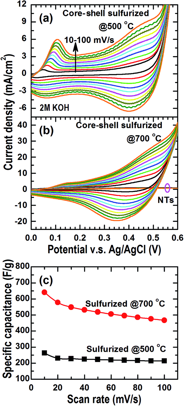 | ||
| Fig. 6 Cyclic voltammetry profiles measured using the sulfurization generated hybrid nanostructures on a Ti foil as an electrode in a typical three-electrode setup. Ag/AgCl and KOH (2 M) were used as the reference electrode and the electrolyte, respectively. The sulfurization was carried at (a) 500 °C and (b) 700 °C; (c) the specific capacitance calculated from the cyclic voltammetry profiles. For comparisons, the profiles of a sulfurized TiO2 nanotubes sample on a Ti foil substrate are also displayer in (b). | ||
It is worth noting that the sulfurized TiO2 nanotubes on a Ti foil under the same conditions as those of the Co(CO3)0.5(OH)·0.11H2O–TiO2 core–shell nanowires do not exhibit the pseudocapacitor behavior in the same cyclic voltammetry measurements [see the profiles at about I(V) = 0 in Fig. 6(b)]. This result indicates that the pseudocapacitor behavior of the hybrid nanostructures is mainly due to the redox reactions of the cobalt sulfide nanoparticles while the TiO2 nanotubes, although in anatase phase, are more likely working as supports/templates to increase the electrode–electrolyte interfaces and facile charge transfer during the charging/discharging process.
4. Conclusion
In conclusion, crystalline Co(CO3)0.5(OH)·0.11H2O nanowires have been synthesized on various substrates using a facile hydrothermal method at 90 °C. Atomic-layer deposition of TiO2 on the obtained Co(CO3)0.5(OH)·0.11H2O nanowires at 25 °C does change their topography but conformally coats an amorphous TiO2 shell on the nanowires, forming Co(CO3)0.5(OH)·0.11H2O–TiO2 core–shell nanowires. A further step of chemical etching in diluted HCl solution can effectively remove the Co(CO3)0.5(OH)·0.11H2O nanowire cores to form amorphous TiO2 nanotubes. Post-growth thermal vapor sulfurizations convert the Co(CO3)0.5(OH)·0.11H2O–TiO2 core–shell nanowires into hybrid nanostructures with crystalline TiO2 nanotubes decorated by cobalt sulfide nanoparticles. The decoration firmness of the nanoparticles on the nanotubes can be increased by increasing the sulfurization temperature. Recrystallization of the TiO2 nanotubes is observed and mainly caused by the annealing effect upon the thermal vapor sulfurization. The sulfurization resultant hybrid nanostructures on Ti foils have shown the typical pseudocapacitor behavior in a diluted KOH electrolyte. A specific capacitance of 650 F g−1 in 2 M KOH electrolyte is obtained at a scan rate of 10 mV s−1. This value is in the same ranges of the those hybrid nanostructures obtained based on Co(CO3)0.5(OH)·0.11H2O–TiO2 core–shell nanowires either by calcination or by combining the core-etching and the TMOs/TMCs deposition (e.g., see ref. 11 and 12).Acknowledgements
The authors would like to thank Ansah-Antwi K. K. for his help in recording the SEM images and Lim Poh Chong for his help in XRD data collection.References
- H. Chen, T. N. Cong, W. Yang, C. Tan, Y. Li and Y. Ding, Progress in electrical energy storage system: A critical review, Prog. Nat. Sci., 2009, 19, 291 CrossRef CAS PubMed.
- L. Mai, X. Tian, X. Xu, L. Chang and L. Xu, Nanowire electrodes for electrochemical energy storage devices, Chem. Rev., 2014, 114, 11828 CrossRef CAS PubMed.
- A. S. Aricò, P. Bruce, B. Scrosati, J.-M. Tarascon and W. van Schalkwijk, Nanostructured materials for advanced energy conversion and storage devices, Nat. Mater., 2005, 4, 336 CrossRef PubMed.
- G. Q. Zhang, H. B. Wu, H. E. Hoster, M. B. Chan-Park and X. W. Lou, Single-crystalline NiCo2O4 nanoneedle array grown on conductive substrates as binder-free electrodes for high-performance supercapacitors, Energy Environ. Sci., 2012, 5, 9453 CAS.
- Y. Feng, T. He and N. Alonso-Vante, In situ free-surfactant synthesis and ORR-electrochemistry of carbon-supported Co3S4 and CoSe2 nanoparticles, Chem. Mater., 2008, 20, 26 CrossRef CAS.
- Q. Liu and J. Zhang, A general and controllable synthesis of ComSn (Co9S8, Co3S4, and Co1−xS) hierarchical microspheres with homogenous phases, CrystEngComm, 2013, 15, 5087 RSC.
- Y. Ji, X. Liu, W. Liu, H. Zhang, M. Yang, X. Wang, X. Zhao and S. Feng, A facile template-free approach for the solid-phase synthesis of CoS2 nanocrystals and their enhanced storage energy in supercapacitors, RSC Adv., 2014, 4, 50220 RSC.
- D. Ghosh and C. K. Das, Hydrothermal growth of hierarchical Ni3S2 and Co3S4 on a reduced graphene oxide hydrogel@Ni foam: A high-energy-density aqueous asymmetric supercapacitor, ACS Appl. Mater. Interfaces, 2015, 7, 1122 CAS.
- N. Mahamood, C. Zhang, J. Jiang, F. Liu and Y. Hou, Multifunctional Co3S4/grapheme composites for lithium ion batteries and oxygen reduction reaction, Chem.–Eur. J., 2013, 19, 5183 CrossRef PubMed.
- Q. Wang, L. Jiao, H. Du, Y. Si, Y. Wang and H. Yuan, Co3S4 hollow nanospheres grown on grapheme as advanced electrode materials for supercapacitors, J. Mater. Chem., 2012, 22, 21387 RSC.
- J. Jiang, J. Luo, J. Zhu, X. Huang, J. Liu and T. Yu, Diffusion-controlled evolution of core–shell nanowire arrays into integrated hybrid nanotube arrays for Li-ion batteries, Nanoscale, 2013, 5, 8105 RSC.
- X. Xia, Z. Zeng, X. Li, Y. Zhang, J. Tu, N. C. Fan, H. Zhang and H. J. Fan, Fabrication of metal oxide nanobranches on atomic-layer-deposited TiO2 nanotube arrays and their application in energy storage, Nanoscale, 2013, 5, 6040 RSC.
- H. F. Liu and D. Z. Chi, Magnetron-sputter deposition of Fe3S4 thin films and their conversion into pyrite (FeS2) by thermal sulfurization for photovoltaic applications, J. Vac. Sci. Technol., A, 2012, 30, 04D102 Search PubMed.
- H. F. Liu, K. K. Ansah Antwi, Y. D. Wang, L. T. Ong, S. J. Chua and D. Z. Chi, Atomic layer deposition of crystalline Bi2O3 thin films and their conversion into Bi2S3 by thermal vapor sulfurization, RSC Adv., 2014, 4, 58724 RSC.
- X. Xia, C. Zhu, J. Luo, Z. Zeng, C. Guan, C. F. Ng, H. Zhang and H. J. Fan, Synthesis of free-standing metal sulfide nanoarrays via anion exchange reaction and their electrochemical energy storage application, Small, 2014, 10, 766 CrossRef CAS PubMed.
- H. F. Liu, K. K. Ansah Antwi, S. J. Chua and D. Z. Chi, Vapor-phase growth and characterization of Mo1−xWxS2 (0 ≤ x ≤ 1) atomic layers on 2-inch sapphire substrates, Nanoscale, 2014, 6, 624 RSC.
- H. F. Liu, K. K. Ansah Antwi, N. L. Yakovlev, H. R. Tan, L. T. Ong, S. J. Chua and D. Z. Chi, Synthesis and phase evolutions in layered structure of Ga2S3 semiconductor thin films on epiready GaAs (111) substrates, ACS Appl. Mater. Interfaces, 2014, 6, 3501 CAS.
- C. Zhao, D. Li and Y. Feng, Size-controlled hydrothermal synthesis and high electrocatalytic performance of CoS2 nanocatalysts as non-precious metal cathode materials for fuel cells, J. Mater. Chem. A, 2013, 1, 5741 CAS.
- H. M. Pathan and C. D. Lokhande, Deposition of metal chalcogenides thin films by successive ionic layer adsorption and reaction (SILAR) method, Bull. Mater. Sci., 2004, 27, 85 CrossRef CAS.
Footnote |
| † Electronic supplementary information (ESI) available. See DOI: 10.1039/c5ra07444d |
| This journal is © The Royal Society of Chemistry 2015 |