Evaluation of methionine and tryptophan derivatised vehicles: Met-ac-TE3A/Trp-ac-TE3A for tumor imaging

Sweta Singhab, Anjani K. Tiwari*a, Raunak Varshneya, Rashi Mathura, Puja P. Hazaria, B. Singhb and Anil K. Mishra*a
aDivision of Cyclotron and Radiopharmaceutical Sciences, Institute of Nuclear Medicine and Allied Sciences, Brig. S. K. Mazumdar Road, Timarpur, Delhi-110054, India. E-mail: akmishra63@gmail.com; dcrs_inmas@rediffmail.com; Fax: +91 1123919509; Tel: +91 23905117
bDepartment of Chemistry, Banaras Hindu University, Varanasi-221005, India

Received 20th April 2015 , Accepted 24th April 2015

First published on 24th April 2015


Abstract

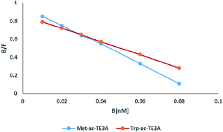
Two novel amino acid (methionine and tryptophan) appended 1,4,8,11-tetraazacyclotetradecane triacetate (TE3A) compounds Met-ac-TE3A and Trp-ac-TE3A were synthesized and evaluated for imaging applications. The pharmacokinetics of these compounds was analyzed by 99mTc labeled tracer methods. In vitro human serum stability of 99mTc labeled Met-ac-TE3A/Trp-ac-TE3A was found to be 96.5% and 96.0% after 24 h respectively. Blood kinetics of both the labeled probes on normal rabbits showed biphasic clearance. The tumor (EAT cell line) grafted in balb/c mice were readily identifiable in the gamma images. Biodistribution revealed significant tumor uptake and good contrast in the EAT tumor bearing mice and also showed high tumor/muscles ratio which is a requisite condition to work as SPECT-radiopharmaceutical for tumor imaging. To look its future applicability for therapy using M+2 and M+3 metal ions, we performed thermodynamic stability constants of complexes derived from Met-ac-TE3A and Trp-ac-TE3A with CuII and LnIII metal ions.


1. Introduction

Radiotracer based imaging probes are essential for the diagnosis of disease, monitoring and targeted therapy in the field of neuroimaging, oncology, neurooncology and infections using positron emission tomography (PET) and single photon emission computed tomography (SPECT). Radiolabeled amino acids has been proved a diverse and useful class of PET and SPECT tracers that records amino acid transportation and uptake exhibited by many tumor cells i.e. brain, neuroendocrine, prostate cancer.1–4 Increase rate for amino acid uptake is one of the earliest and most important events associated with proliferation.5

Recent studies have shown that of IDO (indoleamine 2,3-dioxygenase) is overexpressed in a variety of human tumors, including lung tumors resulting increase of abnormal tryptophan metabolism via the kynurenine pathway. L-Tryptophan is a substrate of IDO.6–8 Intracellular IDO activity may result in the trapping of polar tryptophan (or its derivative) metabolites or indirect accumulation via intracellular L-tryptophan depletion. There are many evidences that show fast growing progression of tumors because of a failure of the immune system to maintain control over budding tumors. Various studies indicate that the consumption of tryptophan is critical factor in progressive tumor.9,10 The PET radiotracer, [11C] methyl-L-tryptophan (AMT) is well suited for such studies; AMT is not a substrate for protein synthesis11 but can be metabolized by IDO because of the low substrate specificity of this enzyme.12

Similarly the major metabolic function of methionine is in protein synthesis and conversion to S-adenosylmethionine (Adomet), which is required in multiple metabolic pathways. Methionine (Met) dependence has been shown in vitro in a number of human cell lines of different cellular origin, and the dependence may reflect the overall imbalance in transmethylation. Metabolic defects in cancer cells often manifest in the inability to grow in media where Met has been replaced by its precursor homocysteine.

It was therefore considered valuable to focus our attention on the development of specific amino acid based imaging system that can be utilized for imaging techniques viz. PET, SPECT and magnetic resonance imaging (MRI) for tumour targeting by conjugation of amino acid with a suitable chelate. Currently a variety of cyclic and acyclic polyamino-polypcarboxylates are being evaluated as vehicles for variety of radiopharmaceuticals and MR agents. These polyamino-polycarboxylates have capability to adopt an organized conformation in the complex formation with metal ions. Two of the most important chelators studied were DOTA and TETA.13 In continuation of our work for design of novel agent having these vehicles, we previously synthesised DTPA-bis(methionine)14 and DO3A-Act-Met15 for imaging applications.

Here, we report the synthesis of two new ligand, synthesis of (11-((3-(1H-indol-3-yl)-1-methoxy-oxopropan-2-yl)amino)-2-oxoethyl)-1,4,8,11-tetraazacyclo tetradecane-(1,4,8-triyl)triacetic acid, Trp-ac-TETA and 2,2,2,-(11-((3-(1H-indol-3-yl)-1-methoxy-oxopropan-2-yl)amino)-2-oxoethyl)-1,4,8,11-tetraazacyclotetradecane-(1,4,8-triyl)triacetic acid, Met-ac-TETA. After wet synthesis and spectroscopic characterisation both the molecules were evaluated in vitro and in vivo after coordinating with suitable metal ion of biomedical interest.

2. Experimental

2.1. Ligand synthesis and characterization

2.1.1. General procedure for chloroacetylation. The methyl ester of L-methionine/L-tryptophan was dissolved in water (50 mL). The chloroacetyl chloride (1.2 eq.) solution in dichloromethane (50 mL) and K2CO3 solution in water (1.2 eq.) were added slowly to the stirring solution at 0 °C using a pressure equalized dropping funnel. The resulting reaction mixture was stirred at room temperature overnight. After completion of the reaction, the pale yellow viscous oil was washed with H2O (2 × 20 mL), 0.1 M HCl (2 × 20 mL) and brine (2 × 20 mL). The organic layer was dried over MgSO4, filtered and the solvent was removed under reduced pressure to yield pure chloroacetylated product.
2.1.2. General procedure for amino acid conjugation. The tris(carbobutoxymethyl)-1,4,8,11-tetraaza-cyclotetradecane 1 was dissolved along with K2CO3 (3 eq.) in 60 mL of dry CH3CN under nitrogen atmosphere. Chloroacetylated amino acid (3 eq.) was added drop wise to the above reaction mixture at 0 °C. The mixture was stirred for 10 min and afterwards refluxed at 70 °C for 3 days under nitrogen. After completion of the reaction (monitored by TLC, 9[thin space (1/6-em)]:[thin space (1/6-em)]1, chloroform[thin space (1/6-em)]:[thin space (1/6-em)]methanol), the reaction mixture was cooled, filtered, and evaporated under reduced pressure to give crude oily residue. The compound was purified by column chromatography (methanol[thin space (1/6-em)]:[thin space (1/6-em)]chloroform) to give the compounds.
2.1.3. General procedure of deprotection. The compound was dissolved in trifluoroacetic acid (3 mL) and stirred at room temperature for another 16 h. The solvent was evaporated and residue was dissolved in 2 mL of MeOH, followed by addition of 50 mL of diethyl ether drop wise and stirred for 1 h at room temperature. The compound was dried, dissolved in water, and neutralized to pH 7 by the addition of 1 M NaOH. The crude product was purified by preparative HPLC to give final compound.
2.1.4. Synthesis of 1,4,7-tri(tert-butoxymethane)-1,4,8,11-tetraazacyclotetradecane (1). The 1,4,8,11 tetraazacyclotetradecane (1.00 g, 5.00 mmol) was dissolved in dry acetonitrile (25 mL) under nitrogen atmosphere at RT. After stirring the solution at 0 °C for 10 min, NaHCO3 (1.26 g, 15.00 mmol) was added drop wise to the solution and stirred for 30 min. tert-Butylbromoacetate (2.92 g, 15.00 mmol) was added slowly in 3 h at 0 °C. The reaction was stirred at room temperature for 48 h and then refluxed at 65–70 °C for 8 h. The completion of the reaction was checked by TLC (9[thin space (1/6-em)]:[thin space (1/6-em)]1; dichloromethane[thin space (1/6-em)]:[thin space (1/6-em)]methanol). After the completion of the reaction, the reaction mixture was filtered and evaporated to dryness. The crude compound was purified by column chromatography (silica gel, 9[thin space (1/6-em)]:[thin space (1/6-em)]1 chloroform[thin space (1/6-em)]:[thin space (1/6-em)]methanol) to give 1 (t-Bu-TE3A) as white powder (2.03 g, 75%). 1H NMR (400 MHz, CDCl3, TMS): δH = 1.45 (3 s, 27H; C(CH3)3), 1.62–1.63 (m, 4H), 1.99 (brs, 1H), 2.55–3.40 (m, 22H); 13C-NMR (100 MHz, CDCl3): δC = 23.52, 24.37 (CH2), 29.01, 29.07, 29.12, (C(CH3), 47.06, 47.53, 49.86, 50.87, 51.88, 53.38, 55.59, 56.70, 56.89, 81.72, 82.15, 82.40 (C(CH3)3), 171.53, 172.47, 172.62 (CO). m/z (ESI) found 543.9 [M + H]+, C28H54N4O6, calculated 542.7.
2.1.5. Synthesis of methyl-2-(2-chloroacetamido)-4-(methylthio) butanoate (2). According to the general procedure methyl ester of methionine (1.00 g, 6.13 mmol), chloroacetyl chloride (0.82 g, 7.36 mmol) and K2CO3 (1.03 g, 7.36 mmol) to afford 1 (0.69 g, 80%) as a colorless oil. 1HNMR (400 MHz, CDCl3, TMS): δH = 2.11 (s, 3H; CH3–S–), 2.18–2.20 (m, 2H), 2.51–2.55 (m, 2H), 3.79 (s, 3H; CH3–O), 4.08 (s, 2H; CH2–Cl), 4.73–4.75 (m, 1H; –CH–NH–); 13CNMR (100 MHz, CDCl3): δC = 15.48 (CH3–S–), 29.83 (CH2–S), 31.29 (CH2), 42.43 (CH3–O), 51.82 (CH2), 52.76 (CH2–NH–), 165.93 and 171.72 (CO). m/z (ESI) found 262.5 (M + Na+), C8H14ClNO3S, calculated 238.5.
2.1.6. Synthesis of methyl-2-(2-chloroacetamido)-3-(1H-indol-3-yl) propanoate (3). By using general procedure the hydrochloride of methyl ester of tryptophan (1.00 g, 3.93 mmol), chloroacetyl chloride (0.53 g, 4.71 mmol) and K2CO3 (0.65 g, 4.71 mmol) as reactant to afford 3 (1.05 g, 78%) as a colorless oil. 1HNMR (400 MHz, CDCl3, TMS): δH = 3.38 (d, 2H, J = 5.6 Hz), 3.69 (s, 3H, –OCH3), 3.96 (s, 2H), 4.92–4.97 (m, 1H), 6.99 (s, 1H), 7.14–7.28 (m, 2H), 7.34 (d, 1H, J = 8.0 Hz), 7.58 (d, 1H, J = 7.6 Hz); 13C-NMR (100 MHz, CDCl3): δC = 27.52, 42.47, 52.58, 53.17, 109.59, 111.32, 118.53, 119.80, 122.41, 122.78, 127.42, 136.12, 165.72 (CO), 171.68 (CO), m/z (ESI) found 296.0 (M + 2H+), C14H15ClN2O3, calculated 294.08.
2.1.7. Synthesis of tri-tert-butyl-2,2,2-(11-(3-((1-methoxy-4-(methylthio)-1-oxobutan-2yl)amino)-2-oxoethyl)-1,4,8,11-tetraazacyclotetradecane-(1,4,8-triyl)triacetate (4). According to the general procedure the tris(carbobutoxymethyl)-1,4,8,11-tetraaza-cyclotetradecane 1 (0.40 g, 0.70 mmol), K2CO3 (0.51 g, 3.60 mmol) and methyl-2-(2-chloroacetamido)-4-(methylthio)butanoate (0.52 g, 2.2 mmol) to give crude oily residue. The compound was purified by column chromatography (silica gel, 95[thin space (1/6-em)]:[thin space (1/6-em)]5 chloroform[thin space (1/6-em)]:[thin space (1/6-em)]methanol) to give the 4 (0.41 g, 75%) as a brown solid. 1HNMR (400 MHz, CDCl3, TMS): δH = 1.39 (s, 27H; C(CH3)3), 1.58–1.59 (m, 4H), 2.04–2.15 (m, 5H, SCH3 and methionine proton), 2.46–2.69 (m, 19H), 3.18 (s, 4H), 3.25 (s, 2H), 3.74 (s, 2H), 3.68 (s, 3H, OCH3), 4.58–4.63 (m, 1H); 13C-NMR (100 MHz, CDCl3): δC = 15.38 (CH3), 25.27, 25.65, 28.15 (C(CH3)3), 30.24, 31.20, 42.38, 50.12, 50.20, 50.52, 51.10, 51.47, 51.76, 52.25, 52.38, 53.00, 56.18, 56.31, 56.36, 58.55, 80.60, 80.65, 80.78 (C(CH3)3), 170.50, 170.83, 171.03, 172.10, 172.54, (CO). m/z (ESI) found 747.8 (M + H+), 769.7 (M + Na+), C36H67N5O9S, calculated 746.0.
2.1.8. Synthesis of tri-tert-butyl-2,2,2-(11-((3-(1H-indol-3-yl)-1-methoxy-1-oxopropan-2yl)amino)-2-oxoethyl)-1,4,8,11-tetraazacyclotetradecane-(1,4,8-triyl)triacetate (5). The general procedure described above was followed using tris(carbobutoxymethyl)-1,4,8,11-tetraaza-cyclotetradecane 1 (0.50 g, 0.92 mmol), K2CO3 (0.38 g, 2.76 mmol) and methyl-2-(2-chloroacetamido)-3-(1H-indol-3-yl)propanoate 3 (0.81 g, 2.76 mmol) as a reactant to give crude oily residue. The compound was purified by column chromatography (silica gel, 95[thin space (1/6-em)]:[thin space (1/6-em)]5 chloroform[thin space (1/6-em)]:[thin space (1/6-em)]methanol) to give the compound as a brown solid (0.56 g, 75%).1HNMR (400 MHz, CDCl3, TMS): δH = 1.44–1.47 (m, 31H), 2.42–2.65 (m, 15H), 2.99–3.24 (m, 11H), 3.68 (s, 3H; CH3–O–), 4.82–4.87 (m, 1H; CH–NH–), 7.08–7.52 (m, 5H; indolylprotons); 13CNMR, (100 MHz, CDCl3): δC = 25.12, 25.38, 28.20 (CH3)3, 42.41(OCH3), 50.81, 51.11, 51.17, 51.53, 51.71, 51.97, 52.23, 52.26, 52.49, 52.61, 52.75, 53.29, 55.84, 56.41, 80.75, 80.87, 80.96, (C(CH3)3), 109.38, 111.57, 118.38, 119.39, 121.98, 123.29, 127.44, 136.43, 165.97, 170.76 (CO), 171.14 (CO), 171.19 (CO), 172.38 (CO). m/z (ESI) found 802 (M + 2H+), C42H68N6O9, calculated 800.0.
2.1.9. Synthesis of 2,2,2-(11-(3-((1-methoxy-4-(methylthio)-)-1-oxobutan-2yl)amino)-2-oxoethyl)-1,4,8,11-tetraazacyclotetradecane-(1,4,8-triyl)triacetic acid (6). The general procedure described above was followed using compound 4 (0.40 g, 0.53 mmol). The compound was dried, dissolved in water, and neutralized to pH 7 by the addition of 1 M NaOH. The crude product was purified by preparative HPLC to give final compound 6 (0.22 g, 70%.). 1HNMR (400 MHz, D2O, TMS): δH = 1.79–2.07 (m, 9H, S–CH3; 4H cyclam ring and 2H methionine), 2.85–3.16 (m, 16H), 3.16–3.96 (m, 11H; cyclam, OCH3), 4.44–4.47 (m, 1H; CH); 13C-NMR (100 MHz, D2O): δC = 13.88 (s, CH3), 21.82, 24.18, 29.17, 48.72, 50.51, 50.99, 51.50, 51.84, 52.01, 52.34, 52.59, 52.95, 53.09, 55.21, 54.20, 54.59, 56.28, 56.38, 162.59, 171.72, 172.81, 173.60, 174.93 (CO). High resolution mass spectrometer (HRMS) (FAB) calculated for C24H43N5O9S, 577.6521; observed: 577.5511.

Chemical purity: >99% by HPLC: Capcell Pack UG80 C18 column, 4.6 mm i.d. × 250 mm; MeCN/H2O/Et3N, 75/25/0.01 (v/v/v); flow rate, 1.0 mL min−1; λuv, 254 nm); retention time (tR), 9.7 min (ESI Fig. 1).

2.1.10. Synthesis of (11-((3-(1H-indol-3-yl)-1-methoxy-oxopropan-2-yl)amino)-2-oxoethyl)-1,4,8,11-tetraazacyclo tetradecane-(1,4,8-triyl)triacetic acid (7). According to the general procedure compound 5 (0.50 g, 0.62 mmol) was deprotected with and 3 mL of TFA. The compound was dried, dissolved in water, and neutralized to pH 7 by the addition of 1 M NaOH. The crude product was purified by preparative HPLC to give final compound. 1HNMR (400 MHz, D2O, TMS): δH = 1.64–1.73 (m, 4H), 2.50–3.98 (m, 30H), 6.99–7.50 (m, 5H; indolyl protons); 13CNMR (100 MHz, D2O): δC = 21.50, 26.88, 35.21, 44.47, 50.58, 51.72, 51.96, 52.29, 52.41, 52.62, 53.02, 53.42, 54.10, 54.36, 55.79, 56.20, 109.32, 111.94, 118.42, 119.52, 122.07, 124.58, 126.90, 135.93, 162.73(CO), 163.08(CO), 173.27(CO).

High resolution mass spectrometer (HRMS) (FAB) calculated for C30H44N6O9, 632.4132; observed: 632.3190.

Chemical purity: >99% by HPLC: Capcell Pack UG80 C18 column, 4.6 mm i.d. × 250 mm; MeCN/H2O/Et3N, 80/20/0.01 (v/v/v); flow rate, 1.0 mL min−1; λuv, 254 nm; retention time (tR), 8.1 min (ESI Fig. 2).

2.2. Determination of protonation and stability constant

The protonation constant of [Trp-ac-TE3A] and [Met-ac-TE3A] and stability constants of the complexes formed with EuIII and CuII have been determined by pH potentiometric titrations (Table 1). Potentiometric measurements were carried out with an automatic titration system consisting of Metrohm 713 pH meter equipped with a Metrohm A.60262.100 electrode 800 Dosino autoburet. The acidity constants of both the compound were determined potentiometrically by titrating 25.0 mL of aqueous 2.5 mM HCl in the presence of 0.01 M both substituted TE3A with 0.1 M NMe4OH.
Table 1 Protonation constants (log[thin space (1/6-em)]β ± SD, n = 3) of the ligands DTPA, DOTA and Trp-ac-TE3A/Met-ac-TE3A at 25 °C
Protonation constants DTPA30a DOTA30a Trp-ac-TE3A Met-ac-TE3A
0.1 M KCl 0.1 M KCl 0.1 M KCl 0.1 M KCl
log[thin space (1/6-em)]K1H 10.48 11.14 10.40 10.25
log[thin space (1/6-em)]K2H 8.60 9.69 8.10 8.80
log[thin space (1/6-em)]K3H 4.28 4.84 4.10 4.00
log[thin space (1/6-em)]K4H 2.6 3.9 2.65 2.52
log[thin space (1/6-em)]K5H 2.0  


2.3. Radiolabeling of Met-ac-TE3A/Trp-ac-TE3A with 99mTc

Met-ac-TE3A/Trp-ac-TE3A (1 mg) was dissolved in a shielded vial and stannous chloride (300 μL; 1 mg dissolved in N2 purged 1 mL of 10% acetic acid) was added followed by addition of freshly eluted (<1 h) 99mTechnetium pertechnetate (82 MBq; 200 μL). The pH of the reaction mixture was adjusted to 7 with 0.1 M Na2CO3 and purged with N2, and shaken to mix. The vial was allowed to stand for 15 min at room temperature (25 °C).

2.4. Radiochemical purity

The number of ligand molecules involved in complexation with 99mTc was determined by ascending instant thin layer chromatography on ITLC-SG (Paul Gelman,USA) strips using 100% acetone as developing solvent and simultaneously in pyridine/acetic acid/water (PAW) (3[thin space (1/6-em)]:[thin space (1/6-em)]5[thin space (1/6-em)]:[thin space (1/6-em)]1.5) and saline. Each TLC was cut in 0.5 cm segments and counts of each segment were taken. By using this method percentage of free Na99mTcO4−, reduced/hydrolysed 99mTc, and the complex formed between 99mTc and Met-ac-TE3A/Trp-ac-TE3A conjugate could be calculated. Met-ac-TE3A/Trp-ac-TE3A conjugate remained at the origin, and free technetium traveled with the solvent front in acetone.

The radio conjugate was then purified using a C-18 reversed-phase extraction cartridge, which was preconditioned with 10 mL methanol and subsequently activated with 30% methanol to make prepare it more appropriate in terms of purity. The cartridge was successively rinsed with 5 mL distilled water, and radiolabeled conjugate was eluted in 5 mL of 5% ethanol.

2.5. In vitro studies

2.5.1. Human serum stability assay. The above freshly prepared technetium radiocomplexes (300 μCi of radioactivity) were incubated in 0.9% saline solution (1 mL) at 37 °C in a humidified incubator maintained at 5% carbon dioxide, 95% air. Then the sample was centrifuged at 400 rpm and the serum was filtered through 0.22 micron syringe filter into sterile plastic culture tubes. Complexes were stable in dilute saline solution. The 99mTc complexes of amino acid analogues were added to 425 μL of male AB type human serum and incubated at 37 °C for 48 h then analyzed by ITLC-SG to assess any dissociation based on the Rf. Percentage of free pertechnetate at a particular time was determined using saline and acetone as mobile phase, represents percentage dissociation of the complex at particular time in serum.

In tumor cell both the ligands were found intact even after 6 h and no other polar metabolite compound was found in hplc chromatograms.

2.5.2. Cell binding studies. The specificity of 99mTc[Met-ac-TE3A] to bind with tumor cells was examined by receptor binding assays. U-87MG was grown in normal DMEM, 10% and 5% FBS, respectively. Monolayer cultures of the cell lines were washed with HBSS and were then incubated for 2 h at 37 °C prior to the experiment. Binding experiments was conducted at 37 °C. Further these cells were incubated for 30 min with increasing concentrations (0.01 nM–10 μM) of 99mTc[Met-ac-TE3A] in the absence and presence (100-fold excess) of unlabeled methionine to estimate the total binding and nonspecific binding respectively. At the end of each experiment, the cells were washed with cold PBS four times. The cell-associated radioactivity was determined by gamma scintillation counting. Scatchard plot analysis was done. The same procedure was applied for the other ligand.
2.5.3. MTT assay. MTT [3-(4,5-dimethylthiazole-2-yl)-2,5-diphenyl-2H-tetrazolium bromide] assay was used to determine the cytotoxicity. Exponentially growing MCF-7 and U-87MG cells were plated in a 96-well microtitre plate at a uniform cell density of 4000 cells per well, 24 h before treatment. Cells were treated with varying concentrations of drug (μM–mM range) for 2 h and MTT assays were performed at 24, 48, 72 and 96 h post treatment. After the treatment, negative control and treated cells were incubated with MTT at a final concentration of 0.05 mg mL−1 for 2 h at 37 °C and the medium was removed. After cell lyses, formazan crystals were dissolved using 150 μL of DMSO. Optical density of the extracts (150 μL) was measured at 570 nm (reference filter: 690 nm). Percentage of cell viability compared to negative control (mean ± SD of triplicate cultures) indicated the mitochondrial activity. % of viability = [OD (570–630 nm) test product/OD (570–630 nm) negative control] × 100%.

2.6. In vivo studies

2.6.1. Blood kinetics. The blood clearance study was performed in albino New-Zealand rabbits weighing approximately 2.5–3.0 kg after administration of 10 MBq of the 99mTc labeled compounds in 0.3 ml via the ear vein. At different time intervals about 0.5 ml blood samples were withdrawn from the dorsal vein of other ear starting from 5 min to 24 h and the radioactivity in circulation was calculated, assuming total blood volume as 7% of the body weight.
2.6.2. Scintigraphy. Tumor imaging was performed in EAT (Ehrlich ascites tumor) cell line implanted tumor bearing balb/c mice after receiving Intravenous i.v. (tail vein) injections of 100 μL of the labeled conjugate (0.1 mCi activity) of each 99mTc[Met-ac-TE3A] and 99mTc[Trp-ac-TE3A]. SPECT scans were acquired 4, 24, and 48 h post injection.
2.6.3. Biodistribution. The in vivo biodistribution was assessed by female balb/c mice (n = 3 per group, 20–22 g) implanted with EAT tumor in hind leg. Each group received i.v. (tail vein) injections of approximately 100 μCi (0.1 mCi activity) of 99mTc labeled Met-ac-TE3A/Trp-ac-TE3A. Animals were euthanized at the designated times and selected tissues were removed, weighed, and counted on a gamma counter to determine 99mTc-complex distribution. Uptake of the radiotracer in each tissue was calculated and expressed as percentage injected dose per gram of the tissue (%ID g−1).

3. Result and discussion

3.1. Synthesis and radiochemistry of Met-ac-TE3A/Trp-ac-TE3A

In an attempt to develop target specific macrocyclic vehicle, two novel ligands Trp-ac-TETA, 6 and Met-ac-TE3A,7 were synthesized as Scheme 1. The cyclam was first converted into 1 by the reaction of tert-butylbromoacetate on 1,4,8,11-tetraazacyclotetradecane in 75% yield using a modified procedure reported in literature.16 The slow addition of tert-butylbromoacetate at 0 °C and purification by column chromatography led to appreciable increase in the final yield. Methyl-2-(2-chloroacetamido)-4-(methylthio) butanoate (2) was prepared by the reaction of methyl ester of methionine and chloroacetyl chloride in presence of base K2CO3. Similarly methyl-2-(2-chloroacetamido)-3-(1H-indol-3-yl) propionate (3) was also obtained as a colorless oil. These two intermediates were treated with 1 in presence of base to give tri-tert-butyl-2,2,2-(11-(3-((1-methoxy-4-(methylthio)-)-1-oxobutan-2yl)amino)-2-oxoethyl)-1,4,8,11-tetraaza-cyclotetradecane-(1,4,8-triyl)triacetate (4) and tri-tert-butyl-2,2,2-(11-((3-(1H-indol-3-yl)-1-methoxy-1-oxopropan-2yl)amino)-2-oxoethyl)-1,4,8,11-tetraaza-cyclotetradecane-(1,4,8-triyl)triacetate (5), which were further deprotected to get final product (6) and (7). 1H NMR spectra of 6 was showing α-H at 4.82–4.87 ppm and heterocyclic ring of tryptophan between 7.08 to 7.52 ppm while for 7 characteristic multiplet of amino acid backbone α-H at 4.58–4.63 ppm and S–CH3 at 2.11 ppm. In the 13C spectrum, the peak at 165 ppm represented the carbonyl carbon of COOH in both 6 and 7 and peak at 80 ppm tertiary carbon disappeared after deprotection of COOH group.
image file: c5ra07138k-s1.tif
Scheme 1 Synthesis of Met-ac-TE3A/Trp-ac-TE3A. Reagents and conditions: general synthetic pathway of Met-ac-TE3A and Trp-ac-TE3A. (a) K2CO3, tBu-bromoacetate,ACN, 65–70 °C (b) chloroacetyl chloride, K2CO3, CH2Cl2[thin space (1/6-em)]:[thin space (1/6-em)]H2O, RT (c) K2CO3, ACN, 65–70 °C (d) TFA, RT.

The compounds were radiolabeled with 99mTc using SnCl2 as reducing agent. All the labeling parameters such as pH, concentration of reducing agent etc. were standardized, to achieve the maximum labeling efficiency. In vitro and in vivo stability of the labeled complexes was checked and the complexes were found to be stable for 24 h under physiological conditions. The labeling yield was found to be greater than 95%, as determined chromatographically by different solvent systems. The reaction mixture was kept in saline for different time intervals to carry out in vitro stability studies. Percentage radiolabeling was calculated for 0, 2, 4, 6, and 24 h. The metal binding assay confirmed the radiochemical purity to be >99%. Radiochromatogram showed a single peak for both the ligands which was found intact over 6 h.

3.2. pH-potentiometric studies

The metal complexes of macrocyclic ligands must be chemically stable under physiological conditions to ensure a desired biodistribution of the conjugates. This is also important to avoid the release of potentially toxic metal ions and transchelation of endogenous metal ions. Thus, such complexes should show high thermodynamic stability as well as strong kinetic inertness to dissociation, and the latter is generally favored in macrocyclic ligand. The prerequisite condition for synthesized ligands 6 and 7 to be used as Cu based PET radiopharmaceuticals and possible agents for lanthanide complexes in MR and other optical uses; it should form a highly stable complex with these metals in vitro. The values of the overall protonation constants and stability constants of their copper and europium complexes are presented in (Tables 1 and 2) respectively and compared with other known vehicles DTPA and DOTA.
Table 2 Stability constants (log[thin space (1/6-em)]β ± SD, n = 3) of the ligands DOTA and Trp-ac-TE3A and Met-ac-TE3A with the metal ions EuIII and ZnII and CuII (0.10 M KCl, at 25 °C)a
Stability constant DOTA30b Trp-ac-TE3A Met-ac-TE3A
0.1 M KCl 0.1 M KCl 0.1 M KCl
a Calculated in separate reaction condition.
log[thin space (1/6-em)]βEuL 24.5 18.9 17.6
log[thin space (1/6-em)]βZnL 18.1 16.8 15.3
log[thin space (1/6-em)]βCuL 20.4 19.2
log[thin space (1/6-em)]βCu2L 22.2 6.2 5.1


As anticipated, first and second protonation constants of the ligand were high, viz. log[thin space (1/6-em)]K1 and log[thin space (1/6-em)]K2 were 10.40 and 8.1 for Trp-ac-TE3A and 10.25 and 8.80 for Met-ac-TE3A. In general these two protonation constant are assigned to two trans group of macrocyclic backbone. Only mononuclear complexes could be found with copper, something that is understandable in view of the metal-to ligand ratio of 1[thin space (1/6-em)]:[thin space (1/6-em)]1 that was used. The metal ligand ratios were 2[thin space (1/6-em)]:[thin space (1/6-em)]1, 1[thin space (1/6-em)]:[thin space (1/6-em)]1 and 1[thin space (1/6-em)]:[thin space (1/6-em)]2. Titration for each ratio was carried out at least three times. It was likely that the presence of an excess of metal ions would lead to the formation of dinuclear complexes, but low stability constants of such species diminishes such possibility.

The values of the stability constants with copper was high for both the ligands and differs from those determined for Zn2+ and Eu3+ ions (Table 2). The values are comparable with those found for other similar ligands and are mainly determined by overall basicity of the nitrogen atoms (pK1 + pK2). The detail natures of the species are also represented in species distribution curve (ESI Fig. 3 and 4).

3.3. In vitro studies

3.3.1. In vitro human serum stability assay. The stability of the amino acid conjugates were assessed in human serum with 99mTc labeled molecules. 99mTc methionine was showing major transcomlexation, 66% while 58% transcomplexation was observed in 99mTc tryptophan after 1 h. 99mTc[Met-ac-TE3A] showed 2.5% transcomplexation in serum at 1 h. It was stable up to 24 h, as only 3.5% of the radiolabeled drug dissociated in serum at 24 h. 99mTc [Trp-ac-TE3A] showed only 2.7% transcomplexation at 1 h. 99mTc [Trp-ac-TE3A] was also found to stable up to 24 h, as only 4% radiolabeled drug dissociated in serum at 24 h similar to 99mTc[DTPA-bis(methionine)].14 These results validated the use of these ligands for further analysis as below 5–6% transcomplexation is found permissible for quantification studies.
3.3.2. Cell binding studies. The ability of both the conjugates 99mTc[Trp-ac-TE3A] and 99mTc[Met-ac-TE3A] to bind LAT1 transporters on the surface of tumor cell line U-87MG was examined by competitive binding assay (Fig. 1). Nonspecific binding was determined by using 100-fold excess of unlabeled methionine/tryptophan. Scatchard analysis showed relatively good binding affinity for 99mTc[Met-ac-TE3A] (Kd = 0.106 nm) in comparison to 99mTc[Trp-ac-TE3A] (Kd = 0.145 nm). Kd value were found below 0.1 nM for Met-ac-TE3A (0.09 nM) and was slightly higher for Trp-ac-TE3A (0.12 nM). Though the binding data were found very appropriate for both ligands but in comparison Met-ac-TE3A was found better for application.
image file: c5ra07138k-f1.tif
Fig. 1 Scatchard plot for Met-ac-TE3A and Trp-ac-TE3A.
3.3.3. MTT assay. The cell viability was determined by using MTT assay on MCF-7 breast adenocarcinoma cells and U87 glioma cells at different concentrations (μM to mM range). The range of concentrations were determined on the basis of our previous studies for such vehicle based ligand system for SPECT.14–16 Significant toxicity was not observed at lower concentrations for Met-ac-TE3A, which showed concentration-dependent cytotoxicity. It was observed that at a concentration of 0.1 mM Met-ac-TE3A at 2 h treatment resulted in lyses of 40% of U-87MG cells. Vice versa 10 μM of the same when incubated with MCF-7 cell line for only 2 h showed 16% cells death (ESI Fig. 5). In case of Trp-ac-TE3A, 7% and 25% cells were killed when incubated with U87 and MCF-7 cell line respectively at 0.1 mM concentration. Trp-ac-TE3A was not showing any significant toxicity at lower concentration against both the cell lines (ESI Fig. 6).

3.4. In vivo studies

3.4.1. Blood kinetics. The blood clearance profile of both 99mTc [Met-ac-TE3A] and 99mTc [Trp-ac-TE3A] showed high uptake initially with the diagnostically useful target-to-nontarget ratio. The blood clearance of both the radiocomplex in rabbits followed biphasic trend with a rapid clearance at initial phase and a slow clearance in second phase. The biological half-life of 99mTc [Met-ac-TE3A] was obtained to be t1/2 (fast): 2 h; t1/2 (slow): 14 h and 30 min. Almost similar trend was exhibited by 99mTc [Trp-ac-TE3A]. Only 16% activity persisted in circulation after 1 h post administration which reduced to 0.82% by 24 h. The biological half life 99mTc[Trp-ac-TE3A] was found to be t1/2 (fast) 1 h 15 min and t1/2 (slow) 22 h 15 min (Fig. 2). This showed relatively more retention initially when compared with acyclic system 99mTc [DTPA-bis(methionine)],14 which showed t1/2 (fast) 36 min. This reflects the additional use of this system in those tumors where tracer take time to reach because it gives more time to quantify activity initially compare to acyclic system.
image file: c5ra07138k-f2.tif
Fig. 2 Blood clearance profile of 99mTc [Met-ac-TE3A] and 99mTc [Trp-ac-TE3A] administered through ear vein in normal rabbit.
3.4.2. Scintigraphy. The in vivo scintigraphy studies were carried out in EAT (Ehrlich ascites tumor) cell line implanted tumor bearing balb/c mice by imaging the animals at 4 h. The mice injected with 99mTc [Met-ac-TE3A] exhibited uptake in tumor with a gradually increasing trend reaching maximum in 4 h while it was relatively low for 99mTc[Trp-ac-TE3A] (Fig. 3). The possible explanation is that methionine derivative has very high affinity as compared to tryptophan derivative. Blocking studies was also performed to see the selectivity of these vehicles by using unlabeled methionine and tryptophan (1 mg kg−1 and 3 mg kg−1) for respective ligands. Fig. 4 showed that on blocking the uptake decrease by 60% by 1 mg kg−1 unlabeled methionine and decreases around 75% with the use of 3 mg kg−1 for 99mTc [Met-ac-TE3A], which demonstrates the stability of this compound also because the metabolized and subpart of this molecule is not able to show such amount of selectivity. In case of 99mTc[Trp-ac-TE3A] it decrease only 25% which itself expresses that tryptophan analogue has low selectivity for tumor cells. This blocking study explains the difference between in vitro and in vivo results of these two compounds that the affinity may be the similar for both but the change in selectivity may play major role for uptake after a substantial time period after injection.
image file: c5ra07138k-f3.tif
Fig. 3 Whole-body γ image of balb/c mice with subcutaneous EAT tumor above the right hind leg at 4 h after i.v. injection of 99mTc [Met-ac-TE3A] and 99mTc [Trp-ac-TE3A].

image file: c5ra07138k-f4.tif
Fig. 4 Blocking studies for Met-ac-TE3A and Trp-ac-TE3A.
3.4.3. Biodistribution. Biodistribution of 99mTc [Met-ac-TE3A] revealed high tumor uptake in the EAT tumor bearing mice as compared to 99mTc [Trp-ac-TE3A]; tumor to muscle ratio reached maximum at 4 h post injection in the both the cases. The concentration for both the radiolabeled compound was rapidly cleared from blood confirmed by the data of blood kinetic study. Biodistribution data of both the compounds showed low accumulation of radioactivity in the stomach which precludes the presence of free pertechnetate, indicative of the in vivo stability of radiotracers. However both the compounds showed very high accumulation in liver, related to the positive net charge of the complex. Significant accumulation of radioactivity was found in the kidney (6.1 ± 0.25 %ID g−1 at 1 h and 1.3 ± 0.12 %ID g−1 at 24 h after injection),which indicated renal route as a possible way for excretion. 99mTc [Met-ac-TE3A] and 99mTc [Trp-ac-TE3A] showed maximum tumor uptake at 4 h (Fig. 5 and 6). The modest accumulation of radioactivity was also observed in non target organ i.e. heart, spleen and stomach.
image file: c5ra07138k-f5.tif
Fig. 5 Tissue distribution data (% ID g−1 organ) of 99mTc [Met-ac-TE3A] in at 1 h, 2 h, 4 h and 24 h after i.v. administration in tumor bearing mice (n = 3).

image file: c5ra07138k-f6.tif
Fig. 6 Tissue distribution data (% ID g−1 organ) of 99mTc [Trp-ac-TE3A] in at 1 h, 2 h, 4 h and 24 h after i.v. administration in tumor bearing mice (n = 3).

4. Conclusion

We have designed and developed possible biocompatible probes which can diagnose the tumor non-invasively and also provide a platform for the further development of multimodal imaging tools. Two novel amino acid based ligands Met-ac-TE3A and Trp-ac-TE3A were successfully synthesized and well characterized through spectroscopic techniques. In the preliminary studies they have shown good prospect as SPECT agent with 99mTc, which may extrapolated for 64Cu and 67Cu. The high stability of the compound with metal ions EuIII and CuII further expands its possible application from diagnosis to therapy as well as MR agent.

Acknowledgements

We thank Dr R. P. Tripathi, Director INMAS, for providing necessary facilities. The work was supported by Defence Research and Development Organization, Ministry of Defence, under R&D project INM-311.

References

  1. C. Huang and J. McConathy, J. Nucl. Med., 2013, 54, 1007–1010 CrossRef CAS PubMed.
  2. K. J. N. Isselbacher, Engl. J. Med., 1972, 286, 929–933 CrossRef CAS PubMed.
  3. K. J. N. Isselbacher, Proc. Natl. Acad. Sci. U. S. A., 1972, 69, 585–589 CrossRef CAS.
  4. T. Miyagawa, T. Oku and H. Uehara, J. Cereb. Blood Flow Metab., 1998, 18, 500–509 CrossRef CAS PubMed.
  5. D. Comar, J. C. Cartron, M. Maziere and C. Marazano, Eur. J. Nucl. Med., 1976, 1, 11–14 CrossRef CAS.
  6. B. Långstrom, G. Antoni and P. Gulberg, J. Nucl. Med., 1987, 28, 1037–1040 Search PubMed.
  7. K. Ishiwata, W. Vaalburg, P. H. Elsinga, A. M. J. Paans and M. G. Woldring, J. Nucl. Med., 1988, 29, 1419–1427 CAS.
  8. J. K. Chung, K. Kim, S. K. Kim, Y. J. Lee, S. Paek, J. S. Yeo, J. M. Jeong, D. S. Lee, H. W. Jung and M. C. Lee, Eur. J. Nucl. Med. Mol. Imaging, 2002, 29, 176–182 CrossRef CAS.
  9. T. Singhal, T. K. Narayanan, V. Jain, J. Mukherjee and J. Mantil, Mol. Imag. Biol., 2008, 10, 1–18 CrossRef PubMed.
  10. K. Higashi, A. C. Clavo and R. L. Wahl, J. Nucl. Med., 1993, 34, 773–779 CAS.
  11. D. H. Munn and A. L. Mellor, Trends Mol. Med., 2004, 10, 15–18 CrossRef CAS PubMed.
  12. C. Uyttenhove, L. Pilotte, I. Theate and J. Benoit, Nat. Med., 2003, 9, 1269–1274 CrossRef CAS PubMed.
  13. A. Mishra, J. Pfeuffer, R. Mishra, J. Engelmann, A. K. Mishra, K. Ugurbil and N. K. Logothetis, Bioconjugate Chem., 2006, 17, 773–780 CrossRef CAS PubMed.
  14. P. P. Hazari, G. Shukla, V. Goel, K. Chuttani, N. Kumar, R. Sharma and A. K. Mishra, Bioconjugate Chem., 2010, 21, 229–239 CrossRef CAS PubMed.
  15. K. G. Kadiyala, A. Datta, J. Tanwar, A. Adhikari, B. S. Kumar, K. Chuttani, M. Thirumal and A. K. Mishra, Pharm. Res., 2015, 32(3), 955–967 CrossRef CAS PubMed.
  16. J. K. Uppal, P. P. Hazari, C. K. Raunak, M. Allard, N. K. Kaushik and A. K. Mishra, Org. Biomol. Chem., 2011, 9, 1591–1599 CAS.

Footnote

Electronic supplementary information (ESI) available. See DOI: 10.1039/c5ra07138k

This journal is © The Royal Society of Chemistry 2015
Click here to see how this site uses Cookies. View our privacy policy here.