DOI:
10.1039/C5RA06935A
(Paper)
RSC Adv., 2015,
5, 56872-56884
Discovery of a low affinity thyrotropin-releasing hormone (TRH)-like peptide that exhibits potent inhibition of scopolamine-induced memory impairment in mice†
Received
17th April 2015
, Accepted 23rd June 2015
First published on 23rd June 2015
Abstract
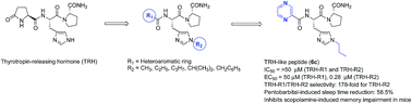
TRH-like peptides were synthesized in which the critical N-terminus residue L-pGlu was replaced with various heteroaromatic rings, and the central residue histidine with 1-alkyl-L-histidines. All synthesized TRH-like peptides were evaluated in vitro as agonists in HEK mTRH-R1 and HEK mTRH-R2 cell lines, an expressing receptor binding assay (IC50), and cell signaling assay (EC50). The analeptic potential of the synthesized peptides was evaluated in vivo by using the antagonism of a pentobarbital-induced sleeping time. The peptides 6a, 6c and 6e were found to activate TRH-R2 with potencies (EC50) of 0.002 μM, 0.28 μM and 0.049 μM, respectively. In contrast, for signaling activation of TRH-R1, the same peptides required higher concentration of 0.414 μM, 50 μM and 19.1 μM, respectively in the FLIPR assay. The results showed that these peptides were 207, 178 and 389-fold selective towards TRH-R2 receptor subtype. In the antagonism of a pentobarbital-induced sleeping time assay, peptide 6c showed a 58.5% reduction in sleeping time. The peptide 6c exhibited high stability in rat blood plasma, a superior effect on the scopolamine-induced cognition impairment mice model, safe effects on the cardiovascular system, and general behavior using a functional observation battery (FOB).
Introduction
Thyrotropin-releasing hormone (TRH) is the first hypothalamic peptide identified by Guillemin et al.1 and Schally et al.2 TRH is a key factor responsible for proper brain–body coordination, synthesized mainly in the hypothalamus and acts as a neurohormone, neurotransmitter and a neuromodulator. As a neurohormone, TRH stimulates the release of thyrotropin (thyroid-stimulating hormone, TSH) and prolactin from the anterior pituitary gland. As a neurotransmitter and neuromodulator in the central nervous system (CNS), it modulates various systems and exerts a variety of extrahypothalamic effects.3 TRH executes its activity in rodents through two subtypes of G-protein coupled (7-transmembrane-spanning) receptors, TRH receptor type 1 and 2 (TRH-R1 and TRH-R2). These receptors activate the same signaling pathways, mediated primarily by coupling to Gq/11 proteins with the subsequent activation of phosphoinositide specific phospholipase C.4,5 Although TRH-R1 and TRH-R2 show identical binding affinities and indistinguishable potencies for TRH and some TRH-derived peptides, they exhibit different basal signaling activities and different rates of internalization.6,7 It is to be noted that in humans only a single type of TRH receptor is reported that is more similar to TRH-R1 than TRH-R2.8 The two TRH receptors show clear differences in their anatomical distribution suggesting distinct biological roles. TRH-R1 has been shown to mediate endocrine and CNS functions. No function has as yet been shown to be mediated by TRH-R2; however, as it is highly expressed in several brain regions, and it is expected to mediate neurotransmitter effects.9,10 The administration of TRH causes a number of CNS effects including arousal, antidepressant activity, anxiolytic effects, increase in locomotor activity, antagonism of pentobarbital (PB)-induced sedation, thermoregulation, and cardiovascular and gastrointestinal autonomic functions.11 The CNS effects have been described in mice in which TRH-R1 or TRH-R2 receptors were “knocked-out”. TRH-R1 knockout mice were found to exhibit increased depression-like and increased anxiety-like behaviors. Whereas female, but not male, TRH-R2 knockout mice exhibited moderately increased depression-like and reduced anxiety-like phenotypes.12,13 Also, TRH-R1 but not TRH-R2 knockout mice were found to be mildly hypothyroid. Several previous studies described TRH-like peptide that exhibited neuroprotective activity in various animal models. Most of the reported analogues have low binding affinity for the TRH receptors and retain TSH secretory activity. They are also limited by their poor selectivity for the two types of TRH receptors, poor stability, poor permeability and access to the CNS, and lesser efficacy with some unwanted side effects. In recent studies, the susceptibility of TRH to degradation by TRH-DE is recognized to be a major factor undermining the investigation of the neurobiological functions and its therapeutic use, and a large number of TRH analogues and their various CNS applications are reported.14,15 Recently Kelly et al. reported a set of compounds that display ability to both inhibit TRH-DE and bind preferentially to central TRH receptors. This dual pharmacological activity within one molecular entity was found through selective manipulation of peptide stereochemistry.16,17 Earlier synthesized TRH-like peptides have been modified at all of the three amino acids of TRH, i.e. pGlu, His and Pro.18 In this report, we synthesized TRH-like peptide that involve simultaneous replacement of the critical residue L-pGlu by a panel of hetero ring-containing carboxylic acids and histidine by 1-alkyl-L-histdines, in order to gain insight into the essential structure features of the native peptide (Fig. 1). The synthesized tripeptides were evaluated for receptor binding assay, and cell signaling assay. Further, we selected one CNS active TRH-like peptide for the stability analysis in rat blood plasma, effect on scopolamine-induced cognition impairment mice model and effect on cardiovascular system and general behavior study using Functional Observation Battery (FOB). The peptide exhibits superior CNS activity resulting in its identification as a new TRH-like peptide possessing good cognitive enhancing activity in the scopolamine-induced memory impairment with safe cardiovascular and CNS-behavioral profile in the mouse model.
 |
| Fig. 1 General structure of the synthesized TRH-like peptides. | |
Results and discussion
Basis of this study is the earlier reported TRH-like peptides in which the N-terminus L-pGlu residue is replaced with a non-natural amino acid, including taltirelin (TAL, TA-0910), CG-3703, YM-14673 and RGH-2202. TAL {[1-methyl-(S)-4,5-dihydroorotyl]-L-His-L-ProNH2} produces CNS effects at about 100 times lower doses than TRH, and also showed eight times longer duration of antagonistic action on PB-induced sleep than TRH.14,15,18 It is the first centrally acting TRH-like peptide that received approval in Japan for use as a drug for the treatment of adult spinal muscular atrophy under the trade name Ceredist®.15 Previously, Matsuoka et al.19 showed that TAL displayed higher activity in stimulating CNS effects than TRH in rodents. The differences in the activities of TAL and TRH in the CNS have been attributed to the higher stability of TAL in blood against TRH-degrading enzyme (TRH-DE), and its greater lipophilicity than TRH that account for its increased penetration across the blood–brain-barrier.20–22 Thirunarayanan et al.23 recently described the pharmacology of TAL at the human TRH receptor expressed in heterologous cells at which TAL appeared to act as a super agonist. Scalabrino et al.17 in a recent study have described compounds showing potent dual-action and the lead compound, pGlu-Asn-Pro-D-Tyr-D-TrpNH2, was effective in vivo in producing and potentiating central actions of TRH without evoking release of TSH. Specifically, this peptide displayed high stability and combined potent inhibition of TRH-DE (Ki = 151 nM) with high affinity binding to central TRH receptors (Ki = 6.8 nM). Moreover, intraperitoneal injection of this peptide mimicked and augmented the effects of TRH on behavioral activity in rat. Kelly et al. later reported that pGlu-Asn-Pro-D-Tyr-D-Trp-NH2 binds to [3H][3-Me-His2]TRH-labeled sites in rat hippocampus and cortex but not pituitary or heterologous cells expressing TRH-R1 or TRH-R2.24 Kelly et al. recently reported that the same peptide bound selectively with high affinity to native TRH receptors in human hippocampal tissues. They noted that these receptors are pharmacologically distinct from TRH receptors in human pituitary, thus providing the possibility of a new TRH receptor subtype.25 It is possible that the inconsistent effects of administered TRH and its analogues in humans may have been caused by their rapid degradation and poor blood–brain barrier penetration.26
In TRH, the first residue L-pGlu is responsible for almost half of peptide binding energy and higher sensitivity towards TRH-DE, while modification on the histidine residue is known to increase the lipophilicity.27,28 As indicated above, pGlu retains half of the peptide binding energy, therefore, we explored its replacement with some simple hetero-ring containing residues to identify its most suitable counterpart for improving stability, CNS activity and reducing hormonal activity.29–31 The other objective of the replacement of the L-pGlu residue at the N-terminus was enhancing the hydrogen bonding capability at the N-terminus to note the effect on the receptor sub type selectivity. The central His residue was modified in such a way to substantially increase the hydrophobicity of the designed peptides. The structural variations performed, include modification of His with the incorporation of bulkier alkyl groups such as methyl, ethyl, propyl, i-propyl, and benzyl at the C-2 position of the ring, to investigate the effect of change in hydrophobicity at the side-chain of the involved amino acid.
Chemistry
The synthetic strategy adopted to synthesize the TRH-like peptides was described in Scheme 1. Firstly, the, N-α-Boc-1-alkyl-L-histidines (2a–e), precursors for TRH-like peptides, were synthesized using a earlier reported procedure in a single step from N-α-Boc-His-OH (1).32
 |
| Scheme 1 Generalized scheme for peptide synthesis. | |
Further, intermediates 2a–e upon coupling reaction with L-ProNH2 (3) in the presence N,N-diisopropylcarbodiimide (DIC) and N-hydroxy-5-norbornene-2,3-dicarboximide (HONB) as the auxiliary nucleophile in DMF afforded N-α-Boc-L-His(1-alkyl)-L-ProNH2 (4a–e). The removal of the Boc group in dipeptides (4a–e) was accomplished by reaction with CF3CO2H (40% solution in CH2Cl2) to produce dipeptide salts (5a–e). The peptides 5a–e were neutralized with a solution of 7 N NH3 in CH3OH for 10 min to obtain L-His(1-alkyl)-L-ProNH2, which upon immediate coupling reaction with various hetero ring-containing carboxylic acids [pyrazine-2-carboxylic acid (2-Pyz), 5-methylpyrazine-2-carboxylic acid (5-Mpyz), 3-aminopyrazine-2-carboxylic acid (3-Apyz), piperidine-2-carboxylic acid (2-Pip), 6-hydroxypicolinic acid (6-Hpic) and 5-hydroxypyrazine-2-carboxylic acid (6-Hpyz)] in the presence of DIC and 1-hydroxybenzotriazole (HOBt) as the auxiliary nucleophile at 4 °C in DMF for 36 h afforded the desired peptides 6a–w.
In vitro pharmacology at TRH-R1 and TRH-R2
The primary screening through receptor binding assay of all synthesized peptides were performed at a single dose in duplicate in HEK293 EM cells stably expressing TRH-R1 or TRH-R2 by incubating the cells with 10 μM dose of the test peptides (Fig. 2). Afterwards, all peptides were examined for their functional signaling at multiple doses in HEK293 EM cells stably expressing TRH-R1 or TRH-R2, for calculating EC50. The extent of agonist behavior was then determined by measuring signaling of calcium mobilization using FLIPR assay, and the data are reported as EC50 values (μM) in Table 1. It was observed that none of peptides have effective competition with [3H]Me-His-TRH at 10 μM (Fig. 2), and some of the peptides exhibited weak signaling for TRH-R1 or TRH-R2, but many of the peptides showed positive calcium signaling towards TRH-R2; however, in most cases potency was no more than moderate when compared to that shown by TRH. For example, peptide 6a (R1 = 2-Pyz, R2 = CH3) displayed 207-fold selectivity towards TRH-R2 with potency (EC50 = 0.002 μM), while another peptide 6b (R1 = 2-Pyz, R2 = C2H5) showed 42-fold selectivity toward TRH-R2 receptor. Peptide 6c (R1 = 2-Pyz, R2 = C3H7) displayed promising signaling activity for TRH-R2, and lower for TRH-R1 (EC50 = 0.28 μM and 50 μM, respectively), resulting in a 178-fold relative selectivity toward TRH-R2. Moreover, peptide 6d [R1 = 2-Pyz, R2 = CH(CH3)2] exhibited high potency to TRH-R1 (EC50 = 0.001 μM) and was equipotent to the parent peptide in toward TRH-R1. Interesting observations were made for peptide 6e (R1 = 2-Pyz, R2 = CH2C6H5) that exhibited moderate potency (TRH-R1
:
EC50 = 19.1 μM; TRH-R2
:
EC50 = 0.049 μM) and produced 389-fold selectivity in comparison to the parent peptide in signaling potency toward TRH-R2. The peptide 6f, where pGlu was replaced with a 5-methylpyrazinyl moiety (R1 = 5-Mpyz, R2 = CH3) exhibited 22-fold selectivity toward TRH-R2 receptor in signaling potency, while 6g (R1 = 5-Mpyz, R2 = C2H5) produced 75-fold selectivity to TRH-R2 and displayed agonist activity at TRH-R2 with potency (EC50 = 0.037 μM).
 |
| Fig. 2 Receptor binding assay. Cells transfected with TRH-R1 or TRH-R2 were incubated with 4 nM [3H]Me-His-TRH in the absence or presence of single dose 10 μM of peptide at 0 °C. All data represent the arithmetic mean of results obtained error bars represent the ±SD of duplicate experiments. | |
Table 1 Results of in vitro receptor binding (IC50 in μM) and receptor signaling potencya (EC50 in μM)

|
S. no. |
R1 |
R2 |
IC50 (μM) |
EC50 (μM) FLIPR assay |
TRH-R1 |
TRH-R2 |
TRH-R1 |
TRH-R2 |
Fold selectivity (R1/R2) |
For binding assays, cells transfected with TRH-R1 or TRH-R2 were incubated with 4 nM [3H]Me-His-TRH in the absence or presence of various doses of unlabeled TRH analogues for 4 h at 0 °C. For signaling, transient changes in [Ca2+] induced by ligands were measured using the FLIPRTETRA® system at multiple doses. All experiments were performed three times in duplicate or triplicate. “—” Not tested. |
6a |
2-Pyz |
CH3 |
>50 |
>50 |
0.414 |
0.002 |
207 |
6b |
2-Pyz |
C2H5 |
>50 |
>50 |
10.48 |
0.248 |
42.2 |
6c |
2-Pyz |
C3H7 |
>50 |
>50 |
50 |
0.28 |
178.5 |
6d |
2-Pyz |
CH(CH3)2 |
>50 |
>50 |
0.001 |
0.030 |
0.03 |
6e |
2-Pyz |
CH2C6H5 |
>50 |
>50 |
19.10 |
0.049 |
389.7 |
6f |
5-Mpyz |
CH3 |
>50 |
>50 |
0.27 |
0.012 |
22.5 |
6g |
5-Mpyz |
C2H5 |
>50 |
>50 |
2.81 |
0.037 |
75.9 |
6h |
5-Mpyz |
C3H7 |
>50 |
>50 |
4.46 |
0.467 |
9.55 |
6i |
5-Mpyz |
CH(CH3)2 |
>50 |
>50 |
0.012 |
0.004 |
3.0 |
6j |
5-Mpyz |
CH2C6H5 |
>50 |
>50 |
0.085 |
0.032 |
2.6 |
6k |
3-Apyz |
CH3 |
>50 |
>50 |
1.31 |
0.061 |
21.4 |
6l |
3-Apyz |
C2H5 |
>50 |
>50 |
2.87 |
0.095 |
30.2 |
6m |
3-Apyz |
C3H7 |
>50 |
>50 |
0.78 |
0.011 |
70.9 |
6n |
3-Apyz |
CH(CH3)2 |
>50 |
>50 |
0.007 |
0.001 |
7 |
6o |
3-Apyz |
CH2C6H5 |
>50 |
>50 |
0.058 |
0.019 |
3.05 |
6p |
2-Pip |
CH3 |
>50 |
>50 |
3.55 |
0.195 |
18.2 |
6q |
2-Pip |
CH(CH3)2 |
>50 |
>50 |
0.75 |
0.030 |
25 |
6r |
5-Hpic |
CH3 |
>50 |
>50 |
1.31 |
0.015 |
87.3 |
6s |
5-Hpic |
C2H5 |
>50 |
>50 |
3.18 |
0.072 |
44.1 |
6t |
5-Hpic |
C3H7 |
>50 |
>50 |
0.12 |
0.437 |
0.27 |
6t |
5-Hpic |
CH2C6H5 |
>50 |
>50 |
4.04 |
0.172 |
23.4 |
6u |
6-Hpyz |
CH3 |
— |
— |
— |
— |
— |
6v |
6-Hpyz |
C2H5 |
>50 |
>50 |
— |
2.228 |
2.22 |
6w |
6-Hpyz |
CH2C6H5 |
>50 |
>50 |
0.11 |
0.436 |
0.25 |
TRH |
pGlu |
H |
0.026 |
0.007 |
0.001 |
0.00052 |
1.9 |
A clear observation was made for the alkyl group placement at the N-1 position of histidine residue. The increase in the size of group results in increased selectivity toward TRH-R2 up to a level, but further increase in the group size with groups such as n-propyl, i-propyl and benzyl resulted in decreased selectivity. For example, 6i [R1 = 5-Mpyz, R2 = CH(CH3)2] and peptide 6j (R1 = 5-Mpyz, R2 = CH2C6H5) displayed about 3-fold and 2.6-fold selectivity for TRH-R2. Peptide analogues 6k–l containing a 3-aminopyrazinyl moiety at the N-terminus (R1 = 3-Apyz, R2 = CH3/C2H5) exhibited equal selectivity but low signaling potency toward TRH-R1 and TRH-R2 receptors with 21–30-fold selectivity towards TRH-R2 receptor. Peptide 6m (R1 = 3-Apyz, R2 = C3H7) exhibited 70-fold selectivity toward TRH-R2 receptor, but moderate potency toward TRH-R2. Interestingly, 6n exhibited good signaling potency (EC50) of 0.007 μM and 0.001 μM for TRH-R1 and TRH-R2, respectively. TRH-like peptides, wherein pGlu residue was replaced with 2-piperidinyl ring also exhibited results along the same lines. For example, peptide 6p (R1 = 2-Pip, R2 = CH3) exhibited 18-fold selectivity toward TRH-R2 receptor, whereas 6q [R1 = 2-Pip, R2 = CH(CH3)2] displayed signaling potencies (EC50) of 0.75 μM and 0.030 μM to TRH-R1 and TRH-R2, respectively. Similarly, peptides 6r (R1 = 5-Hpyz, R2 = CH3), 6s (R1 = 5-Hpyz, R2 = C2H5) and 6t (R1 = 5-Hpyz, R2 = CH2C6H5) exhibited moderate signaling for TRH-R1 and TRH-R2. The peptide 6r showed signaling potency (EC50) of 0.015 μM at TRH-R2, which is 87-fold selective toward TRH-R2. Peptide 6s exhibited an EC50 value of 0.072 μM at TRH-R2, which is 44-fold selective for TRH-R2. Finally, TRH-like peptides 6u–w, in which pGlu residue is replaced with 6-hydroxypiperzinyl group exhibited moderate activation potencies.
The results of the in vitro data showed that none of the peptide tested appear to bind to known TRH receptors expressed in HEK cells, while they show selectivity for TRH-R2 over TRH-R1 in inhibiting calcium-dependent fluorescence response in the FLIPR assay. With the exception of 6d, all of the compounds exhibited EC50 values greater than TRH. These receptor-based results are consistent as found earlier, when analogue where ring NH of pGlu was replaced by a methylene group did not show binding affinity, confirming that the ring NH group of pGlu is necessary for the binding of the peptide to TRH receptors, while the modulation of the His residue at the N-1 position imparts selectivity toward TRH-R2.14a–b
In vivo reversal of pentobarbital-induced sleeping time assay of the peptides
The sleeping time was recorded as the time elapsed from the onset of loss of righting reflex until it returned (Table 2). Some of peptides showed a significant decrease in sleeping time as compared with the control group. The observed potentiation of pentobarbital-induced sleep activity may be caused by alterations in receptor interactions due to the specific properties of the substitution on the His residue of the TRH-like peptides. Most of the tested peptides showed positive results when evaluated in vivo for antagonism of pentobarbital-induced sleeping time in mice.
Table 2 Results of reversal of pentobarbital-induced sleeping time assaya
S. no. |
R |
R1 |
Sleeping duration in min (mean ± SEM) |
(%) Reduction in time (mean ± SEM) |
Pentobarbital was injected (50 mg kg−1 i.p.) 10 min after i.v. Injection of the TRH/TRH analogues. Sleep was recorded as the time elapsed from the onset of loss to regaining of the righting reflex. Six to eight Swiss albino mice (body weight 20–30 g) were used in each group. Values are expressed as the mean SEM. p < 0.05 relative to control using one-way ANOVA followed by post hoc Tukey test. p < 0.05 relative to TRH using one-way ANOVA followed by post hoc Tukey test. |
6a |
CH3 |
2-Pyz |
82.7 ± 4.2 |
11.8 |
6b |
C2H5 |
2-Pyz |
81.1 ± 7.1 |
13.5 |
6c |
C3H8 |
2-Pyz |
39.0 ± 7.1 |
58.5 |
6d |
CH(CH3)2 |
2-Pyz |
57.1 ± 0.7 |
39.1 |
6e |
CH2C6H5 |
2-Pyz |
93.5 ± 4.4 |
0.32 |
6f |
CH3 |
5-Mpyz |
65.6 ± 4.9 |
30.0 |
6g |
C2H5 |
5-Mpyz |
51.6 ± 7.7 |
44.9 |
6h |
C3H7 |
5-Mpyz |
65.5 ± 4.5 |
30.1 |
6i |
CH(CH3)2 |
5-Mpyz |
19.1 ± 2.2 |
79.6 |
6j |
CH3 |
3-Apyz |
51.1 ± 8.5 |
45.5 |
6k |
C2H5 |
3-Apyz |
31.5 ± 6.7 |
54.6 |
6l |
C3H7 |
3-Apyz |
47.0 ± 11.0 |
49.8 |
6m |
CH(CH3)2 |
3-Apyz |
85.1 ± 5.5 |
9.2 |
6n |
CH2C6H5 |
3-Apyz |
67.6 ± 6.1 |
27.93 |
Control |
— |
— |
93.8 ± 3.5 |
0.0 |
TRH |
H |
pGlu |
53.9 ± 3.7 |
42.5 |
The sleeping time after administration of peptides 6c and 6i were 39.0 ± 7, and 19.1 ± 2.2 min, respectively. The decrease in sleeping time caused by these peptides was significantly better from control (93.8 ± 3.5 min) and TRH-treated group (53.9 ± 3.7 min). Analogues 6c and 6i were found to be the most potent analogues, effecting decreases in sleep time by 58.5% and 79.6%, relative to control. In continuation, we also observed interesting result with peptide 6k (31.5 ± 6.7 min) showing 54.6% reduction in sleep time. The outcome of these results show that TRH-like peptides 6c and 6i are more potent than TRH in their ability to decrease pentobarbital-induced sleeping time in mice. The promising analeptic potential, along with the ease in the synthesis, and specificity to TRH-R2 receptor in the signaling assay prompted us to undertake further biological evaluation of 6c. It may be noted that earlier studies indicated the possibility that novel TRH receptors may mediate the CNS actions of TRH-like peptides, and it can be postulated that the CNS effects of the peptides reported herein may be mediated through such a novel TRH receptor subtype.23–25
Stability study of TRH and TRH analogue 6c in plasma
To check the stability profile of 6c over TRH, we have studied its stability in plasma using HPLC.4 The peptide 6c was found to be more stable in plasma than TRH; TRH degraded by 80% within 3 h whereas 6c remained 60% intact up to 18 h of incubation in the plasma at 37 °C, and therefore is considered more stable compared to TRH in the blood plasma (Fig. 3).
 |
| Fig. 3 Ex vivo stability study of 6c and TRH in plasma sample of rat at different time intervals (values represent the % remained in plasma). | |
Permeability prediction
The hydrophobicity of a drug molecule can be used to predict its cellular permeability. More the hydrophobicity of the molecule more will be the accessibility in the brain. The hydrophobicity of TRH and 6c was predicted by calculating C
log
P values using Chemoffice 6.0 software. The predicted C
log
P value of 6c is −0.6425 and that of TRH is −2.791, which confirms more lipid solubility of 6c compare to TRH. Further the stability studies of 6c have indicated it to be more stable in plasma as compared to TRH, which represents its good stability profile and lower tendency for early degradation by degrading enzymes.
Behavioral experiments
Scopolamine-induced memory impairment experiments were with the passive avoidance and Morris water maze test in which studies were conducted with five groups, namely, control (saline), positive control (rivastigmine), scopolamine, TRH and investigational peptide 6c.
Effect of 6c on scopolamine-induce memory impairment
Effect of peptide 6c at different dose on reference memory. Peptide 6c in Morris water maze learning showed decrease in latency time at dose level of 1.9 and 3.9 mg kg−1, but at the dose level of 7.8 mg kg−1 it failed to do so (Fig. 4). In the amnesic mice treated with 6c (1.9 and 3.9 mg kg−1), the mean escape latency of 111.2 ± 4.7 s and 114.8 ± 5.2 s was observed on day 1, which fell to 72.6 ± 9.6 s and 53.1 ± 6.8 s by day 4, respectively. Peptide 6c at a dose of 3.9 mg kg−1 showed significant reduction of the escape latency in scopolamine-treated mice on day 2, 3 and 4, and at 1.9 mg kg−1 on day 2 and 3, but not on day 4. On the other hand, 6c (7.8 mg kg−1) failed to improve learning at all stages. The area under curve of the escape latencies in four-day trial of 6c at 1.9 mg kg−1 was 261.3 ± 25.6 s, and at 3.9 mg kg−1 was 217.8 ± 20.5 s, which was significantly different from scopolamine (337.5 ± 12.2 s) treated group (Fig. 5).
 |
| Fig. 4 Antagonism of scopolamine-induced memory impairment by peptide 6c at different doses in Morris water maze on latency time to reach the platform. Values represent mean ± SEM of total escape latencies for daily trials; n = 8–10, ***p < 0.001. 6c (1.9, 3.9 and 7.8 mg kg−1 equivalent to 5, 10 and 20 μmol kg−1) vs. scopolamine; ###p < 0.001 scopolamine vs. saline, one way ANOVA followed by Dunnet test. | |
 |
| Fig. 5 Area under curve of the escape latencies in four day study of peptide 6c at different doses in daily trials in Morris water maze. Values represent mean ± SEM. ###p < 0.001 scopolamine vs. saline ***p < 0.001 vs. scopolamine. | |
Effect of peptide 6c on reference memory. The efficacy of the TRH-like peptide 6c (3.9 mg kg−1) was investigated in scopolamine-induced memory impairment model through Morris water maze test. It was compared with TRH and the positive control, rivastigmine. Control mice rapidly learned the platform location but scopolamine-treated mice fail to find the platform. Scopolamine-treated mice took significantly longer time to find the platform from day 2 onwards, in which escape latency (106.8 ± 4.9 s) on day 1 was reduced to (66.2 ± 3.8 s) on day 4, as compared to saline (91.8 ± 4.1 s) on day 1 reduced to (31.4 ± 2.0 s) on day 4. In the amnesic mice treated with 6c (3.9 mg kg−1), the mean escape latency of (102.9 ± 3.2 s) observed on day 1 fell to (34.2 ± 2.3 s) by day 4. The peptide 6c showed significant reduction of the escape latency in scopolamine treated mice on day 2, 3 and 4. On the other hand, TRH significantly reduce the escape latency on day 3 and 4 only in which the escape latency on day 1 (102.9 ± 3.9 s) fell to (46.2 ± 1.0 s) on day 4. Rivastigmine, a cholinesterase inhibitor at a dose of 1.5 mg kg−1, also reverse the scopolamine-induced amnesia in which the latency time of 101.7 ± 5.5 s on day 1 was reduced to 36.2 ± 3.4 s on day 4 (Fig. 6). The mean and maximum swimming speed of the animal in the water maze did not differ significantly between groups (Fig. 7). The area under curve of the escape latencies, which shows the overall comparison was calculated for all groups in a four days trials, in which 6c (148.3 ± 7.0 s) and rivastigmine (170.1 ± 9.1 s) significantly differ from scopolamine (246 ± 12.5 s) treated group (Fig. 8).
 |
| Fig. 6 Antagonism of scopolamine-induced memory impairment by TRH and 6c in Morris water maze. Values represent mean ± SEM. *p < 0.05 vs. scopolamine; values represent mean ± SEM of total escape latencies for daily trials; n = 8–10, *p < 0.05, ***p < 0.001, 6c and TRH vs. scopolamine; ###p < 0.001, #p < 0.05 scopolamine vs. saline, one way ANOVA followed by Dunnet test. | |
 |
| Fig. 7 Antagonism of scopolamine-induced memory impairment by TRH and 6c in Morris water maze. (a) Mean speed of mice during test. (b) Maximum speed of mice during test and values represent mean + SEM of average speed and maximum speed for daily trials; n = 8–10. Values were analyzed by one way ANOVA followed by Dunnet test. | |
 |
| Fig. 8 Antagonism of scopolamine-induced memory impairment by TRH and 6c in area under curve of the escape latencies in four day, daily trials and values represent mean ± SEM. *p < 0.05 vs. scopolamine; values represent mean ± SEM of total escape latencies for daily trials; n = 8–10, *p < 0.05, ***p < 0.001, 6c and TRH vs. scopolamine; ###p < 0.001, #p < 0.05 scopolamine vs. saline, one way ANOVA followed by Dunnet test. | |
Effect of 6c on working memory. The Morris water maze-learning task is used to assess the hippocampal dependent spatial learning ability. The reductions of latency time (time to reach the platform) from day to day reflect learning with respect to reference or long-term memory, which is not the case with scopolamine. Effect of peptide 6c on working memory was analyzed by comparing the escape latencies in trial 1 and trial 2, on each day for four days. Scopolamine-treated amnesic group did not show any significant difference between two trials. On the other hand, control mice on day 2 and 3 showed significant difference on day 1, 2 and 3. In the TRH treated mice, the escape latencies significantly decrease in trial 2 compared to trial 1 on all four days (Fig. 9). Escape latencies in trial 1 and 2 in mice treated with rivastigmine showed significant effect on day 1, 2 and 3. The escape latencies in trial 1 and 2 with 6c (3.9 mg kg−1) showed significant effect on day 1, 2 and 4. The treated mice of rivastigmine and 6c showed significantly reduction in the latency time on day 1, 2, 3 and 4. In this study 6c was found to ameliorate the scopolamine-induced memory impairment similar to rivastigmine.
 |
| Fig. 9 The effect of TRH and 6c on trial 1 and 2 in scopolamine (1 mg kg−1) induced amnesia in Morris water maze. Values represent mean ± SEM. ***p < 0.001, **p < 0.01, *p < 0.05 significantly differ from values in trial 1. | |
Passive avoidance task
The effect of TRH and 6c on the scopolamine-induced memory impairment was evaluated using passive avoidance test. In this task, no significant difference was found in the mean initial trial latency between control, scopolamine, TRH, 6c (1.8, 3.9 and 7.8 mg kg−1) and rivastigmine groups (Fig. 10). Scopolamine treated group showed significant difference in retention trial (39.9 ± 7.8 s) as compared to control group (252.4 ± 27.3 s). A significant group effect was observed during retention trial for 6c at 1.8 and 3.9 mg kg−1 (195.5 ± 37.6 s) and (148.8 ± 27.9 s) as compared to scopolamine. The step through latency of scopolamine treated mice was significantly shorter than that of saline treated control mice. In rivastigmine (positive control) plus scopolamine treated mice, step through latency, (235.1 ± 27.6 s), was significantly (p < 0.001) greater than for scopolamine treated mice. The peptide 6c (7.8 mg kg−1) did not show (68.3 ± 32.0 s) any significant increase in retention latency as compared to scopolamine (Fig. 10).
 |
| Fig. 10 Effect of TRH and 6c (1.9, 3.9 and 7.8 mg kg−1) on scopolamine induced memory impairment in passive avoidance task. Memory was impaired by scopolamine (1 mg kg−1). Data represents mean ± SEM, n = 8–13, ***p < 0.001 vs. scopolamine, ###p < 0.001 vs. saline, by one way ANOVA followed by Dunnet test. | |
Effect of 6c on cerebral blood flow
TRH and 6c were found to improve cerebral blood flow in anesthetized mice (n = 4–5). TRH and 6c (3.9 mg kg−1) have significantly increased the cerebral blood flow (93.4 ± 2.9%) and (42.7 ± 12.6%), respectively as compared with saline (5.7 ± 1.3%), p < 0.001 (Fig. 11).
 |
| Fig. 11 Effect of TRH and 6c on cerebral blood flow in anesthetized mice n = 4–5 per group. Data represents the mean ± SEM of the % increase in cerebral blood flow, ***p < 0.001 vs. saline. Baseline values for cerebral blood flow in mice were 70 + 10 perfusion units. | |
Effect of 6c on mean arterial blood pressure
TRH has shown dose dependently increase in mean arterial pressure (MAP) at the dose of 7.2 mg kg−1 and also significant (p < 0.05) increase in MAP (17.8 ± 1.2) compared with saline (2.1 ± 0.8) whereas peptide 6c did not cause any change (2.8 ± 0.9) in cardiovascular parameters at a dose of 7.8 mg kg−1 (Table 3).
Table 3 Effect of 6c and TRH on cardiovascular systema
S. no. |
Treatment |
ΔMAP (mm Hg) |
ΔMean systolic BP (mm Hg) |
ΔMean diastolic BP (mm Hg) |
ΔHeart rate (BPM) |
Mice (4–5) were used to study the effect of TRH and 6c on CVS. Values are expressed as mean ± SEM, n = 4–5, *p < 0.05 compared with saline (one way ANOVA followed by Dunnet Test). Basal recordings (blood pressure: Sys BP 100–105; mean BP 82–85; dia BP 74–78; heart rate: 500–600 BPM). |
1 |
Saline |
2.1 ± 0.8 |
2.3 ± 1.2 |
2.4 ± 0.6 |
10.5 ± 2 |
2 |
Noradrenaline |
42.8 ± 2.2* |
54.1 ± 1.7* |
37.2 ± 3.2* |
56.6 ± 9* |
3 |
TRH (3.6 mg kg−1) |
3.8 ± 1.4 |
5.5 ± 2.6 |
2.6 ± 1.1 |
20 ± 4 |
4 |
TRH (7.2 mg kg−1) |
17.8 ± 1.2* |
19.6 ± 2.4* |
12.6 ± 2* |
95 ± 6* |
5 |
6c (3.9 mg kg−1) |
2.6 ± 0.9 |
3.8 ± 1.4 |
2.6 ± 0.5 |
11 ± 2 |
6 |
6c (7.8 mg kg−1) |
2.8 ± 0.9 |
3.0 ± 0.5 |
2.3 ± 0.7 |
26 ± 8 |
Effect of 6c on functional observation battery (FOB)
Home cage observations. Home cage observations are made while the animal is undisturbed in the cage.
Spontaneous activity. The spontaneous activity 6c treated mice at all dose levels do not significantly change as compared to saline and TRH.
Respiration. It is found that TRH and peptide 6c did not affect normal respiration after administration. The breathing pattern of control and treated mice remained normal throughout the study period.
Posture. The normal posture was observed in saline, TRH and 6c treated animals right from the beginning.
Home cage removal and excitability.
Excitability. Measures of reactivity (excitability) assess the animal's level of responsiveness to nonspecific stimuli such as handling or being placed in an open field. Peptide 6c did not show increase excitability as compare to saline and TRH of animal to auditory, visual, olfactory and prick response was noted down.
Other parameters. Other parameters such as, salivation and lacrimation was normal in all groups at all time intervals. Piloerection, ptosis and exophthalmia were absent.
Open fields activity.
Spontaneous activity in open field. The TRH and 6c did not increase spontaneous activity in open field as compared to saline.
Gait and posture. Changes in posture and movements reflect alterations in the central and peripheral nervous systems and as such are sensitive indicators of neurological dysfunction. Gait descriptions refer to movements of the limbs as the rat ambulates. Postural descriptions refer to the placement of the body and spinal curvature. The gait and posture of the TRH and 6c and vehicle treated animals was normal till three hours.
Arousal. Arousal or alertness is inferred from watching the animal in the open field. It can be thought of as the attentiveness or vigilance of the animal. Observing the locomotion, rearing and sniffing behaviors of the animal can infer arousal; the animal treated with TRH and 6c shows moderate arousal. It did not differ with control animals.
Sensory responses
Sensory reactivity tests measure the responses to stimuli of different sensory modalities. In these parameters the responsiveness. A scale of 1 to 5 was used to determine the response of animals to these stimuli. The scoring was done as, 1 = no reaction or response; 2 = slight or sluggish reaction (flinch or startle as evidence of perception); 3 = obvious reaction (locomotor orientation as evidence of perception); 4 = clear reaction or response (more intense startle or locomotion); 5 = exaggerated reaction (may jump, bite, or attack). Mice treated with 6c showed increase in somatosensory response at all dose level at some time point. Peptide 6c (1.9 mg kg−1) showed significant increase in somatosensory response. It did not show that much significant increase in auditory, visual and olfactory response. Other sensory responses including pinna reflex, extensor thrust reflex, palpebral reflex, visual placing, surface righting, aerial righting, tail pinch response and pupil reaction was present in all groups at all time points.
Neuromuscular response
This parameter was recorded by measuring landing hind limb foot splay. The animal was dropped from a height of about 30 cm thrice. A dye was applied underneath the hind foot, which serves to mark the distance between the hind limbs when animal lands over a piece of paper. The stretching limits of the normal control animals do not differ significantly throughout the study period. Peptide 6c did not show any difference in hind limb foot splay distance as compare to control. Tables 4 and 5 available in ESI† provides details of FOB.
Conclusion
A new class of TRH-like peptides has been synthesized by the solution phase peptide synthesis protocol. We have observed interesting receptor binding and signaling assay results for some of the tested peptide. For example, peptides 6a–c, 6e, 6g, 6m and 6s have displayed high selectivity towards TRH-R2 in the FLIPR assay. In the pentobarbital-induced sleeping time model, twenty-four peptides were investigated for their CNS activity. One of the promising TRH-like peptide 6c was selected for further pharmacological evaluation. The C
log
P calculation of 6c predicts more lipid solubility for the peptide, when compare to TRH. The plasma stability studies of 6c confirmed its enhanced stability in plasma. Since previous research indicates that an N-terminus pGlu appears to be essential for the catalytic activity of TRH-DE and Ceredist is not metabolized by TRH-DE, thus 6c is unlikely to be metabolized by this enzyme. Peptide 6c at the doses of 1.9 mg kg−1 and 3.9 mg kg−1 was found effective in scopolamine-induced memory impairment model, using Morris water maze and passive avoidance test. It is pertinent to mention that it can also be speculated that peptide 6c exhibits CNS activity by binding to a novel TRH receptor subtype, and is the topic of further investigations. Peptide 6c was also tested for cardiovascular activity and was found to be safe in the absence of any effect on the CVS. The peptide 6c was also found to have CNS safety profile, as it does not show any abnormal behavior in Functional Observation Battery (FOB). This study suggests that 6c is a CNS active TRH-like peptide that possesses cognitive-enhancing activity in the scopolamine-induced memory impairment mice model similar to rivastigmine.
Experimental sections
Material and methods
All Amino acids, and coupling reagents were purchased from either Chem-Impex International or Nova Biochem (Merck Ltd). All solvents used for synthesis were of analytical grade. The peptides were synthesized using the t-Boc solution phase synthesis protocol.
Synthesis of peptides and purification
General method for the synthesis of N-α-Boc-1-alkyl-L-His-OH (2a–e). N-α-Boc-1-alkyl-L-histidines (2a–e) were synthesized using a earlier reported procedure in a single step from N-α-Boc-His-OH (1).32
General method for the synthesis of N-α-Boc-L-His(1-alkyl)-L-ProNH2 (4a–e). To a solution of respective N-α-1-alkyl-L-histidine (2a–e, 1 mmol) and L-ProNH2 (1 mmol) in DMF was added HONB (1.1 mmol) and DIC (1.1 mmol). The resulting mixture was stirred at 4 °C for 36 h. After the completion of reaction, the solvent was removed under reduced pressure, and the residue thus obtained was purified by flash column chromatography using a stationary phase of neutral alumina and a mobile phase of 6% MeOH in CH2Cl2 to afford 4a–e.33
General method for the synthesis of R1-L-His(1-alkyl)-L-ProNH2 (6a–w). The respective dipeptides (4a–e) were dissolved in 40% CF3CO2H in CH2Cl2 (5 mL) and stirred for 30 min at 0 °C. The resulting dipeptide salt (5a–e) were neutralized by added a solution of 7 N methanolic ammonia solution (5 mL), and stirring the mixture for 10 min at ambient temperature. The solvent was evaporated under reduced pressure to afford free dipeptide, which was used in the next coupling step without isolation. The free dipeptide (1 mmol) was dissolved in DMF (5 mL) and cooled to 4 °C. The requisite hetero ring-containing carboxylic acid (1 mmol), DIC (1.1 mmol) and 1-hydroxybenzotriazole HOBt (1.1 mmol) was added and the resulting mixture stirred at 4 °C for 36 h. The solvent was removed under reduced pressure and residue was purified by flash column chromatography using a stationary phase of neutral alumina and a mobile phase of 0–7% CH3OH in CH2Cl2 to afford 6a–w.
In vitro molecular pharmacology
Material and methods. Dulbecco's modified Eagle's medium (DMEM) and fetal bovine serum were purchased from Biosource (Rockville, MD, USA). TRH (pGlu-His-ProNH2) and 1-Me-His-TRH [pGlu-His(1-(τ)-methyl)-ProNH2] were purchased from Sigma (St. Louis, MO, USA). [3H]Me-His-TRH (0.25 mCi) was purchased from Perkin Elmer (Waltham, MA, USA).
Cell culture and transfection. The generation of HEK-EM 293 (human embryonic kidney) cells stably expressing murine TRH-R1 or TRH-R2 was described earlier by Engel et al.24 HEK-EM cells stably expressing all TRH receptors were grown in DMEM containing 10% fetal bovine serum, 100 units per mL penicillin, 10 mg per mL streptomycin and 200 mg per mL hygromycin B (Invitrogen, Carlsbad, CA) at 37 °C in a humidified 5% CO2 incubator.
Receptor binding assay. All peptides were examined for both their affinity for mTRH-R1 and mTRH-R2 and their ability to serve as agonists and their selectivity for the receptors. Twenty-four hour before the experiment (human embryonic kidney cells 293) HEK293 cells stably expressing mTRH-R1 or mTRH-R2, were plated in appropriate cell culture plate 24 well plates. 300
000 cells per well were seeded and incubated overnight at 37 °C. The following day, the cell monolayer was washed 3 times and the plate and buffer were placed on ice for at least 45 min prior the experiment. The plates were kept at 0–4 °C and washed once with 1 mL per well of ice cold Hank's Balanced Salt Solution (HBSS) and with 10 mM 4-(2-hydroxyethyl)-1-piperazineethanesulfonic acid (HEPES), without disturbing the cell monolayer. The compounds were added and mixed with [3H]Me-His-TRH (100 μL), TRH analogues (250 μL) at 4 °C before adding in plate (cold), all three-fold dilution in a single well totaling 350 μL (in duplicate). The plates were kept in the cold room in an ice tray up to 4 h and then the supernatant was aspirated and cells were carefully washed three times by adding 1 mL ice cold buffer per well (without disturbing monolayer of cells). Later, 0.4 N NaOH solution (1 mL) was added to it, and shaken for 30 min on the shaking platform for cell lysis. In small scintillation vial, 4 mL of scintillation fluids was taken and dispensed with 0.7 mL of lysate. It was shaken using a vortex mixer for 5 s, and placed in the scintillation counter. Affinities, reported as IC50 (μM), were determined as described earlier; briefly by measuring the concentration of the peptides required to displace 50% of 4 nM [3H]Me-His-TRH from the mTRH receptor.31
Fluorescent imaging plate reader (FLIPR) (Ca+2) assay. HEK293 cells stably expressing mTRH-R1 or mTRH-R2 were seeded in black-walled, clear-bottomed 96-well plates (Corning, NY) at a density of 6 × 10⁴ cells per well in DMEM (10% FBS, 1% penicillin–streptomycin, 0.1% hygromycin) media and incubated for 24 h at 37 °C and 6% CO2. On the following day, the culture media was replaced with 100 μL of HBSS supplemented with 20 mM HEPES, pH 7.5 and the cells were loaded with 100 μL of calcium 3 fluorescent dye (Molecular Devices, Sunnyvale, CA) for 1 h at room temperature before addition of compounds. Transient changes in intracellular (Ca2+) induced by ligands were measured. Changes in fluorescence were detected at the emission wavelength of 515–575 nm using FLIPR calcium assay kit according to the manufacturer's recommendation.34 This kit includes a calcium sensitive dye that is taken into the cytoplasm of the cell during incubation. The kit masking technology remains outside the cell and blocks background fluorescence. Upon ligand binding to the receptor, calcium released into the cytoplasm of the cell, the dye binds to the intracellular calcium and becomes fluorescent; fluorescence is measured by the FLIPRTETRA® high throughput cellular screening system (Molecular Devices, Sunnyvale, CA). The agonistic responses of ligands were assessed immediately upon their addition in a concentration range from 0.0001 μM to 30 μM. Responses were measured as peak fluorescent intensity minus basal fluorescent intensity at each peptide concentration and are presented as % of the maximum response. Data are reported as EC50 (μM) values.
In vivo reversal of pentobarbital-induced sleeping time assay
Animals. Male Swiss albino mice (20–30 g) were procured from the Central Animal Facility of the Institute. Animals were fed a normal pellet diet (Ashirwad Industries, Chandigarh, India) and purified water ad libitum and maintained at 22 ± 2 °C and 50 ± 5% relative humidity with a 12 hour dark/12 hour light cycle (lights on 06:00–18:00 hours). The Institutional Animal Ethics Committee NIPER approved all animal experimental protocols and experiments were conducted in accordance with the guidelines of CPCSEA, Government of India, for animal care and experimentation.
Drugs and solutions preparation. Pentobarbital sodium was procured from Sigma, (USA), scopolamine hydrobromide from Tocris Biosciences (USA) and rivastigmine was a gift sample from Dr Reddy's Lab, India. All compounds were dissolved in saline.
Analeptic activity. Analeptic activity of TRH-like peptides was studied using the antagonism of the pentobarbital (PB)-induced sleeping time method in albino Swiss mice as described elsewhere as it is a well known model to study the effectiveness of TRH and analogues on central nervous system.35–38 Vehicle (5 mL kg−1), TRH or TRH like-peptides (10 μmol kg−1 equivalent to 3.6 mg kg−1 of TRH) were injected into mice (n = 8–10 per group) via the tail vein. Ten minutes after injection, each mouse received PB (50 mg kg−1, i.p.). Sleep duration was recorded as the time elapsed from the onset of loss to regain of the righting reflex. Based on this study, the peptide, which showed the maximum analeptic effect, was selected for further studies (Table 2).
Analeptic activity = (1 − S/C) × 100 |
(S = sleeping time in compound treated, C = sleeping time in vehicle treated group).
Stability assay (ex vivo in plasma). Animals were sacrificed and truncated blood was collected in freezed heparin containing centrifuge tube, immediately centrifuged at 10
000 rpm for 5 min for plasma. Then 1 mL of plasma was incubated with 500 nmol of TRH or analogue and kept at 37 °C in water bath, 50 μL aliquots were taken at different intervals i.e. at 0, 15, 30, 60, 90 and 180 min and immediately mixed with 200 μL of acetonitrile and centrifuged at 10
000 rpm for 5 min. Supernatant were collected and run in gradient mobile phase containing 0.1% TFA in water (A) and 0.08% TFA in acetonitrile (B). Gradient flow containing A and B in ratio of 98
:
2 for 3 min, reduced to 50
:
50 in 12 min, further increased to 98
:
2 in 14 min and remained same for 20 min for 6c and for TRH 99
:
1 for 4 min, reduced to 90
:
10 in 10 min, further reduced to 50
:
50 in 16 min, then increased to 99
:
1 in 20 min and remained same for 24 min, was used. Samples were injected using Reno dyne manual injector at the volume of 20 μL. Compounds were analyzed using UV-VIS detector (Waters 2489) at the 210 nm and TRH at 254 nm.
Passive avoidance test. Training and testing of passive avoidance performance were carried out with an apparatus (Ugo Basile, Type-7551, Italy) consisted of a rectangular box (52 cm × 30 cm × 35 cm) divided into two compartments, a light chamber illuminated with a 40 W bulb and a dark chamber, separated by an automatically operated sliding door. Amnesia was induced by scopolamine (1 mg kg−1, i.p.). TRH and analogues were given 20 min after scopolamine treatment.39 Mice were initially placed in the light chamber and 10 s later the door between compartments was opened. When mice entered the dark compartment, the door automatically closed and an electrical foot shock 0.5 mA lasting for 5 s was delivered through the stainless steel rods (acquisition trial). 8–10 mice were used per treatment. The time taken by the mice to step into the dark compartment is recorded as initial trial latency (ITL). Mice, which were failed to enter the dark compartment in 90 s, are not considered for further experiments. After twenty-four hours, retention trial is performed and latency to step into the dark compartment is recorded as retention trial latency (RTL) to a maximum of 300 s.
Cerebral blood flow. Mice (n = 4–5 per group) were anaesthetized with urethane (1.5 g kg−1, i.p.). Jugular vein was cannulated for administration of saline, TRH and 6c (3.9 mg kg−1). Cerebral blood flow (CBF) was measured using a laser-Doppler blood flow meter (Periflux 4001, Perimed, Sweden). Laser Doppler probe was positioned (1 mm posterior and 2 mm lateral to bregma), without damaging duramater. CBF was calculated as averaged values in perfusion units (PU) 10 min before and after TRH and 6c administration. Baseline values for cerebral blood flow in mice were 70 ± 10 perfusion units. Basal value was taken as 100% and results are presented as % changes from the basal value.40,41
Cardiovascular parameters. Mean arterial blood pressure (MABP) was recorded using the Power Lab System (AD Instruments, Australia). Briefly, mice (n = 4 or 5 per group) were anesthetized with urethane (1.5 g kg−1, i.p.), TRH and 6c, and the carotid artery was cannulated for the recording of blood pressure. The effect of TRH and 6c (3.9 mg kg−1) on MABP was recorded.
Behavioral experiments. Scopolamine-induced memory impairment was studied with the passive avoidance and Morris water maze test in which studies were conducted with five groups, namely, control (saline), positive control (rivastigmine), scopolamine, TRH and 6c.
Morris water maze (cued test version). A spatial memory test was performed by the method of Morris with minor modifications. Water maze consists of a large circular pool (80 cm in diameter, 30 cm in height and 25 cm deep, filled with water) painted black from inside and a submerged platform, painted black, was placed inside this pool 1 cm below the surface of water. The position of the platform was kept same in between the entire trials. In the water maze experiments, the day prior to the experiment was dedicated to swim training for 60 s, in the absence of the platform. In the days following, the mice were given two trial sessions each day for four consecutive days. Mice climb this platform to escape the necessity of swimming. For four consecutive days, mice were given two acquisition trials per day with an inter-trial interval of 20 min. Mice were allowed to locate the platform for a maximum of 120 s and those fail were drifted towards it and then permitted to stay on it for a maximum of 10 s. Scopolamine (1 mg kg−1, i.p.) was given 30 min prior to test and TRH and test analogue just 10 min prior the test. Rivastigmine was given 5 min after scopolamine treatment.42–45 During the experiments, the pool was videotaped and scores for the escape latency (time taken to find the hidden platform), mean swimming speed and the maximum swimming speed were recorded by Any-Maze software (Stoelting, USA).
Functional observational battery. Functional observational battery is a systematic study performed to determine the effect of any disease or drug treatment on different functions of central nervous system/functions. This consists of measurement of various behavioral parameters in the home cage, hand held and open field cages.40 The scoring patterns are a modified form of Irwin's scoring.40 Various functional parameters, which were studied, result are represent into Tables 4 and 5.†
Statistical analysis. All the values are expressed as mean ± S.E.M. Statistical analysis was performed using Sigma Stat 2.0 statistical software. Statistical significance for multi group was assessed by using one-way ANOVA followed by Dunnet test. Median was determined in FOB and Kruskal–Wallis one way analysis of variance on ranks followed by Dunnet test assessed statistical significance. p < 0.05 is considered as statistically significant.
Abbreviations
TRH | Thyrotropin-releasing hormone |
pGlu | Pyroglutamic acid |
His | Histidine |
ProNH2 | Prolinamide |
HOBt | 1-Hydroxybenzotriazole |
DIC | N,N′-Diisopropylcarbodiimide |
HPLC | High-pressure liquid chromatography |
HONB | N-Hydroxy-5-norbornene-2,3-dicarboximide |
IC50 | Inhibitory concentration that affords 50% of competitive inhibition binding |
EC50 | Therapeutic effective concentration that affords 50% response |
HEK293 | Human embryonic kidney 293 cells |
TRH-Rs | Thyrotropin releasing hormone receptors |
FLIPR | Fluorescent imaging plate reader |
IP-1 | Inositol-1-phosphate |
HPT | Hypothalamic-pituitary-thyroid |
TSH | Thyrotropin-stimulating hormone |
CBF | Cerebral blood flow |
PKC | Protein kinase C |
GPCR | G-protein coupled receptor |
BLAST | Basic local alignment search tool |
Erap1 | Endoplasmic reticulum aminopeptidase 1 |
Acknowledgements
Dr Chhuttan L. Meena acknowledges the support provided by Dr Marvin C. Gershengorn NIDDK, NIH, USA for this study. This study was supported by Rajiv Gandhi National Fellowship Award (RGNF) 2006, University grant commission (UGC), New Delhi, India and Fulbright Nehru Doctrol and Professional Research Fellowship (FN-DPR), United State India Education Foundation (USIEF, 1493/DPR/2011–2012).
Notes and references
- J. Boler, F. Enzmann, K. Folkers, C. Y. Bowers and A. V. Schally, Biochem. Biophys. Res. Commun., 1969, 37, 705 CrossRef CAS.
- R. Burgus, T. F. Dunn, D. W. Desiderio, V. Wylie and R. Guillemin, Nature, 1970, 226, 321 CrossRef CAS PubMed.
- E. A. Nillni and K. A. Sevarino, Endocr. Rev., 1999, 20, 599 CAS.
- M. C. Gershengorn, Annu. Rev. Physiol., 1986, 48, 515 CrossRef CAS PubMed.
- Z. Drastichova and J. Novotny, Cell Struct. Funct., 2012, 37, 1 CrossRef CAS.
- Y. Sun and M. C. Gershengorn, Endocrinology, 2002, 143, 2886 CrossRef CAS PubMed.
- W. Wang and M. C. Gershengorn, Endocrinology, 1999, 140, 4916 CrossRef CAS PubMed.
- V. Matre, H. E. Karlsen, M. S. Wright, A. K. Fjeldheim and O. S. Gabrielsen, Biochem. Biophys. Res. Commun., 1993, 195, 179 CrossRef CAS PubMed.
- A. Horita, Life Sci., 1998, 62, 1443 CrossRef CAS.
- T. Mitsuma, H. Rhue, M. Kayama, Y. Mori, Y. Yokoi, K. Adachi, R. Ikai, A. Nakamura, A. Nakayashiki, T. Nogimori, J. Sakai and Y. Hirooka, Endocr. Regul., 1999, 33, 135 CAS.
- A. Horita, M. A. Carino and H. Lai, Annu. Rev. Pharmacol. Toxicol., 1986, 26, 311 CrossRef CAS PubMed.
- Y. Sun, B. Zupan, B. M. Raaka, M. Toth and M. C. Gershengorn, Neuropsychopharmacology, 2009, 34, 1601 CrossRef CAS PubMed.
- H. Zeng, B. A. Schimpf, A. D. Rohde, M. N. Pavlova, A. Gragerov and J. E. Bergmann, Mol. Endocrinol., 2007, 21, 2795 CrossRef CAS PubMed.
-
(a) J. H. Perlman, A. O. Colson, R. Jain, B. Czyzewski, L. A. Cohen, R. Osman and M. C. Gershengorn, Biochemistry, 1997, 36, 15670 CrossRef CAS PubMed;
(b) R. Jain, J. Singh, J. H. Perlman and M. C. Gershengorn, Bioorg. Med. Chem., 2002, 10, 189 CrossRef CAS;
(c) N. Kaur, X. Lu, M. C. Gershengorn and R. Jain, J. Med. Chem., 2005, 48, 6162 CrossRef CAS PubMed;
(d) N. Kaur, V. Monga, J. S. Josan, X. Lu, M. C. Gershengorn and R. Jain, Bioorg. Med. Chem., 2006, 14, 5981 CrossRef CAS PubMed;
(e) A. O. Colson and M. C. Gershengornn, Mini-Rev. Med. Chem., 2006, 6, 221 CrossRef CAS;
(f) N. Kaur, V. Monga, X. Lu, M. C. Gershengorn and R. Jain, Bioorg. Med. Chem., 2007, 15, 433 CrossRef CAS PubMed;
(g) V. Monga, C. L. Meena, N. Kaur and R. Jain, Curr. Med. Chem., 2008, 15, 2718 CrossRef CAS;
(h) K. S. Khomane, P. P. Nandekar, B. Wahlang, N. Shaikh, Y. B. Pawar, C. L. Meena, A. T. Sangamwar, R. Jain, K. B. Tikoo and A. K. Bansal, Mol. Pharmaceutics, 2012, 9, 2458 CrossRef CAS PubMed;
(i) P. Bagul, K. S. Khomane, S. S. Kesharwani, P. Pragyan, P. P. Nandekar, C. L. Meena, A. K. Bansal, R. Jain, K. B. Tikoo and A. T. Sangamwar, J. Mol. Recognit., 2014, 27, 609 CrossRef CAS PubMed.
- K. S. Khomane, C. L. Meena, R. Jain and A. K. Bansal, Expert Opin. Ther. Pat., 2011, 21, 1673 CrossRef CAS PubMed.
- J. A. Kelly, G. R. Slator, K. F. Tipton, C. H. Williams and K. Bauer, J. Biol. Chem., 2000, 275, 16746 CrossRef CAS PubMed.
- G. A. Scalabrino, N. Hogan, G. R. Slator, D. J. Gregg, C. M. Fitchett, S. M. Draper, G. W. Bennett, P. M. Hinkle, K. Bauer, C. H. Williams, K. F. Tipton and J. A. Kelly, Neuropharmacology, 2007, 7, 1472 CrossRef PubMed.
-
(a) R. Bílek, M. Bicíková and L. Safarík, Physiol. Res., 2011, 60, 207 Search PubMed;
(b) I. Fukuchi, T. Asahi, K. Kawashima, Y. Kawashima, M. Yamamura, Y. Matsuoka and K. Kinoshita, Arzneim.-Forsch., 1998, 48, 353 CAS.
- K. Kinoshita, M. Yamamura, M. Suzuki and Y. Matsuoka, CNS Drug Rev., 1998, 4, 25 CrossRef CAS PubMed.
- G. L. Olson, H. Cheung, E. Chiang, V. S. Madison, J. Sepinwall, G. P. Vincent, A. Winokur and K. A. Gary, J. Med. Chem., 1995, 38, 2866 CrossRef CAS.
- M. Yamamura, K. Kinoshita, H. Nakagawa and R. Ishida, Jpn. J. Pharmacol., 1991, 55, 69 CrossRef CAS.
- L. D. Rutledge, J. H. Perlman, M. C. Gershengorn, R. Garland, G. R. Marshall and K. M. Moeller, J. Med. Chem., 1996, 39, 1571 CrossRef CAS PubMed.
- N. Thirunarayanan, B. M. Raaka and M. C. Gershengorn, Front. Endocrinol., 2012, 12, 1 Search PubMed.
- N. Hogan, K. M. O'Boyle, P. M. Hinkle and J. A. Kelly, Neurosci. Lett., 2008, 431, 26 CrossRef CAS PubMed.
- J. A. Kelly, N. T. Boyle, N. Cole, G. R. Slator, M. A. Colivicchi, C. Stefanini, O. L. Gobbo, G. A. Scalabrino, S. M. Ryan, M. Elamin, C. Walsh, A. Vajda, M. M. Goggin, M. Campbell, D. C. Mash, S. M. O'Mara, D. J. Brayden, J. J. Callanan, K. F. Tipton, L. Della Corte, J. Hunter, K. M. O'Boyle, C. H. Williams and O. Hardiman, Neuropharmacology, 2015, 89, 193 CrossRef CAS PubMed.
- M. Suzuki, H. Sugano, K. Matsumoto, M. Yamamura and R. Ishida, J. Med. Chem., 1990, 33, 2130 CrossRef CAS.
- P. M. Hinkle, A. E. Pekary, S. Senanayaki and A. Sattin, Brain Res., 2002, 935, 59 CrossRef CAS.
- S. Engel, S. Neumann, N. Kaur, V. Monga, R. Jain, J. Northup and M. C. Gershengorn, J. Biol. Chem., 2006, 281, 13103 CrossRef CAS PubMed.
- A. O. Colson, J. H. Perlman, A. Smolyar, M. C. Gershengorn and R. Osman, J. Biophys., 1998, 74, 1087 CrossRef CAS.
- J. H. Perlman, C. N. Thaw, L. Laakkonen, C. Y. Bowers, R. Osman and M. C. Gershengorn, J. Biol. Chem., 1994, 269, 1610 CAS.
- S. Engel, A. P. Skoumbourdis, J. Childress, S. Neumann, J. R. Deschamps and C. J. Thomas, J. Am. Chem. Soc., 2008, 130, 5115 CrossRef CAS PubMed.
- N. Kaur, V. Monga and R. Jain, Tetrahedron Lett., 2004, 45, 6883 CrossRef CAS PubMed.
- V. Monga, C. L. Meena, S. Rajput, C. Pawar, S. S. Sharma, X. Lu, M. C. Gershengorn and R. Jain, ChemMedChem, 2011, 6, 531 CrossRef CAS PubMed.
- N. Thirunarayanan, E. A. Nir, B. M. Raaka and M. C. Gershengorn, Neuropsychopharmacology, 2013, 38, 950 CrossRef CAS PubMed.
- N. Sah, S. K. Rajput, J. N. Singh, C. L. Meena, R. Jain, S. K. Sikdar and S. S. Sharma, Neuroscience, 2011, 199, 74 CrossRef CAS PubMed.
- D. H. Kim, D. Y. Kim, Y. C. Kim, J. W. Jung, S. Lee, B. H. Yoon, J. H. Cheong, Y. S. Kim, S. S. Kang, K. H. Ko and J. H. Ryu, Life Sci., 2007, 80, 1944 CrossRef CAS PubMed.
- T. Ogasawara, Y. Nakagawa, Y. Ukai, M. Tamura and K. Kimura, Pharmacol., Biochem. Behav., 1995, 51, 929 CrossRef CAS.
- J. Wu, S. H. Yoon, W. M. Wu and N. Bodor, J. Pharm. Pharmacol., 2002, 54, 945 CrossRef CAS PubMed.
- M. Miyamoto, Y. Nagai, S. Narumi, Y. Saji and Y. Nagawa, Pharmacol., Biochem. Behav., 1982, 17, 797 CrossRef CAS.
- L. O. Koskinen, Peptides, 1989, 10, 933 CrossRef CAS.
- L. O. Koskinen, Br. J. Pharmacol., 1986, 87, 509 CrossRef CAS PubMed.
- N. Hubler, B. Gottschling, M. Jacobs, F. Von Landenberg and M. Hewicker-Trautwein, Toxicol. Appl. Pharmacol., 2005, 208, 266 CrossRef PubMed.
- S. Irwin, Psychopharmacologia, 1968, 13, 222 CrossRef CAS.
- R. Morris, J. Neurosci. Methods, 1984, 11, 47 CrossRef CAS.
- S. K. Rajput, M. A. Siddiqui, V. Kumar, C. L. Meena, A. B. Pant, R. Jain and S. S. Sharma, Peptides, 2011, 32, 1225 CrossRef CAS PubMed.
Footnote |
† Electronic supplementary information (ESI) available: Complete spectral data, 1H, 13C, HPLC of all peptides and tables depicting effect of 6c on functional observation battery (FOB). See DOI: 10.1039/c5ra06935a |
|
This journal is © The Royal Society of Chemistry 2015 |
Click here to see how this site uses Cookies. View our privacy policy here.