Synthesis of rattle-type magnetic mesoporous Fe3O4@mSiO2@BiOBr hierarchical photocatalyst and investigation of its photoactivity in the degradation of methylene blue

Wei Liab, Yi Tianab, Peitao Liab, Baoliang Zhangab, Hepeng Zhangab, Wangchang Geng*ab and Qiuyu Zhang*ab
aDepartment of Applied Chemistry, College of Science, Northwestern Polytechnical University, Xi'an 710072, China. E-mail: qyzhang@nwpu.edu.cn; w.geng@nwpu.edu.cn; Fax: +86-029-88431675
bKey Laboratory of Space Physics and Chemistry, Ministry of Education, Northwestern Polytechnical University, P. O. Box 624, Xi'an 710072, China

Received 16th April 2015 , Accepted 13th May 2015

First published on 13th May 2015


Abstract

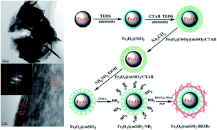
A rattle-type magnetic mesoporous Fe3O4@mSiO2@BiOBr hierarchical photocatalyst was successfully synthesized by a facile solvothermal method under the orientation of the surface amino-groups of rattle-type magnetic mesoporous Fe3O4@mSiO2 microspheres. Then, this photocatalyst was characterized via X-ray diffraction, transmission electron microscopy, field-emitting scanning electron microscopy, Fourier transform-infrared spectroscopy, X-ray photoelectron spectroscopy and vibrating sample magnetometry. Due to the presence of an inner cavity and orderly mesoporous opening structure, this novel photocatalyst exhibits superior adsorption and transfer performance to organic contaminants in aqueous systems. In particular, the complex between BiOBr and SiO2 had significantly increased absorption ability to visible-light due to the some extent of the direct contact of the interfaces of the two materials. Studies show that the assembly capacity of BiOBr nanosheets plays an important role in enhancing the photoactivity. Even though methylene blue is a relatively stable organic contaminant, it can still be decomposed completely by this novel photocatalyst in a very short amount of time (about 120 min). Encouragingly, the photoactivity of this novel photocatalyst is far higher (about 2.6 times) than that of pure BiOBr photocatalyst for its unique structure. According to the radical trapping experiments, the photogenerated holes (h+) and superoxide radicals (O2˙) are considered to be the main active species that drive the photodegradation under visible-light irradiation. Due to the unique structures and fast interfacial charge transfer, this novel photocatalyst is absolutely a superior alternative visible-light-driven photocatalyst.


1 Introduction

Semiconductor photocatalysis is an effective method for eliminating most types of environmental contaminants and for producing hydrogen.1–5 Among the various semiconductor photocatalysts, bismuth oxyhalides are being extensively investigated in recent years due to their superior properties and potential applications. Due to the favourable visible-light absorption ability of most of the bismuth oxyhalides, these photocatalysts have been studied in varying degrees by many researchers. For instance, Weng et al.6 reported that single-crystalline BiOCl nanosheets exposing (010) facets exhibit higher photoactivity due to more surface complexity and more terminal bismuth atoms on the surface of BiOCl (010). Shi et al.7 fabricated BiOBr hierarchical nanostructures by a polyvinyl pyrrolidone (PVP) assisted hydrothermal method. They found that the concentration of PVP in the reaction mixture played a key role in controlling the structures and morphologies because PVP could selectively attach to the (012) plane of the BiOBr hierarchical nanostructures and insulate the growth of this plane. However, as a semiconductor with a wide band gap, the absorption ability of a pure bismuth oxyhalide to visible-light is very limited for degrading organic contaminants in an aqueous system. Therefore, many efforts have been devoted for developing functional bismuth oxyhalide based visible-light photocatalysts by incorporating metal ions or coupling with other semiconductors. For instance, Yu et al.8 prepared the noble metal (Rh, Pd, Pt)/BiOX(Cl, Br, I) composite photocatalysts, and their study showed that appropriate noble metal depositions can effectively enhance the visible-light absorption and photoactivity of bismuth oxyhalide based photocatalysts due to the increased separation rate of photogenerated electron–hole pairs and the role of plasmon photocatalysis. Furthermore, Jiang et al.9 prepared visible-light-induced BiOBr photocatalysts by chemical reduction, photoreduction and thermal reduction. They assumed that the solvothermal reduction method was relatively simple and efficient, and that the 3% Ag/Ti-doped BiOBr photocatalysts exhibited higher photoactivity and excellent durability. Beyond that, Wei et al.10 prepared a BiOBr–TiO2–graphene composite, and this material also exhibited superior photoactivity towards the photodegradation of rhodamine B under visible-light irradiation.

To effectively improve the separation of the photocatalyst from the catalysis system, preparing the photocatalyst with appropriate particle size or good magnetic response are the most effective methods. In particular, good magnetic response can accomplish the simple magnetic separation and regeneration of the photocatalyst. For instance, Zhang et al.11 prepared a magnetic BiOBr@SiO2@Fe3O4 photocatalyst, and their research revealed that this recyclable magnetic photocatalyst exhibited superior photoactivity than that of commercial TiO2 under the visible-light irradiation. Furthermore, Guo et al.12 prepared a plasmonic photocatalyst of Ag–AgI/Fe3O4@SiO2 by a deposition–precipitation and photoreduction method, and this photocatalyst exhibited efficient photoactivity in the degradation of rhodamine B and 4-chlorophenol and could be easily recovered due to its paramagnetic property.

Beyond that, the superior adsorption ability of the photocatalyst to organic contaminant molecules also plays an important role in its degradation performance. To the best of our knowledge, mesoporous silica shows very high specific surface area due to its orderly mesoporous opening structure.13,14 Combining the mesoporous structure with a rattle-type structure undoubtedly can significantly improve the mass transport ability due to the presence of inner cavities and connected mesopores, and it can indirectly enhance the degradation ability to some extent. Based on this idea, we designed and synthesized a novel rattle-type magnetic mesoporous Fe3O4@mSiO2@BiOBr hierarchical photocatalyst by a facile solvothermal synthesis procedure under the orientation of the surface amino-groups of rattle-type magnetic Fe3O4@mSiO2 microspheres. Due to the presence of the inner cavity and orderly mesoporous opening structure, this hierarchical photocatalyst exhibited very high adsorption ability to organic contaminant molecules. Simultaneously, the direct contact between the BiOBr semiconductor and mesoporous silica effectively improved the interfacial charge transfer and enhanced the suppression of the rapid recombination of photogenerated electron–hole pairs. Thus, this novel rattle-type magnetic mesoporous Fe3O4@mSiO2@BiOBr hierarchical photocatalyst exhibited highly superior photoactivity for the degradation of methylene blue in an aqueous system. Moreover, the main active species were also investigated by radical trapping experiments, and the h+ and O2˙ radicals were demonstrated to be the main active species that drive the photodegradation under the irradiation of visible light.

2 Experimental section

2.1 Reagents and materials

All the chemicals were purchased from J&K Chemical and were used without further treatment.

2.2 Preparation of rattle-type magnetic mesoporous Fe3O4@mSiO2 microspheres

The magnetic mesoporous Fe3O4@SiO2@mSiO2/CTAB microspheres with double shell structure were prepared by optimizing our previous method.15 Subsequently, the rattle-type magnetic mesoporous Fe3O4@mSiO2/CTAB composite microspheres were successfully synthesized after an etching process in the presence of CTAB. Briefly, 5 g of dried aforementioned microspheres was redispersed into 500 mL of distilled water. After being treated by ultrasonication for 15 min, 10 g of anhydrous Na2CO3 was added into this system, and the reaction system was stirred vigorously for 10 h at 50 °C. After the reaction was completed, the sample was separated from the system by applying an external magnetic field and was washed several times with distilled water with the aid of ultrasonic techniques. Lastly, the ultimate sample was dried for 12 h via a vacuum freeze-drying device. Here, it is denoted as R–Fe3O4@mSiO2.

2.3 Preparation of rattle-type magnetic mesoporous Fe3O4@mSiO2@BiOBr hierarchical photocatalyst

The aforementioned R–Fe3O4@mSiO2 microspheres were firstly modified with γ-aminopropyltriethoxysilane (APTES). Then, BiOBr sheets were orientally assembled on the surface of this composite material through the complexation between the amino-groups and Bi3+ ions. Briefly, 1.5 g of rattle-type magnetic mesoporous Fe3O4@mSiO2 microspheres was ultrasonically dispersed into 200 mL of acetone, and 4–6 drops of ammonia solution (25 wt%) was added. Then, the system was stirred vigorously for 15 min. Subsequently, the temperature was adjusted to 60 °C, and 4 mL of APTES was added into the aforementioned system. Then, this system was stirred for 12 h at a speed of 250 rpm. After the reaction was completed, the sample was collected from the system by applying an external magnetic field and washed several times with acetone and distilled water. Ultimately, the sample was dried for 12 h via the vacuum freeze-drying device, and the amino-functionalized rattle-type magnetic mesoporous silica microspheres (R–Fe3O4@mSiO2–NH2) were successfully obtained. Then, 0.3 g of R–Fe3O4@mSiO2–NH2, 0.5 g of KBr, 0.1 g of PVP and varying stoichiometric amounts of Bi(NO3)3·5H2O were added into 40 mL of EG. Then, this system was treated for 15 min under ultrasound to obtain a uniformly dispersed system. Subsequently, the aforementioned suspension was transferred to a Teflon-lined stainless steel autoclave with a capacity of 50 mL and kept for 8 h at 180 °C. After the reaction was completed, the system was allowed to cool naturally to room temperature. Then, the sample was separated by externally applying a permanent magnet and washed several times with ethanol and distilled water, respectively. Finally, the sample was dried for 12 h via the vacuum freeze-drying device, and the ultimate product was successfully obtained. Beyond that, the samples prepared under different temperatures or reaction times were also obtained by the aforementioned method, and a sample was prepared in the absence of PVP by the same method. Herein, the products are denoted as R–Fe3O4@mSiO2@BiOBr-α-β (α = 0, 0.2, 0.4, 0.6, 0.8 and 1.0 g, and β = 2, 4 and 6 h).

2.4 Characterization

The Fourier transform-infrared (FT-IR) spectra of the samples were recorded on a Perkin-Elmer 580BIR spectrophotometer using the KBr pellet technique. X-ray powder diffraction (XRD) analysis was performed on a Bruker AXS D8-advance X-ray diffractometer with Cu Kα radiation. The morphologies and sizes of the samples were characterized using transmission electron microscopy (TEM, JEOL JEL2010) and field-emitting scanning electron microscopy (FESEM, JEOL-JSM-6700 F). X-ray photoelectron spectroscopy (XPS) data were collected to examine the chemical states of the multi-component photocatalyst with an Axis Ultra instrument (Kratos Analytical, Manchester, U.K.) under ultrahigh vacuum conditions (<10−6 Pa) and using a monochromatic Al Kα X-ray source (1486.6 eV). N2 adsorption/desorption isotherms were obtained on a TriStar II 20 apparatus. The Brunauer–Emmett–Teller (BET) method was used to calculate the specific surface area based on the adsorption branches.

2.5 Photocatalytic performance tests

The photocatalytic activities of the as-prepared R–Fe3O4@mSiO2@BiOBr hierarchical photocatalysts were evaluated by catalyzing the photodegradation of MB in the water system at room temperature under visible-light irradiation with a 500 W xenon lamp (CHF-XM500, light intensity = 600 mW cm−2) located 20 cm away from the reaction solution. To make sure that the photocatalytic reaction was really driven by visible-light, all the UV irradiations with wavelengths less than 420 nm were removed by a glass filter (JB-420). In a typical reaction, 0.1 g of as-prepared photocatalyst was dispersed into 100 mL of an aqueous solution of MB (20 mg L−1). Before the light irradiation, the suspension was stirred for 30 min in the dark to reach the saturated adsorption equilibrium of MB molecules. Then, 5 mL of degradation solution was extracted to determine the concentration of MB in the aqueous solution by UV-vis spectroscopy. In this study, the pure BiOBr photocatalyst was also used as the reference catalyst to catalyze the photodegradation of MB under the same conditions as in the aforementioned operation. The aqueous solution of MB without photocatalyst irradiated by visible light was used as the blank. After the experiment was completed, the catalyst was collected by centrifugal separation and washed several times with ethanol and distilled water. Then, the photodegradation experiment catalyzed by the recovered catalyst was carried out repeatedly according to the aforementioned operational steps.

2.6 Radical trapping experiments

For detecting the main active species during photocatalytic reactivity, holes (h+), photoexcited electrons (e), hydroxyl radicals (˙OH) and superoxide radicals (O2˙) were investigated by adding 1.0 mM KI (a scavenger of photogenerated h+),16 1.0 mM tert-butanol (TBA, a quencher of photoexcited e),17 1.0 mM isopropanol (IPA, a scavenger of ˙OH),18 and 1.0 mM 1,4-benzoquinone (BQ, a scavenger of O2˙),19 respectively. The method was similar to the former photocatalytic activity test.

3 Results and discussion

3.1 Characterization of materials

The synthesis of the magnetic R–Fe3O4@mSiO2-BiOBr photocatalyst is depicted in Fig. 1. To investigate the morphologies of Fe3O4, Fe3O4@SiO2, Fe3O4@SiO2@mSiO2 and R–Fe3O4@mSiO2, TEM characterization was carried out, and Fig. 2 displays the corresponding TEM images. The magnetic Fe3O4 microparticles synthesized by the solvothermal method show a representative cauliflower-like morphology, and their diameter is about 500 nm (Fig. 2a and b). After being coated with a layer of dense silica film, the surface of the particles becomes smoother than that of Fe3O4, and the representative core–shell structure with shell thickness of 35 nm can be observed clearly (Fig. 2c and d). Subsequently, the magnetic mesoporous Fe3O4@SiO2@mSiO2/CTAB microspheres with a double-shell structure were successfully obtained, and the thickness of the outer mesoporous silica coating was determined to be about 50 nm (Fig. 2e and f). After the etching process, the internal dense silica coating was successfully removed under the protection of CTAB, and the representative rattle-type structure can be observed clearly (Fig. 2g and h). In addition, the distinct mesoporous opening structure can be observed after the removal of CTAB in the mesoporous silica shell (Fig. 2h).
image file: c5ra06894k-f1.tif
Fig. 1 Schematic illustration of the synthesis of the magnetic R–Fe3O4@mSiO2-BiOBr photocatalyst.

image file: c5ra06894k-f2.tif
Fig. 2 TEM images of Fe3O4 (a and b), Fe3O4@SiO2 (c and d), Fe3O4@SiO2@mSiO2/CTAB (e and f) and R–Fe3O4@mSiO2 (g and h).

Subsequently, the R–Fe3O4@mSiO2 microspheres were modified by APTES, and the corresponding FTIR characterization was obtained. As shown in Fig. S1a, the characteristic absorption peaks corresponding to the antisymmetric and symmetric stretching vibration of the Si–O–Si bond in the oxygen–silicon tetrahedron can be clearly observed at 1077 and 793 cm−1, respectively, and the characteristic absorption peak of the Fe–O bond can also be observed at 480 cm−1. After the amino-functionalization, the characteristic absorption peaks at 2918 and 2850 cm−1 corresponding to the symmetric and antisymmetric stretching vibrations of νC–H in methyl and methylene appeared (Fig. S1b). Beyond that, a shoulder peak attributed to the stretching vibration of δN–H and γN–H can be observed clearly at 1552 cm−1. This indicates that the R–Fe3O4@mSiO2 microspheres were successfully modified by APTES.

After the amino-functionalization, the BiOBr nanosheets were synthesized on the surface of the R–Fe3O4@mSiO2 microspheres by the orienting role of the amino-groups. Subsequently, the reaction time was chosen as 4 h, and a series of R–Fe3O4@mSiO2@BiOBr hierarchical photocatalysts were synthesized by adjusting the added amount of the bismuth source. After the assembly of the BiOBr nanosheets, the sheet structures, which constitute the BiOBr photocatalyst, can be observed clearly on the surface of the R–Fe3O4@mSiO2 microspheres (Fig. 3a–j). Expectedly, the amounts of BiOBr nanosheets on the surface of the R–Fe3O4@mSiO2 microspheres increased with increase of the bismuth source from 0.2 g to 1.0 g. In particular, when the addition of the bismuth source was 1.0 g, the morphology of the R–Fe3O4@mSiO2@BiOBr photocatalyst exhibited the representative hierarchical structure (Fig. 3i and j). In addition, assembly of the BiOBr nanosheets was also carried out on the surface of magnetic R–Fe3O4@mSiO2 microspheres, which were not functionalized through APTES, and the corresponding TEM and SEM images were obtained and are shown in Fig. 4a and b. Apparently, very few sheet structures can be observed on the surface of the R–Fe3O4@mSiO2 microspheres, which indicates that the amino groups on the surface of the magnetic R–Fe3O4@mSiO2 microspheres indeed play an important role in the oriented growth of the BiOBr nanosheets. Predominantly, the amino groups on the surface of the magnetic R–Fe3O4@mSiO2 microspheres can chelate the Bi3+ ions in the synthesis system, which would produce a strong orientation effect for the growth of BiOBr nanosheets.


image file: c5ra06894k-f3.tif
Fig. 3 TEM and SEM images of the R–Fe3O4@mSiO2@BiOBr hierarchical photocatalysts synthesized through the addition of Bi(NO3)3·5H2O: 0.2 g (a and b), 0.4 g (c and d), 0.6 g (e and f), 0.8 g (g and h) and 1.0 g (i and j). The added amount of PVP is 0.1 g, and the reaction time and reaction temperature is 4 h and 180 °C, respectively.

image file: c5ra06894k-f4.tif
Fig. 4 TEM and SEM images of the R–Fe3O4@mSiO2@BiOBr hierarchical photocatalyst synthesized on the surface of magnetic R–Fe3O4@mSiO2 microspheres, which were not functionalized by APTES. The added amount of Bi(NO3)3·5H2O and PVP is 1.0 g and 0.1 g, respectively. The reaction time and reaction temperature is 4 h and 180 °C, respectively.

In addition, the effect of reaction time was also investigated, and the corresponding TEM and SEM images are shown in Fig. 5. When the reaction time was chosen as 2 h, the independent R–Fe3O4@mSiO2 microspheres and BiOBr hierarchical photocatalyst were observed (Fig. 5a and b). However, when the reaction time was extended to 6 h, more BiOBr nanosheets were synthesized on the surface of the magnetic R–Fe3O4@mSiO2 microspheres (Fig. 5c and d). This indicates that the reaction time also plays an important role in the synthesis of the R–Fe3O4@mSiO2@BiOBr hierarchical photocatalyst. In detail, the assembly of the BiOBr nanosheets on the surface of the amino-functionalized magnetic R–Fe3O4@mSiO2 microspheres obviously depends on the reaction time. When the reaction time is very short, there is not enough time to support the assembly of the BiOBr nanosheets on the surface of the amino-functionalized magnetic R–Fe3O4@mSiO2 microspheres; thus, the R–Fe3O4@mSiO2@BiOBr hierarchical photocatalyst cannot be obtained, and only the independent magnetic R–Fe3O4@mSiO2 microspheres and a small amount of BiOBr hierarchical photocatalyst can be obtained. Instead, when the reaction took a longer time, the bismuth source would be consumed entirely, and more BiOBr nanosheets would be assembled on the surface of the amino-functionalized magnetic R–Fe3O4@mSiO2 microspheres under the orienting effect of the surface amino-groups.


image file: c5ra06894k-f5.tif
Fig. 5 TEM and SEM images of the R–Fe3O4@mSiO2@BiOBr hierarchical photocatalyst synthesized under the reaction time of 2 h (a and b) and 6 h (c and d). The reaction temperature is 180 °C, and the addition amounts of Bi(NO3)3·5H2O and PVP are 1.0 g and 0.1 g, respectively.

XPS spectra for the measurement of the surface compositions and chemical states of this R–Fe3O4@mSiO2@BiOBr-1-4 hierarchical photocatalyst are displayed in Fig. S2, and the XPS spectrum in Fig. S2a reveals that the surface of this photocatalyst consisted of O, C, Bi and Br. As shown in Fig. S2b, the two peaks at 159.2 and 164.5 eV are respectively attributed to Bi 4f7/2 and Bi 4f5/2, which indicates the existence of Bi3+ in BiOBr. The Br 3d5/2 and Br 3d3/2 peaks (Fig. S2c) are associated with the binding energy at 68.4 and 69.4 eV, respectively. The two peaks at 531.5 eV and 530.1 eV should be assigned as the characteristic peaks of O 1s in BiOBr, and the two peaks at 533.7 eV and 532.8 eV should be assigned to the characteristic peaks of O 1s in PVP and other components (such as –OH groups and crystalline H2O) adsorbed on the surface of the BiOBr photocatalyst (Fig. S2d). In addition, the three peaks at 287.9 eV, 285.9 eV and 284.7 eV in Fig. S2e should be attributed to the characteristic peaks of C 1s in PVP adsorbed on the surface of the BiOBr photocatalyst. It has been efficiently demonstrated that the BiOBr photocatalyst was successfully assembled on the surface of the R–Fe3O4@mSiO2 microspheres.

XRD was employed to determine the crystallographic phases of the products, and the corresponding XRD patterns are displayed in Fig. 6. As shown in Fig. 6a, the XRD pattern is well indexed to the standard cubic phase of Fe3O4 (JCPDS 65-3107). Fig. 6b shows that a wide diffraction peak around 24°, corresponding to the amorphous silica, can be observed after the introduction of mesoporous silica. Subsequently, six intense and sharp diffraction peaks at 25.3°, 32.5°, 39.6°, 46.4°, 67.7° and 77.0°, indexed to the tetragonal phase of BiOBr, can be observed clearly (Fig. 6c), indicating that the BiOBr nanosheets assembled on the surface of the amino-functionalized R–Fe3O4@mSiO2 microspheres are well-crystallized. In addition, Fig. 6d displays a high resolution image of the R–Fe3O4@mSiO2@BiOBr-1-4 hierarchical photocatalyst. The lattice fringes with the d-spacing of 0.278 nm and 0.35 nm can be assigned to the (110) and (101) lattice planes of the tetragonal system of BiOBr, respectively. In addition, the FFT image in the inset of Fig. 6d also indicates the aforementioned conclusion.


image file: c5ra06894k-f6.tif
Fig. 6 XRD patterns of Fe3O4 (a), R–Fe3O4@mSiO2 (b) and R–Fe3O4@mSiO2@BiOBr-1-4 (c). (d) HRTEM image of the R–Fe3O4@mSiO2@BiOBr-1-4 hierarchical photocatalyst. The inset is the corresponding FFT image.

It is important to characterize the magnetic properties of the magnetic materials. Fig. 7 displays the VSM curves of the R–Fe3O4@mSiO2 microspheres and R–Fe3O4@mSiO2@BiOBr-1-4 hierarchical photocatalyst. It is easy to see that the saturation magnetization of the aforementioned two materials is about 63 and 40 emu g−1, respectively. After the assembly of BiOBr nanosheets on the surface of the R–Fe3O4@mSiO2 microspheres, the saturation magnetization is decreased to some extent. However, it can still enable the simple separation of the photocatalyst from the degradation system. In addition, the magnification of the low fields (in the inset, Fig. 7) shows that both magnetic particles exhibit the representative feature of superparamagnetism because no hysteresis is observed in the low fields, which is very conducive to the redispersion of the particles.


image file: c5ra06894k-f7.tif
Fig. 7 Magnetization curves of R–Fe3O4@mSiO2 (black curve) and R–Fe3O4@mSiO2@BiOBr-1-4 (red curve) measured at room temperature, and the inset is the corresponding magnetization of the low field (−2000 to 2000 Oe).

3.2 Photocatalytic performance

The photodegradation of MB under visible-light response (λ > 420 nm) was used as a probe to evaluate the performance of the R–Fe3O4@mSiO2@BiOBr photocatalysts. First, the photocatalysts that were prepared using different added amounts of bismuth source and the pure BiOBr photocatalyst were kept for 30 min in the dark under vigorous agitation, and the corresponding adsorption saturation capacities were measured and are displayed in Fig. 8a. Obviously, the adsorption capacity of the photocatalyst for MB improved gradually with increasing amount of bismuth source, except in the case of R–Fe3O4@mSiO2@BiOBr-0.4-4 (∼27.64%), and R–Fe3O4@mSiO2@BiOBr-1-4 photocatalyst exhibited the highest adsorption capacity (∼51.27%) for MB, which may have resulted from the increased amount of BiOBr assembled on the surface of the magnetic R–Fe3O4@mSiO2 microspheres. In addition, the adsorption capacities of the pure BiOBr photocatalyst and R–Fe3O4@mSiO2 microspheres are respectively about 2.58% and 33.74%, which indicates that pure BiOBr exhibits poor adsorption ability to MB molecules, but R–Fe3O4@mSiO2 microspheres show superior adsorption ability to MB molecules due to the existence of the ordered mesoporous structure and the inner cavities. At the same time, this indicates that the introduction of the R–Fe3O4@mSiO2 microspheres with the unique structure had indeed enhanced the adsorption ability of the BiOBr photocatalyst. Subsequently, the photodegradation of MB was initiated under visible light, and the corresponding photodegradation kinetic curves are displayed in Fig. 8b. Then, the degradation kinetics of MB were also investigated by fitting the experimental data to the Langmuir–Hinshelwood models following the equation: −ln(ct/co) = kt (Fig. 8c). Obviously, all the photodegradations of MB by R–Fe3O4@mSiO2@BiOBr-α-4 photocatalysts efficiently obeyed the first order kinetics. It is easy to see that the photoactivities of these photocatalysts are basically consistent with their adsorption capacity. Among the photocatalysts, R–Fe3O4@mSiO2@BiOBr-1-4, which exhibited the highest photoactivity (k = 0.0223 min−1), could degrade about 96% of the MB molecules within 120 min. However, the activity is basically the same between R–Fe3O4@mSiO2@BiOBr-0.4-4 (k = 0.0070 min−1) and R–Fe3O4@mSiO2@BiOBr-0.8-4 (k = 0.0064 min−1). As a whole, the increase of the bismuth source would lead to increased effective contact area with MB molecules due to the increased assembly of BiOBr nanosheets on the amino-functionalized magnetic R–Fe3O4@mSiO2 microspheres. Correspondingly, this would lead to the higher adsorption capacity and enhanced photoactivity. Beyond that, the R–Fe3O4@mSiO2 microspheres and pure BiOBr photocatalyst were chosen as the reference photocatalysts, and the corresponding photoactivities were also investigated by catalyzing the degradation of MB in an aqueous system. Fig. 8d displays the corresponding Langmuir–Hinshelwood models of the photodegradation kinetic curves, and it shows that the R–Fe3O4@mSiO2 microspheres do not exhibit the ability to photodegrade MB under visible-light irradiation. Obviously, the photoactivity of the R–Fe3O4@mSiO2@BiOBr-1-4 hierarchical photocatalyst is far higher (about 5.9 times) than that of the pure BiOBr photocatalyst. This indicates that the introduction of the rattle-type magnetic mesoporous Fe3O4@mSiO2 microspheres with unique structure had indeed significantly enhanced the photoactivity of the BiOBr photocatalyst, which should mainly be caused by the superior adsorption ability to the MB molecules and the fast interfacial charge transfer (Fig. S3).
image file: c5ra06894k-f8.tif
Fig. 8 The saturated adsorptions in the dark (a), the photodegradation kinetic curves of the photocatalysts with differing BiOBr content (b), the corresponding fitted kinetic curves according to the Langmuir–Hinshelwood model (c) and the Langmuir–Hinshelwood model of the photoactivity of R–Fe3O4@mSiO2, pure BiOBr and R–Fe3O4@mSiO2@BiOBr-1-4 (d).

Furthermore, the saturated adsorptions and photoactivities of the R–Fe3O4@mSiO2@BiOBr hierarchical photocatalysts synthesized under the different reaction times (2 h, 4 h and 6 h) were also investigated. Fig. 9a displays the saturated adsorption capacities of the R–Fe3O4@mSiO2@BiOBr-1-2, R–Fe3O4@mSiO2@BiOBr-1-4 and R–Fe3O4@mSiO2@BiOBr-1-6, and it shows that both R–Fe3O4@mSiO2@BiOBr-1-2 and R–Fe3O4@mSiO2@BiOBr-1-4 exhibit higher adsorption capacity than R–Fe3O4@mSiO2@BiOBr-1-6, and the R–Fe3O4@mSiO2@BiOBr-1-2 photocatalyst shows the highest saturated adsorption (∼69.95%) among the three photocatalysts. To explain this phenomenon, these three materials were characterized by N2 adsorption/desorption, and Fig. 9e and f display the corresponding isotherms and pore size distributions, respectively. Beyond that, the summary of all data is also displayed in the table of Fig. 9. Although the BET surface area of R–Fe3O4@mSiO2@BiOBr-1-2 is the smallest (about 131.66 m2 g−1), the independent existence of the BiOBr hierarchical microparticles and R–Fe3O4@mSiO2 microspheres (Fig. 5a and b) would lead to higher adsorption ability for MB molecules due to the combined effect of the BiOBr surface and the orderly mesoporous opening structure of the R–Fe3O4@mSiO2 microspheres. Subsequently, the adsorption ability of the R–Fe3O4@mSiO2@BiOBr photocatalyst would gradually decrease with prolonging of the reaction time, which is caused by the sacrificial effect of the orderly mesoporous opening structure of the R–Fe3O4@mSiO2 microspheres for the increased assembly of BiOBr nanosheets. Furthermore, this also indicates that the orderly mesoporous opening structure of the R–Fe3O4@mSiO2 microspheres plays an important role in enhancing the adsorption ability. Fig. 9b and c display the corresponding photocatalytic degradation kinetic curves and the Langmuir–Hinshelwood models, respectively. It is noteworthy that the R–Fe3O4@mSiO2@BiOBr-1-4 hierarchical photocatalyst exhibited the highest photoactivity at 120 min, and this was lower for R–Fe3O4@mSiO2@BiOBr-1-2 (∼84%, k = 0.0135 min−1) and R–Fe3O4@mSiO2@BiOBr-1-6 (∼81%, k = 0.0128 min−1). Regarding the BET surface area (see the table in Fig. 9), it would gradually increase (131.66 to 161.96 m2 g−1) with prolonging of the reaction time. When the reaction time is 2 h, the photodegradation of MB is mainly caused by the independent BiOBr hierarchical microparticles; thus, the photoactivity of R–Fe3O4@mSiO2@BiOBr-1-2 is lower than that of the R–Fe3O4@mSiO2@BiOBr-1-4 hierarchical photocatalyst. When the reaction time is 6 h, the mass transfer effect of the orderly mesoporous opening structure of the R–Fe3O4@mSiO2 microspheres is weakened significantly by the excess assembly of BiOBr nanosheets; thus, their photoactivity is also lower than that of the R–Fe3O4@mSiO2@BiOBr-1-4 hierarchical photocatalyst, even though their BET surface area is higher. When the reaction time is 4 h, the mass transfer effect of the orderly mesoporous opening structure of the R–Fe3O4@mSiO2 microspheres is not inhibited due to the reasonable assembly of BiOBr nanosheets; thus, the R–Fe3O4@mSiO2@BiOBr-1-4 hierarchical photocatalyst exhibits the highest photoactivity. Fig. 9d displays the corresponding UV-vis spectra of MB solution in the presence of the R–Fe3O4@mSiO2@BiOBr-1-4 hierarchical photocatalyst under visible-light irradiation, and it shows that the maximum absorption peak (662 nm) would gradually weaken with prolonging of the illumination time and would disappear entirely after being irradiated for 120 min, indicating that the MB molecules had been degraded entirely. In addition, the digital photos in the inset of Fig. 9d demonstrate the abovementioned conclusion. After the photocatalytic degradation process, the R–Fe3O4@mSiO2@BiOBr-1-4 hierarchical photocatalyst was also characterized by the TEM instrument, and the corresponding TEM and HRTEM images are displayed in Fig. S4a and b. Both the images show unobvious variation, indicating that this novel hierarchical photocatalyst exhibits superior stability.


image file: c5ra06894k-f9.tif
Fig. 9 The saturated adsorptions in the dark (a), the photodegradation kinetic curves of the photocatalysts prepared under the different reaction times (b), the fitted kinetic curves according to the Langmuir–Hinshelwood model (c) and the corresponding UV-vis spectra of the MB solution under photodegradation using the R–Fe3O4@mSiO2@BiOBr-1-4 hierarchical photocatalyst (d). The inset is the corresponding digital photos of the photodegradation system. The N2 adsorption/desorption isotherms (e) and pore size distribution (f) of the samples, and the table is the summary of the whole data.

3.3 Photocatalytic mechanism

To investigate the main active species involved in MB photodegradation over the R–Fe3O4@mSiO2@BiOBr hierarchical photocatalyst, KI, TBA, IPA and BQ were used as scavengers of photogenerated holes (h+), photoexcited electrons (e), hydroxyl radicals (˙OH) and superoxide radicals (O2˙), respectively. As shown in Fig. 10, when KI or BQ was added into the degradation system, a significant decrease in photoactivity was observed, and the corresponding degradation rate was about 54% or 61% in 120 min. Compared with the degradation system that does not contain any scavenger, the rates demonstrate that both the h+ and O2˙ radicals are the main active species. However, when TBA or IPA was added into the degradation system, the change of the photoactivity would not be obvious, indicating that the e and ˙OH radicals are not the main active species. Therefore, in this photocatalytic degradation system, the possible photocatalytic mechanism is proposed as follows:
 
Photocatalyst + → photocatalyst (h+ + e) (1)
 
O2 + e → O2˙ (2)
 
O2˙ + 2H+ + e → H2O2 (3)
 
H2O2 + e → ˙OH + OH (4)
 
OH + h+ → ˙OH (5)
 
H2O2 + h+ → ˙OH + H+ (6)
 
MB + h+ → MB+ (7)
 
MB+ + O2˙ → degradation products (8)
 
MB + ˙OH → degradation products (9)

image file: c5ra06894k-f10.tif
Fig. 10 Effects of different scavengers on the degradation of MB in the presence of R–Fe3O4@mSiO2@BiOBr-1-4 hierarchical photocatalyst under the visible-light irradiation.

Under visible-light irradiation, the reaction is initiated by the excitation of the photocatalyst, resulting in the promotion of electrons (e) from the valence band (VB) to the conduction band (CB) of the BiOBr semiconductor and the generation of holes (h+) in the VB (eqn (1)). Then, the electrons react with O2 molecules to generate O2˙ radicals (eqn (2)), and O2˙ radicals can react with H+ ions and electrons to generate H2O2 molecules (eqn (3)). Subsequently, H2O2 molecules react with electrons to generate ˙OH radicals and OH ions (eqn (4)). Simultaneously, OH ions react with h+ to generate ˙OH radicals (eqn (5)), and H2O molecules react with h+ to generate ˙OH radicals and H+ ions (eqn (6)). In addition, MB can react with h+ to generate the activated MB+ (eqn (7)), and then activated MB+ ions react with O2˙ radicals to generate the ultimate degradation products (H2O and CO2) (eqn (8)). At the same time, MB can also directly react with ˙OH radicals to generate the ultimate degradation products (eqn (9)). According to the radical trapping experiments, the h+ and O2˙ radicals are the main active species; thus, eqn (1), (2), (7) and (8) are the main degradation routes.

4 Conclusion

In this study, a novel rattle-type magnetic mesoporous Fe3O4@mSiO2@BiOBr hierarchical photocatalyst was successfully synthesized by a facile solvothermal method under the orientation of the surface amino-groups of rattle-type magnetic mesoporous Fe3O4@mSiO2 microspheres. Due to the presence of an inner cavity and orderly mesoporous opening structure, this novel photocatalyst exhibits superior adsorption and transfer performance to organic contaminants in aqueous systems. In particular, the complex between BiOBr and SiO2 significantly increased the visible light absorption ability to some extent due to the direct contact of the interfaces of the two materials. Investigations show that the assembly capacity of BiOBr plays an important role in enhancing photoactivity. Even though methylene blue is a relatively stable organic contaminant, it can still be decomposed completely by this novel photocatalyst within a very short amount of time (about 120 min). Encouragingly, the photoactivity of this novel photocatalyst is far higher (about 2.6 times) than that of the pure BiOBr photocatalyst due to its unique structure. According to the radical trapping experiments, the photogenerated holes (h+) and superoxide radicals (O2˙) are considered to be the main active species that drive the photodegradation under visible-light irradiation. Due to the unique structures and fast interfacial charge transfer, this novel photocatalyst is absolutely a superior alternative visible-light-driven photocatalyst.

Acknowledgements

The authors are grateful for the financial support provided by the National Natural Science Foundation of China (no. 51173146 and no. 21201140) and the basic research fund of Northwestern polytechnical university (3102014JCQ01094, 3102014ZD).

References

  1. X. Dang, X. Zhang, W. Zhang, X. Dong, G. Wang, C. Ma, X. Zhang, H. Ma and M. Xue, Ultra-thin C3N4 nanosheets for rapid charge transfer in the core–shell heterojunction of a-sulfur@C3N4 for superior metal-free photocatalysis under visible light, RSC Adv., 2015, 5, 15052–15058 RSC.
  2. S. Liu, M.-Q. Yang and Y.-J. Xu, Surface charge promotes the synthesis of large, flat structured graphene–(CdS nanowire)–TiO2 nanocomposites as versatile visible light photocatalysts, J. Mater. Chem. A, 2014, 2, 430–440 CAS.
  3. X. Ren, P. Ying, Z. Yang, M. Shang, H. Hou and F. Gao, Foaming-assisted electrospinning of large-pore mesoporous ZnO nanofibers with tailored structures and enhanced photocatalytic activity, RSC Adv., 2015, 5, 16361–16367 RSC.
  4. L. J. Zhang, R. Zheng, S. Li, B. K. Liu, D. J. Wang, L. L. Wang and T. F. Xie, Enhanced Photocatalytic H2 Generation on Cadmium Sulfide Nanorods with Cobalt Hydroxide as Cocatalyst and Insights into Their Photogenerated Charge Transfer Properties, ACS Appl. Mater. Interfaces, 2014, 6, 13406–13412 CAS.
  5. J. Jiang, L. Tong, H. Zhou, F. Zhang, J. Ding, D. Zhang and T. Fan, A photochromic nano-system via self-recovery for stable photocatalytic hydrogen evolution by optimizing TiO2 surface energy, RSC Adv., 2015, 5, 15844–15849 RSC.
  6. S. Weng, Z. X. Pei, Z. Zheng, J. Hu and P. Liu, Exciton-Free, Nonsensitized Degradation of 2-Naphthol by Facet-Dependent BiOCl under Visible Light: Novel Evidence of Surface-State Photocatalysis, ACS Appl. Mater. Interfaces, 2013, 5, 12380–12386 CAS.
  7. X. Shi, X. Chen, X. Chen, S. Zhou, S. Lou, Y. Wang and L. Yuan, PVP assisted hydrothermal synthesis of BiOBr hierarchical nanostructures and high photocatalytic capacity, Chem. Eng. J., 2013, 222, 120–127 CrossRef CAS PubMed.
  8. C. Yu, F. Cao, G. Li, R. Wei, J. C. Yu, R. Jin, Q. Fan and C. Wang, Novel noble metal (Rh, Pd, Pt)/BiOX(Cl, Br, I) composite photocatalysts with enhanced photocatalytic performance in dye degradation, Sep. Purif. Technol., 2013, 120, 110–122 CrossRef CAS PubMed.
  9. G. Jiang, R. Wang, X. Wang, X. Xi, R. Hu, Y. Zhou, S. Wang, T. Wang and W. Chen, Novel Highly Active Visible-Light-Induced Photocatalysts Based on BiOBr with Ti Doping and Ag Decorating, ACS Appl. Mater. Interfaces, 2012, 4, 4440–4444 CAS.
  10. X.-X. Wei, C.-M. Chen, S.-Q. Guo, F. Guo, X.-M. Li, X.-X. Wang, H.-T. Cui, L.-F. Zhao and W. Li, Advanced visible-light-driven photocatalyst BiOBr–TiO2–graphene composite with graphene as a nano-filler, J. Mater. Chem. A, 2014, 2, 4667–4675 CAS.
  11. L. Zhang, W. Wang, S. Sun, Y. Sun, E. Gao and Z. Zhang, Elimination of BPA endocrine disruptor by magnetic BiOBr@SiO2@Fe3O4 photocatalyst, Appl. Catal., B, 2014, 148, 164–169 CrossRef PubMed.
  12. J.-F. Guo, B. Ma, A. Yin, K. Fan and W.-L. Dai, Photodegradation of rhodamine B and 4-chlorophenol using plasmonic photocatalyst of Ag–AgI/Fe3O4@SiO2 magnetic nanoparticle under visible light irradiation, Appl. Catal., B, 2011, 101, 580–586 CrossRef CAS PubMed.
  13. H.-S. Huang, K.-H. Chang, N. Suzuki, Y. Yamauchi, C.-C. Hu and K. C.-W. Wu, Evaporation-Induced Coating of Hydrous Ruthenium Oxide on Mesoporous Silica Nanoparticles to Develop High-Performance Supercapacitors, Small, 2013, 9, 2520–2526 CrossRef CAS PubMed.
  14. J. T. Lee, Y. Zhao, S. Thieme, H. Kim, M. Oschatz, L. Borchardt, A. Magasinski, W.-I. Cho, S. Kaskel and G. Yushin, Sulfur-Infiltrated Micro- and Mesoporous Silicon Carbide-Derived Carbon Cathode for High-Performance Lithium Sulfur Batteries, Adv. Mater., 2013, 25, 4573–4579 CrossRef CAS PubMed.
  15. W. Li, B. Zhang, X. Li, H. Zhang and Q. Zhang, Preparation and characterization of novel immobilized Fe3O4@SiO2@mSiO2-Pd(0) catalyst with large pore-size mesoporous for Suzuki coupling reaction, Appl. Catal., A, 2013, 459, 65–72 CrossRef CAS PubMed.
  16. J. Di, J. Xia, S. Yin, H. Xu, L. Xu, Y. Xu, M. He and H. Li, Preparation of sphere-like g-C3N4/BiOI photocatalysts via a reactable ionic liquid for visible-light-driven photocatalytic degradation of pollutants, J. Mater. Chem. A, 2014, 2, 5340–5351 CAS.
  17. F. Li, Q. Wang, J. Ran, Y. Hao, X. Wang, D. Zhao and S. Z. Qiao, Ionic liquid self-combustion synthesis of BiOBr/Bi24O31Br10 heterojunctions with exceptional visible-light photocatalytic performances, Nanoscale, 2015, 7, 1116–1126 RSC.
  18. L. Ye, J. Liu, Z. Jiang, T. Peng and L. Zan, Facets coupling of BiOBr-g-C3N4 composite photocatalyst for enhanced visible-light-driven photocatalytic activity, Appl. Catal., B, 2013, 142, 1–7 Search PubMed.
  19. X. Wang, W. Yang, F. Li, Y. Xue, R. Liu and Y. Hao, In Situ Microwave-Assisted Synthesis of Porous N-TiO2/g-C3N4 Heterojunctions with Enhanced Visible-Light Photocatalytic Properties, Ind. Eng. Chem. Res., 2013, 52, 17140–17150 CrossRef CAS.

Footnote

Electronic supplementary information (ESI) available. See DOI: 10.1039/c5ra06894k

This journal is © The Royal Society of Chemistry 2015
Click here to see how this site uses Cookies. View our privacy policy here.