
A sensitive electrochemical immunosensor for the detection of squamous cell carcinoma antigen by using PtAu nanoparticles loaded on TiO2 colloidal spheres as labels
Hongmin Maa,
Yaoguang Wanga,
Hui Zhangb,
Dan Wua,
Aiping Guoa,
Tao Yanc,
Qin Weia and
Bin Du*a
aKey Laboratory of Chemical Sensing & Analysis in Universities of Shandong, School of Chemistry and Chemical Engineering, University of Jinan, Jinan 250022, China. E-mail: dubin61@gmail.com; Fax: +86 531 82767873; Tel: +86 531 82767873
bDepartment of Municipal and Environmental Engineering, Shandong Urban Construction Vocational College, Jinan 250103, China
cSchool of Resources and Environment, University of Jinan, Jinan 250022, China
First published on 6th July 2015
Abstract
A sensitive sandwich-type electrochemical immunosensor for detection of squamous cell carcinoma antigen (SCCA) was developed by using PtAu nanoparticles loaded on TiO2 colloidal spheres (PtAu/TiO2) as secondary-antibody (Ab2) labels. In this work, nitrogen-doped graphene sheets (N-GS) and chitosan (CS) were applied to modify the glassy carbon electrode (GCE) surface. Then the SCCA primary antibody (Ab1), SCCA and PtAu/TiO2-Ab2 were sequentially modified onto the electrode surface. Electrochemical impedance spectroscopy (EIS) and cyclic voltammetry (CV) were used to characterize the preparation process of the immunosensor and the amperometric response of the immunosensor for catalyzing hydrogen peroxide (H2O2) reduction was also recorded. Under optimal conditions, the immunosensor exhibited a wide linear response to SCCA ranging from 0.001 ng mL−1 to 15 ng mL−1, with a low detection limit of 0.3 pg mL−1. The proposed immunosensor displayed good reproducibility, high sensitivity, and acceptable stability. Therefore, this method has potential application for clinical immunoassays of other biomolecules.
1. Introduction
Highly sensitive detection and accurate analysis of biomarker molecules in human serum are essential for early detection, treatment and management of cancer.1–4 Squamous cell carcinoma antigen (SCCA), a glycoprotein with isoforms ranging from 45 to 55 kD which can be separated from cervical squamous cell carcinoma tissue, was first described as a tumor associated antigen by Kato and Torigoe.5 Initially, SCCA was considered as a serological marker for squamous cell tumors in the cervix. Later, it was found to be associated with other types of cancer with epithelial or endodermal origins, including lung cancer, head and neck cancer, melanomas, and hepatocellular carcinoma.6–9 The concentration of SCCA increases as the patient's condition worsens. So it can be applied as an auxiliary diagnosis index of cervical cancer, lung cancer, head and neck tumor and an index of curative effect, recurrence and metastasis, for prognosis monitoring.Chemiluminescence enzyme immunoassay (CLEIA), radioimmunoassay (RIA), localized surface plasmon resonance (LSPR) and enzyme-linked immunosorbent assay (ELISA) have been used to detect the total SCCA in serum.10–13 Nevertheless, the aforementioned techniques have defects like poor reproducibility, low sensitivity, narrow linear range, high cost and environmental pollution. Therefore, it is necessary to seek simple, sensitive, and low-cost detection techniques for the detection of SCCA. Electrochemical immunosensor especially sandwich-type has gained significant attention recently because of its high sensitivity and selectivity.14–17 For example, Li et al.18 have developed a magneto-controlled electrochemical immunosensor for direct detection of SCCA. Although the detection range and detection limit were acceptable, applying horseradish peroxidase to catalyze the reduction of H2O2 indirectly increased the cost of the detection. Hence, we developed an enzyme-free sandwich-type electrochemical immunosensor to achieve a more sensitive detection of SCCA.
As is well known, the electrode modifying material and the secondary-antibody (Ab2) labels are two of the most important factors affecting the sensitivity of the sandwich-type electrochemical immunosensor. It is well-known that chemical doping with foreign atoms is an effective method to produce local changes to the elemental composition of host materials, thus to improve electronic properties, and manipulate surface chemistry.19,20 Nitrogen-doped graphene (N-GS) is widely used as electrode modifying material in the field of electrochemical immunosensor because of its large surface area, strong conductivity and chemical properties.21–27 At the same time, chitosan (CS) also is one of the most promising materials to work as substrate material due to its excellent biocompatibility, high mechanical strength, good film-forming properties, and low cost.28–30 Most of all, the abundant amino groups in CS can add the loading capacity of biomolecules such as protein, enzyme and antibodies.31,32 For the Ab2 labels, noble metal nanoparticles have been widely used recently. Both Pt nanoparticles and Au nanoparticles exhibit good performances in fabricating immunosensors due to their good electrical conductivity and catalytic performance.33,34
As far as we know, no literatures have reported about the detection of SCCA using Pt and Au nanoparticles with TiO2 as labels. In this study, PtAu nanoparticles loaded on TiO2 colloidal sphere (PtAu/TiO2) was fabricated and applied as the labels of Ab2. The sandwich-type strategy was used to fabricate the immunosensor, with the SCCA primary antibody (Ab1) immobilized onto the N-GS and CS modified electrode through the covalent binding of amino groups. Subsequently, the SCCA and PtAu/TiO2-Ab2 were modified onto the electrode sequentially. Finally the properties of the immunosensor were measured by recording the current responses for catalytic reduction of H2O2. The results showed that the proposed method is credible and shows potential application in the detection of other tumor markers.
2. Experimental section
2.1. Materials and apparatus
SCCA and SCCA antibody (Ab1 and Ab2) were purchased from Shanghai Linc-Bio Science Co. Ltd. K3Fe(CN)6 and K4Fe(CN)6 were purchased from Sinopharm Chemical Reagent Beijing Co., Ltd., China. Bovine serum albumin (BSA, 96–99%) was purchased from Sigma (USA) and used as received. H2PtCl6·6H2O and HAuCl4·4H2O were obtained from Sigma-Aldrich (Beijing, China). Phosphate buffered saline (PBS, 0.1 mol L−1 containing 0.1 mol L−1 NaCl, pH 7.4) was used as an electrolyte for all electrochemical measurements. Ultrapure water was used throughout the experiments. All other chemicals were of analytical reagent grade.Electrochemical measurements were performed on a CHI760D electrochemical workstation (Chenhua Instrument Shanghai Co., Ltd., China). Electrochemical impedance spectroscopy (EIS) was obtained from the impedance measurement unit (IM6e, ZAHNER-Elektrik, Germany). Scanning electron microscope (SEM) images were obtained using a field emission SEM (ZEISS, Germany).
2.2. Preparation of N-GS
The graphene oxide was synthesized by using a modified Hummers method.35 The N-GS was obtained by heat treatment of graphene oxide (GO) in aqueous solution of ammonia.36 In a typical experiment, 140 mg of GO was dispersed in water, then the solution pH was adjusted to 10.0 by 30% of ammonia with magnetic stirring about 10 min at room temperature. Subsequently, 2 mL of hydrazine hydrate was added into the mixture. Afterwards the above mixture was transferred to the high-pressure reaction kettle, maintaining 12 h at 80 °C. The obtained black sediment was filtered and washed to neutral, and the N-GS would be obtained by vacuum drying.2.3. Preparation of Au nanoparticles
Au nanoparticles were prepared by modifying a literature method.37 Typically, 2.0 mL of 0.04 mol L−1 HAuCl4 solution was added to 298 mL of ultrapure water. Then the solution pH was adjusted to 6.0–7.0 by 1.0 mol L−1 NaOH. The solution was stirred and heated to boiling. Consequently, 2.8 mL of 0.1 mol L−1 of sodium citrate was added into the above solution. Then the solution was heated continually until the solution was turned into wine red, and the Au nanoparticles would be obtained.2.4. Preparation of PtAu/TiO2
PtAu/TiO2 was synthesized according to the literature report.38 The procedures were as follows.2.5. Preparation of PtAu/TiO2-Ab2
The preparation procedure of PtAu/TiO2-Ab2 is shown in Scheme 1A. The PtAu/TiO2 (2 mg) was dispersed in 1.0 mL of PBS at pH 7.4. Then it was mixed with 1.0 mL of 10 μg mL−1 of SCCA antibody. The mixture was reacted at 4 °C for 12 h. On the basis of the conjugation of PtAu/TiO2 with SCCA antibody via the covalent bond between Au nanoparticles and amino groups of SCCA antibody,39–41 the PtAu/TiO2-Ab2 was obtained successfully. The resulting PtAu/TiO2-Ab2 was centrifuged and washed with PBS (pH = 7.4), and it was redispersed in 1.0 mL of PBS and stored at 4 °C before use.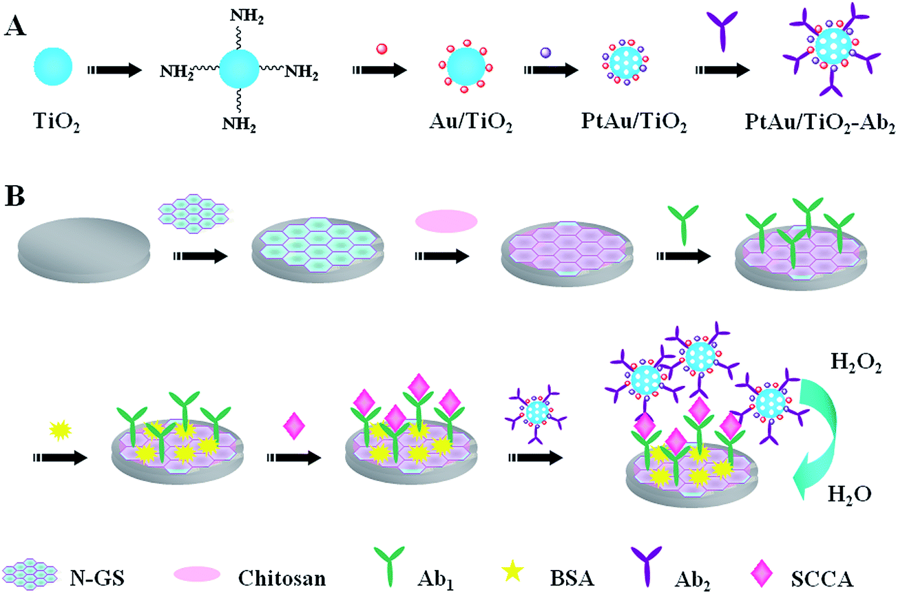 | ||
| Scheme 1 Schematic representation of the preparation of PtAu/TiO2-Ab2 (A) and immunosensor (B). | ||
2.6. Fabrication of the immunosensor
Scheme 1B shows the fabrication process of the immunosensor. Glassy carbon electrode (GCE) was polished with 1, 0.3, and 0.05 μm alumina powder in sequence, and then washed ultrasonically in ethanol for a few minutes and dried in air at room temperature. Afterwards, 6 μL of N-GS solution was initially dropped onto the electrode surface and dried at room temperature. Then 6 μL of 0.5% CS was added onto the electrode surface. As the electrode surface dried, 6 μL of Ab1 (10 μg mL−1) was dropped onto it and incubated for 1 h. After drying and washing, 1% BSA solution was used to eliminate nonspecific binding sites. Subsequently, SCCA buffer solutions at different concentrations were dropped onto the electrode and incubated for 1 h. Then the electrode was washed thoroughly to remove unbound SCCA molecules. Finally, the prepared PtAu/TiO2-Ab2 solution was dropped onto the electrode surface. When the electrode had dried, it was washed, and was then ready for measurements.2.7. Characterization of the immunosensor
A conventional three-electrode system was used for all electrochemical measurements: a GCE (4 mm in diameter) as the working electrode, a saturated calomel electrode (SCE) as the reference electrode, and a platinum wire electrode as the counter electrode. The pH 7.4 PBS (0.1 mol L−1 NaCl as electrolyte) was used for all the electrochemical measurements. EIS was tested in the presence of 5.0 mmol L−1 [Fe(CN)6]3−/4− containing 0.1 mol L−1 KCl. Cyclic voltammetry (CV) was recorded in PBS at 100 mV s−1. For amperometric measurement of the immunosensor, a detection potential of −0.4 V was selected. After the background current was stabilized, 5.0 mmol L−1 H2O2 was injected into the buffer and the current change was recorded.3. Results and discussion
3.1. Characterization of N-GS, TiO2, Au/TiO2 and PtAu/TiO2 nanoparticles
Fig. 1a shows the SEM image of prepared N-GS. As shown in the figure, the shape of N-GS was paper-like: a thin layer with a wrinkled structure. The thin layer and the wrinkled structure can increase the specific surface area of electrode and facilitate the electron transfer. Fig. 1b displays the SEM image of TiO2 precursor. It is obvious that the size of TiO2 is regular with a sphere shape. As can be seen from the SEM image of Au/TiO2 (Fig. 1c), the Au nanoparticles are dispersed on the surface of TiO2 uniformly. Fig. 1d also demonstrates the homogeneous dispersion of PtAu nanoparticles on the surface of TiO2 sphere. It is apparent that the amount of PtAu nanoparticles is more than Au nanoparticles in Fig. 1c. At the same time, the energy dispersive spectroscopy (EDS) (Fig. 1e) analysis of the PtAu/TiO2 nanoparticles shows five elements in the sample: Au, Pt, Al, O and Ti. Obviously, Au, Pt, O and Ti are from PtAu/TiO2 nanoparticles. At the same time, Al comes from tinfoil for SEM characterization. The above consequences prove that the PtAu/TiO2 nanoparticles were synthesized successfully. | ||
| Fig. 1 SEM images of N-GS (a), TiO2 (b), Au/TiO2 (c), PtAu/TiO2 (d) and EDS analysis of PtAu/TiO2 nanoparticles (e). | ||
3.2. Optimization of experimental conditions
Firstly, the operating voltage was chosen for the amperometric measurement of the immunosensor. As displayed in Fig. 2A, the CV curves of PtAu/TiO2 were obtained before (curve a) and after (curve b) the H2O2 was injected. It is obvious that the current changes obviously when the potential is lower than −0.3 V. At the same time, considering the effect of electrochemically reducible compounds like dissolved oxygen,42–44 the potential of −0.4 V was chosen in the succedent tests. | ||
| Fig. 2 The CV curves of PtAu/TiO2 used for choosing the operating voltage (A); effect of the pH (B) and the concentration of N-GS (C) on the response of the immunosensor to 8.0 ng mL−1 of SCCA. | ||
To obtain an optimal electrochemical response, the experimental conditions were optimized. During the immunosensor preparation process, the same concentration of SCCA (8.0 ng mL−1) was used to fabricate the immunosensor. The pH value of the substrate solution has been proven to be an important factor for current response.45,46 As shown in Fig. 2B, pH values of PBS from 4.5 to 9.1 were investigated and the optimal amperometric response was achieved at pH 7.4. The reason could be that highly acidic or alkaline surroundings could damage the immobilized protein. Thus, PBS at pH 7.4 was used as the electrolyte for all electrochemical measurement.
In addition, the concentration of N-GS also affects the current responses of the immunosensor. As can be seen from Fig. 2C, different concentrations of N-GS were tested for the current responses. With the increase of the N-GS concentration, the current response was firstly increased and then decreased. As N-GS possesses the properties of large specific surface area and excellent electron transferability, the current response would significantly increase with increase of the N-GS concentration. When the N-GS was excessive, the current response decreased, which might because the excessive N-GS could lower the electron transfer efficiency. As the maximum current response appeared at around 2.0 mg mL−1, this concentration was selected for the subsequent research.
3.3. Analysis of the immunosensor
EIS has been proven to be one of the most powerful tools for probing the features of surface modified electrodes.47,48 The impedance spectra include a semicircle portion and a linear portion. The semicircle portion at higher frequencies corresponds to the electron transfer limited process, and the semicircle diameter is equal to the electron transfer resistance. The linear portion at lower frequencies represents the diffusion limited process.49–51The Nyquist diagrams of EIS are shown in Fig. 3A. It can be found that the resistance decreased obviously after the N-GS modified onto the electrode (curve b) than the bare GCE (curve a). This can be ascribed to the good electron transfer capability of N-GS. When the electrode surface was modified with CS (curve c), the resistance increased. This may because that CS can form membrane which can hinder the electron transfer on the electrode surface. After modification of Ab1 onto the electrode (curve d), the resistance increased obviously, demonstrating the SCCA antibody was immobilized onto the electrode successfully. Afterwards, when BSA was modified (curve e), the resistance increased continuously. After the capture of SCCA, the resistance continued increasing (curve f), indicating that the protein can block the electron transfer tremendously. Finally, with PtAu/TiO2-Ab2 immobilized onto the electrode, the resistance increased (curve g), which implied that the immunosensor was fabricated successfully.
 | ||
| Fig. 3 Nyquist diagrams of EIS (A) recorded from 0.1 to 105 Hz and CV curves (B) in 5 mmol L−1 Fe(CN)63−/Fe(CN)64− containing 0.1 mmol L−1 KCl solution, (a) GCE; (b) N-GS/GCE; (c) CS/N-GS/GCE; (d) Ab1/CS/N-GS/GCE; (e) BSA/Ab1/CS/N-GS/GCE; (f) SCCA/BSA/Ab1/CS/N-GS/GCE; (g) PtAu/TiO2-Ab2/SCCA/BSA/Ab1/CS/N-GS/GCE. | ||
CV was also used for further characterization of the modification processes of the electrode. Fig. 3B shows the CV curves of different modified electrodes in 5 mmol L−1 Fe(CN)63−/Fe(CN)64− containing 0.1 mmol L−1 KCl solution. The results were in accordance with the EIS test results well, which further proved the successful fabrication of the immunosensor.
Under the optimal conditions, the immunosensors using PtAu/TiO2-Ab2 as labels were used to detect different concentrations of SCCA. The amperometric responses (Fig. 4A) were measured by successively adding H2O2 (5 mmol L−1) to a continuously stirred PBS (pH = 7.4) at −0.4 V. When the current responses were stable (about 100 s), the difference values (from 50 s to 100 s) were applied to plot the working curve (Fig. 4B). Due to the high sensitivity of Pt and Au nanoparticles for catalytic reduction of H2O2 (ref. 52–55) (electrocatalytic reduction equation: H2O2 + 2H+ + 2e− → 2H2O), the immunosensor using PtAu/TiO2-Ab2 as labels was built and characterized. Since the specific interaction of antigen and antibody, the amount of captured PtAu/TiO2-Ab2 labels was in accordance with the SCCA. Therefore, the concentration of SCCA could be detected quantitatively by this proposed immunosensor. The catalytic current increased linearly with the SCCA concentration in the range 0.001–15 ng mL−1, with a detection limit of 0.3 pg mL−1 (S/N = 3). Compared with other reports10,13,18 for the detection of SCCA (Table 1), the proposed immunosensor exhibits a much lower detection limit. The low detection limit may be attributed to several factors. First, the large specific surface area of N-GS can add the loading capacity for the Ab1, and the good electroconductivity and electron transfer capability of N-GS can facilitate the electron transfer on the electrode, leading to a high sensitivity. Second, the membrane that chitosan formed also can add the loading capacity for Ab1. Third, the PtAu/TiO2 as labels can have good biocompatibility, high conductivity, and high electrocatalytic activity toward H2O2.
 | ||
| Fig. 4 (A) Amperometric response of the immunosensor for different concentrations of SCCA at −0.4 V (PBS pH = 7.4, 5 mmol L−1 H2O2), with SCCA concentration (ng mL−1) of (a) 0.001, (b) 0.01, (c) 0.1, (d) 0.5, (e) 1.0, (f) 3.0, (g) 5.0, (h) 8.0, (i) 10, (j) 12, (k) 15; (B) calibration curve of the immunosensor for different concentrations of SCCA. Error bar = RSD (n = 5). | ||
3.4. Reproducibility, selectivity and stability of the immunosensor
To investigate the reproducibility of the immunosensor, a series of five electrodes were prepared for the detection of 8 ng mL−1 SCCA. The relative standard deviation (RSD) of the measurements for the five electrodes was 2.5%, indicating that the precision and reproducibility for the proposed immunosensor were quite good.Selective determination of target analyte plays an important role in analyzing biological samples.56 The amperometric responses of the immunosensor to alpha fetoprotein (AFP), BSA, carbohydrate antigen 125 (CA125), carbohydrate antigen 724 (CA724) were also investigated. Measurements were made on 1 ng mL−1 of SCCA solution containing 100 ng mL−1 of interfering substances using the immunosensor and the results are shown in Fig. 5. The current variation due to the interfering substances was less than 5% of the value for the pure SCCA solution, indicating acceptable selectivity of the immunosensor.
 | ||
| Fig. 5 Amperometric response of the immunosensor to 1 ng mL−1 SCCA (1); 1 ng mL−1 SCCA + 100 ng mL−1 AFP (2); 1 ng mL−1 SCCA + 100 ng mL−1 BSA (3); 1 ng mL−1 SCCA + 100 ng mL−1 CA125 (4); 1 ng mL−1 SCCA + 100 ng mL−1 CA724 (5). Error bar: RSD (n = 5). | ||
Stability of the immunosensors also is a key factor in their application and development.57 The stability of the immunosensor was examined by checking its current response regularly. When the immunosensor was not in use, it was stored in a refrigerator at 4 °C. The current response of the prepared immunosensor decreased 3.7% after seven days. Three weeks later, the current response of the immunosensor using PtAu/TiO2 as labels decreased to about 88% of its initial value. These small decreases of the current responses showed the good stability of the immunosensor.
3.5. Detection of SCCA in serum samples
In order to investigate the possibility of the immunosensor for practical analysis, the detection of SCCA in human serum samples (Provided by the hospital of University of Jinan) was performed using the proposed immunosensor with standard addition methods58–60 (Table 2). Firstly, the content of SCCA in the samples was detected by the proposed immunosensor according to the relation between the current and the concentration. Then a certain concentration of SCCA was added into the previous sample. At last, the final content of SCCA in the samples could be determinated. The RSD was in the range from 3.05% to 4.30% and the recovery was in the range from 99.3% to 101.4%. Thus, the proposed immunosensor could be effectively applied to the clinical determination of SCCA in human serum.| Serum sample | Content of SCCA in the sample (ng mL−1) | The addition content (ng mL−1) | The detection content (ng mL−1) | RSD (%) | Recovery (%) |
|---|---|---|---|---|---|
| 1 | 0.95 | 1.00 | 1.85, 2.02, 2.06, 1.91, 1.98 | 4.30 | 101.4 |
| 2 | 0.86 | 3.00 | 3.91, 3.90, 3.94, 3.65, 3.80 | 3.09 | 99.3 |
| 3 | 0.99 | 5.00 | 5.95, 5.75, 6.21, 5.85, 6.08 | 3.05 | 99.6 |
4. Conclusions
A sandwich-type immunosensor based on PtAu/TiO2 as labels for the sensitive detection of SCCA has been fabricated successfully. The excellent sensitivity of the fabricated immunosensor could be ascribed to following reasons: first, the large specific surface area of N-GS and the membrane that the CS formed could add the loading capacity of Ab1. Second, the good electroconductivity and electron transfer capability of N-GS also enhanced the sensitivity of the immunosensor. Significantly, the PtAu/TiO2 as labels had good catalytic reduction effect toward H2O2, which was beneficial to the detection of SCCA. In general, the proposed immunosensor shows wide linear range (0.001–15 ng mL−1) with a low detection limit (0.3 pg mL−1), good reproducibility and selectivity, acceptable stability as well as excellent sensitivity. Therefore, this simple technique may have a certain potential in the detection of other cancer biomarkers.Acknowledgements
This study was supported by the Natural Science Foundation of China (No. 21175057, 21375047, 21377046), the Science and Technology Plan Project of Jinan (No. 201307010), the Science and Technology Development Plan of Shandong Province (No. 2014GSF120004), and QW thanks the Special Foundation for Taishan Scholar Professorship of Shandong Province and UJN (No. ts20130937)References
- G. Jie, L. Wang and S. Zhang, Chem.–Eur. J., 2011, 17, 641–648 CrossRef CAS PubMed.
- J. Lin and H. Ju, Biosens. Bioelectron., 2005, 20, 1461–1470 CrossRef CAS PubMed.
- K. Liu, J.-J. Zhang, C. Wang and J.-J. Zhu, Biosens. Bioelectron., 2011, 26, 3627–3632 CrossRef CAS PubMed.
- J. Liu, C.-Y. Lu, H. Zhou, J.-J. Xu, Z.-H. Wang and H.-Y. Chen, Chem. Commun., 2013, 49, 6602–6604 RSC.
- H. Kato and T. Torigoe, Cancer, 1977, 40, 1621–1628 CrossRef CAS.
- X. Li, X. Zhang, H. Ma, D. Wu, Y. Zhang, B. Du and Q. Wei, Biosens. Bioelectron., 2014, 55, 330–336 CrossRef CAS PubMed.
- O. Micke, F. Bruns, U. Schäfer, F.-J. Prott and N. Willich, Anticancer Res., 2005, 25, 1663–1666 Search PubMed.
- N. Bandoh, T. Ogino, A. Katayama, M. Takahara, A. Katada, T. Hayashi and Y. Harabuchi, Oncol. Rep., 2010, 23, 933–939 CrossRef CAS.
- X.-Y. Feng, J.-H. Li, J.-Z. Li, Z. Han and R. Xing, Int. J. Biol. Markers, 2009, 25, 93–98 Search PubMed.
- H. Zhang and S. Qi, Clin. Chim. Acta, 2011, 412, 1572–1577 CrossRef CAS PubMed.
- W. Neunteufel, G. Tatra and C. Bieglmayer, Gynecol. Obstet. Invest., 1990, 29, 154–157 CrossRef CAS PubMed.
- Q. Zhao, R. Duan, J. Yuan, Y. Quan, H. Yang and M. Xi, Int. J. Nanomed., 2014, 9, 1097–1104 Search PubMed.
- J. A. Erickson, J. Lu, J. J. Smith, J. A. Bornhorst, D. G. Grenache and E. R. Ashwood, Clin. Chem., 2010, 56, 1496–1499 CAS.
- Z. Zhong, M. Li, D. Xiang, N. Dai, Y. Qing, D. Wang and D. Tang, Biosens. Bioelectron., 2009, 24, 2246–2249 CrossRef CAS PubMed.
- X. Pei, B. Zhang, J. Tang, B. Liu, W. Lai and D. Tang, Anal. Chim. Acta, 2013, 758, 1–18 CrossRef CAS PubMed.
- L. Wang, X. Jia, Y. Zhou, Q. Xie and S. Yao, Microchim. Acta, 2010, 168, 245–251 CrossRef CAS.
- A. Kaushik, P. R. Solanki, A. A. Ansari, S. Ahmad and B. D. Malhotra, Nanotechnology, 2009, 20, 055105–055112 CrossRef PubMed.
- Q. Li, D. Tang, J. Tang, B. Su, G. Chen and M. Wei, Biosens. Bioelectron., 2011, 27, 153–159 CrossRef CAS PubMed.
- Y. Wang, Y. Shao, D. W. Matson, J. Li and Y. Lin, ACS Nano, 2010, 4, 1790–1798 CrossRef CAS PubMed.
- Y. Shao, J. Sui, G. Yin and Y. Gao, Appl. Catal., B, 2008, 79, 89–99 CrossRef CAS PubMed.
- Y. Shao, S. Zhang, M. H. Engelhard, G. Li, G. Shao, Y. Wang, J. Liu, I. A. Aksay and Y. Lin, J. Mater. Chem., 2010, 20, 7491–7496 RSC.
- H. Wang, C. Zhang, Z. Liu, L. Wang, P. Han, H. Xu, K. Zhang, S. Dong, J. Yao and G. Cui, J. Mater. Chem., 2011, 21, 5430–5434 RSC.
- D. Usachov, O. Vilkov, A. Gruneis, D. Haberer, A. Fedorov, V. Adamchuk, A. Preobrajenski, P. Dudin, A. Barinov and M. Oehzelt, Nano Lett., 2011, 11, 5401–5407 CrossRef CAS PubMed.
- J. Gao, B. Du, X. Zhang, A. Guo, Y. Zhang, D. Wu, H. Ma and Q. Wei, Anal. Chim. Acta, 2014, 833, 9–14 CrossRef CAS PubMed.
- S. Deng, J. Lei, Y. Huang, Y. Cheng and H. Ju, Anal. Chem., 2013, 85, 5390–5396 CrossRef CAS PubMed.
- J. Borowiec, R. Wang, L. Zhu and J. Zhang, Electrochim. Acta, 2013, 99, 138–144 CrossRef CAS PubMed.
- R. Feng, Y. Zhang, H. Ma, D. Wu, H. Fan, H. Wang, H. Li, B. Du and Q. Wei, Electrochim. Acta, 2013, 97, 105–111 CrossRef CAS PubMed.
- R. Liang, H. Peng and J. Qiu, J. Colloid Interface Sci., 2008, 320, 125–131 CrossRef CAS PubMed.
- H. Yu, F. Yan, Z. Dai and H. Ju, Anal. Biochem., 2004, 331, 98–105 CrossRef CAS.
- G. Wang, J.-J. Xu, H.-Y. Chen and Z.-H. Lu, Biosens. Bioelectron., 2003, 18, 335–343 CrossRef CAS.
- J.-D. Qiu, R.-P. Liang, R. Wang, L.-X. Fan, Y.-W. Chen and X.-H. Xia, Biosens. Bioelectron., 2009, 25, 852–857 CrossRef CAS PubMed.
- D. Tang, R. Yuan, Y. Chai, J. Dai, X. Zhong and Y. Liu, Bioelectrochemistry, 2004, 65, 15–22 CrossRef CAS PubMed.
- S. Singal, A. Biradar and A. Mulchandani, Appl. Biochem. Biotechnol., 2014, 1–13 Search PubMed.
- H. Wang, H. Li, Y. Zhang, Q. Wei, H. Ma, D. Wu, Y. Li, Y. Zhang and B. Du, Biosens. Bioelectron., 2014, 53, 305–309 CrossRef CAS PubMed.
- D. C. Marcano, D. V. Kosynkin, J. M. Berlin, A. Sinitskii, Z. Sun, A. Slesarev, L. B. Alemany, W. Lu and J. M. Tour, ACS Nano, 2010, 4, 4806–4814 CrossRef CAS PubMed.
- D. Long, W. Li, L. Ling, J. Miyawaki, I. Mochida and S.-H. Yoon, Langmuir, 2010, 26, 16096–16102 CrossRef CAS PubMed.
- A. Guo, D. Wu, H. Ma, Y. Zhang, H. Li, B. Du and Q. Wei, J. Mater. Chem., 2013, 1, 4052–4058 RSC.
- R. Huang, A. Zhu, Y. Gong, Q. Zhang and Q. Liu, Ind. Eng. Chem. Res., 2013, 52, 7432–7438 CrossRef CAS.
- K.-J. Huang, D.-J. Niu, W.-Z. Xie and W. Wang, Anal. Chim. Acta, 2010, 659, 102–108 CrossRef CAS PubMed.
- X. Li, R. Yuan, Y. Chai, L. Zhang, Y. Zhuo and Y. Zhang, J. Biotechnol., 2006, 123, 356–366 CrossRef CAS PubMed.
- C.-X. Lei, F.-C. Gong, G.-L. Shen and R.-Q. Yu, Sens. Actuators, B, 2003, 96, 582–588 CrossRef CAS PubMed.
- J. Wu, J. Tang, Z. Dai, F. Yan, H. Ju and N. E. Murr, Biosens. Bioelectron., 2006, 22, 102–108 CrossRef CAS PubMed.
- K. Singh, Biosens. Bioelectron., 2008, 23, 1595–1601 CrossRef CAS PubMed.
- S. Liu, J. Tian, W. Lei, H. Li, Y. Zhang and X. Sun, Macromolecules, 2010, 43, 10078–10083 CrossRef CAS.
- Y. Wang, Q. Wei, Y. Zhang, D. Wu, H. Ma, A. Guo and B. Du, Nanotechnology, 2014, 25, 055102–055108 CrossRef PubMed.
- R. Yuan, D. Tang, Y. Chai, X. Zhong, Y. Liu and J. Dai, Langmuir, 2004, 20, 7240–7245 CrossRef CAS PubMed.
- S. Carrara, V. Bavastrello, D. Ricci, E. Stura and C. Nicolini, Sens. Actuators, B, 2005, 109, 221–226 CrossRef CAS PubMed.
- M. Tolba, M. U. Ahmed, C. Tlili, F. Eichenseher, M. J. Loessner and M. Zourob, Analyst, 2012, 137, 5749–5756 RSC.
- Z. Cheng, E. Wang and X. Yang, Biosens. Bioelectron., 2001, 16, 179–185 CrossRef CAS.
- J.-J. Zhang, F.-F. Cheng, T.-T. Zheng and J.-J. Zhu, Anal. Chem., 2010, 82, 3547–3555 CrossRef CAS PubMed.
- B. Y. Zhao, Q. Wei, C. Xu, H. Li, D. Wu, Y. Cai, K. Mao, Z. Cui and B. Du, Sens. Actuators, B, 2011, 155, 618–625 CrossRef PubMed.
- Y. Mukouyama, S. Nakanishi, T. Chiba, K. Murakoshi and Y. Nakato, J. Phys. Chem. B, 2001, 105, 7246–7253 CrossRef CAS.
- P. Karam and L. I. Halaoui, Anal. Chem., 2008, 80, 5441–5448 CrossRef CAS PubMed.
- T. C. Nagaiah, D. Schäfer, W. Schuhmann and N. Dimcheva, Anal. Chem., 2013, 85, 7897–7903 CrossRef CAS PubMed.
- P. Landon, P. J. Collier, A. F. Carley, D. Chadwick, A. J. Papworth, A. Burrows, C. J. Kiely and G. J. Hutchings, Phys. Chem. Chem. Phys., 2003, 5, 1917–1923 RSC.
- X. Zhang, F. Li, Q. Wei, B. Du, D. Wu and H. Li, Sens. Actuators, B, 2014, 194, 64–70 CrossRef CAS PubMed.
- G. Lai, F. Yan and H. Ju, Anal. Chem., 2009, 81, 9730–9736 CrossRef CAS PubMed.
- J. M. Pusch, D. Brondani, L. Luza, J. Dupont and I. C. Vieira, Analyst, 2013, 138, 4898–4906 RSC.
- D. Brondani, C. W. Scheeren, J. Dupont and I. C. Vieira, Analyst, 2012, 137, 3732–3739 RSC.
- S. Gomes, J. Nogueira and M. Rebelo, Biosens. Bioelectron., 2004, 20, 1211–1216 CrossRef CAS PubMed.
| This journal is © The Royal Society of Chemistry 2015 |