Synthesis and characterization of a CuS–WO3 composite photocatalyst for enhanced visible light photocatalytic activity

J. Theerthagiria, R. A. Senthila, A. Malathia, A. Selvib, J. Madhavan*a and Muthupandian Ashokkumarc
aSolar Energy Lab, Department of Chemistry, Thiruvalluvar University, Vellore-632 115, India. E-mail: jagan.madhavan@gmail.com; Fax: +91 416 2274748; Tel: +91 416 2274747
bBioremediation Lab, SBST, VIT University, Vellore-632 014, India
cSchool of Chemistry, University of Melbourne, Parkville campus, Melbourne, VIC 3010, Australia

Received 11th April 2015 , Accepted 9th June 2015

First published on 9th June 2015


Abstract

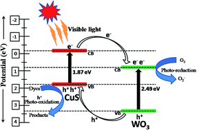
WO3 nanorods and flower-like CuS were synthesized by a hydrothermal process. The visible light driven CuS–WO3 photocatalyst was prepared by adding different weight ratios (10–40%) of CuS on WO3 by a wet impregnation method. The synthesized photocatalysts were characterized by powder X-ray diffraction (XRD), Fourier transform infrared (FT-IR) spectroscopy, Field emission scanning electron microscopy (FE-SEM), High-resolution transmission electron microscopy (HR-TEM), energy dispersive X-ray spectroscopy (EDAX) and UV-vis diffuse reflectance (DRS) spectroscopy. The photocatalytic performance of synthesized photocatalysts was evaluated for the photodegradation of methylene blue (MB) under visible light irradiation. The 10% CuS–WO3 photocatalyst showed higher photocatalytic degradation activity than others which could be due to the increased absorption of light in the visible region and also a lower recombination of charge carriers. Further, the photoelectrochemical measurements carried out for 10% CuS–WO3 revealed the faster migration of photo-induced charge-carriers. A possible reaction mechanism for the enhancement of photocatalytic activity of CuS–WO3 has been proposed.


Introduction

The development of highly ordered architectures of semiconductor photocatalysts has received great attention due to their natural growth with unique morphology and their potential applications for energy and environmental issues.1,2 The pollution of the environment by industrial and agricultural sectors is increasing. The textiles, cosmetics and food industries use organic dyes which are the primary sources for contamination of the environment due to their toxicity and the non-biodegradability of the organic dyes.3 The photocatalytic degradation is an effective way for the removal of organic pollutants from wastewaters. Therefore, it is necessary to develop highly effective, visible light active catalytic materials to solve most of the critical problems related to energy and environment. Over the past few decades, numerous active semiconductor photocatalysts such as metal oxides,4,5 sulfides,6,7 oxynitrides8 and graphitic carbon nitride9 have been synthesized and utilized for the treatment of organic pollutants in aqueous environment. Among these materials, tungsten oxide (WO3) is a well-known and an important semiconductor photocatalyst because of its wider response in the electromagnetic spectrum of solar radiation, mechanical strength and good metal support interactions.1,10 Because of these unique properties, WO3 is recognized as not only a visible light active photocatalyst but also extensively investigated for potential applications in photochromic, electrochromic devices and secondary batteries.11 The photocatalytic activity of WO3 is still limited due to the fast recombination of charge carriers and the conduction band electrons can not be efficiently trapped by O2 to yield superoxide radicals.10 Researchers have modified the band energy positions of WO3 by the addition of low bandgap semiconductor catalysts to reduce the recombination of charge carriers and thereby improving its photocatalytic activity.12–16 The difference in conduction band energy between the two semiconductors reduces the charge recombination and extends its absorption range in the visible region. Further, it leads to a higher yield of photo-generated electrons in the conduction band that contribute to achieve a higher photocatalytic degradation.17

To our knowledge, there have been no reports on the photocatalytic degradation of organic pollutants using CuS–WO3. In this investigation, CuS is chosen to dope WO3 because of its interesting optical, electrical properties and also it is available at low cost. CuS exhibits low reflectance in visible and relatively high reflectance in the near-infrared region, which makes it a prime material for solar energy absorption.18

The morphological features of the photocatalyst such as nanorods, nanowires and flower-like have been preferred to improve the photocatalytic activity. In order to control the morphology of CuS and WO3 photocatalysts, hydrothermal route was employed as well the organic additives like cetyl trimethylammonium bromide (CTAB) was used as a surfactant.

In this work, WO3 nanorods and flower-like CuS were successfully synthesized by hydrothermal route and different weight percentage CuS-doped WO3 photocatalysts were prepared via a simple wet impregnation method. The visible light photocatalytic performance of the synthesized photocatalysts was evaluated by the photodegradation of methylene blue (MB). A possible reaction mechanism for the photodegradation of MB over CuS–WO3 has been proposed.

Experimental section

Materials

Copper(II) nitrate trihydrate, thiourea, CTAB, tungstic acid, hydrogen peroxide (30% w/v of H2O2), Triton X-100 and sodium sulphate were purchased from SDFCL, India. Nitric acid and deionised water were obtained from Spectrum reagents and chemicals Pvt. Ltd. MB was obtained from Sigma-Aldrich. All reagents were used without further purification.

Synthesis of photocatalysts

Synthesis of flower-like CuS. In a typical synthesis, copper nitrate trihydrate (1 g), thiourea (0.945 g) and CTAB (0.452 g) were dissolved in 30 mL of distilled water. Then, the solutions were mixed homogeneously under constant stirring and then transferred into a Teflon-lined stainless-steel autoclave with a volume capacity of 150 mL. The content was finally diluted with water to a volume of 50% of autoclave capacity. The hydrothermal reaction was carried out in hot air oven at 130 °C for 12 h and then allowed to cool to room temperature naturally. The obtained black precipitate was collected, washed with deionized water and ethanol several times to remove impurities. Finally, the prepared sample was dried in hot air oven at 60 °C for 6 h.
Synthesis of WO3 nanorods. Tungstic acid (2 g) and CTAB (0.895 g) were dissolved in appropriate amount of distilled water. Then, the solution was transferred into a autoclave. The hydrothermal reaction was carried out in hot air oven at 120 °C for 12 h and then allowed to cool to room temperature. The obtained yellow color precipitate was collected, washed with deionized water and ethanol several times to remove impurities, and dried in hot air oven at 60 °C for 6 h.

The different weight percentage of CuS added WO3 were prepared as follows: 10, 15, 20, 30 and 40% (0.05, 0.1, 0.15, 0.2, 0.3 and 0.4 g) weight ratios of CuS were added separately to 0.5 g of WO3 and dissolved in 15 mL of ethanol. The resulting solution was continuously stirred and subsequently heated to evaporate the solvent at 60 °C. Then, the obtained CuS doped WO3 photocatalysts were dried in hot air oven for about 5 h.

Characterization of photocatalysts

X-ray diffraction (XRD) patterns of the synthesized photocatalysts were recorded using a X-ray diffractometer (Mini Flex II, Japan) with Cu Kα radiation (λ = 0.154 nm) at a scan speed of 3°/min. The phase purity was ascertained using X-ray diffraction. The Fourier transform infrared (FT-IR) spectra were recorded to study the interaction among CuS and WO3 with a wavenumber ranging from 4000–400 cm−1 using an JASCO 460 plus FT-IR instrument. The morphology of the synthesized photocatalysts were examined by field emission scanning electron microscopy (FE-SEM) with an FEI QUANTA-200 system at an accelerating voltage of 30 kV. The distribution of particles was examined using high-resolution transmission electron microscopy (HR-TEM) (JEOL-JEM 1011, Japan). Elemental analysis was examined by using EDAX-Bruker Nano GmbH, X Flash Detector, model 5010. UV-vis diffuse reflectance spectra (DRS) were recorded using a Shimadzu 2100 spectrophotometer in the range of 200–800 nm.

Photocatalytic degradation studies

The photocatalytic degradation of MB was studied under visible light irradiation to evaluate the photocatalytic efficiency of synthesized photocatalysts. Visible light irradiation was provided by a 100 W tungsten-halogen lamp. The distance between the sample and lamp was 20 cm, and the wavelength range of light source was ∼380–800 nm. Typically, a known amount of photocatalyst was added to a 75 mL of dye solution. Prior to light irradiation, the suspension was magnetically stirred in the dark to establish adsorption–desorption equilibrium of MB onto the photocatalyst. During irradiation, 4 mL of aliquots were collected at regular intervals and the photocatalyst was removed by filtration through a 0.45 μm membrane filter (Pall Corporation). The concentration of MB at different time intervals were determined at its characteristic absorption wavelength, viz., 664 nm using a Shimadzu UV-2450 UV-vis spectrophotometer.

Photoelectrochemical measurements

The photoelectrochemical properties were investigated with a CHI608E electrochemical workstation in a conventional three electrode system with a Pt-wire as a counter electrode, Ag/AgCl (in saturated KCl) as a reference electrode and synthesized catalyst coated on FTO conducting glass as a working electrode and the electrolyte was 0.1 M Na2SO4 solution. In a typical working electrode preparation, 5 mg of the catalyst was ground with 10 μL of Triton X-100 and 20 μL of deionised water to make slurry. The slurry was coated with conducting side of FTO glass surface by doctor blade method. The active area of the electrode was 0.5 × 0.5 cm2 and for a uniform coating scotch tape was used as a spacer. Then, it was dried in hot air oven at 100 °C for about 5 h.

Results and discussion

XRD studies

The XRD patterns of the synthesized CuS, WO3 and CuS–WO3 photocatalysts are shown in Fig. 1. The diffraction peaks of the synthesized CuS are indexed to the data of the hexagonal phase of CuS (JCPDS no. 06-0464) and are in good agreement with the previous report by Cheng et al.19 The XRD pattern of pure WO3 displayed monoclinic crystal structure corresponding to the (002), (020), (200), (120), (112), (022), (202), (222), (004), (400) and (420) diffraction at 2θ = 23.1°, 23.7°, 24.4°, 26.7°, 28.8°, 33.7°, 34.2°, 41.9°, 47.5°, 49.9° and 55.4° (JCPDS no. 43-1035). The XRD pattern of 10–40% weight ratio of CuS–WO3 are shown in Fig. 1(c)–(f), which demonstrates that the XRD pattern of WO3 is not changed due to the presence of CuS. Further, when the amount of CuS is increased to 20%, the intensity of the peaks are slightly decreased but it is interesting to note that the CuS peaks do not appear because it is present in below detectable limit of XRD.20 Also, It can be observed that a peak corresponding to CuS appears with an increase in CuS content, which confirms the formation of CuS–WO3 composite material.
image file: c5ra06512g-f1.tif
Fig. 1 XRD pattern of CuS, WO3 and CuS–WO3 photocatalysts.

FT-IR spectroscopy studies

The FT-IR spectra of pure CuS, WO3 and CuS–WO3 are recorded in the region of 500–4000 cm−1 and the results are shown in Fig. 2. In the FT-IR spectrum of pure CuS, the absorption peaks at 608 cm−1 and 1074 cm−1 are assigned to Cu–S stretching modes and bending modes of H–O–H of absorbed water molecules in sulfide products.21 In the spectrum of WO3, the broad band at 793 cm−1 corresponds to the stretching vibration modes of O–W–O bonds. The absorption peaks at 1616 and 1469 cm−1 corresponds to the asymmetric and stretching vibration mode of CH3–N+ bond of CTAB in the sample.22 The characteristic peak at 3418 cm−1 may be due to the stretching frequency of the O–H group.23 The new distinct peak observed at 1065 cm−1 for the CuS doped WO3 photocatalysts corresponds to asymmetric valence S[double bond, length as m-dash]O vibration.24 The FT-IR results clearly indicate the existence of CuS in the WO3 photocatalyst.
image file: c5ra06512g-f2.tif
Fig. 2 The FT-IR spectra of pure CuS, WO3 and CuS–WO3 photocatalysts.

Morphology and elemental analysis

The shape and morphology of the synthesized photocatalysts were examined by FE-SEM. The typical SEM images obtained for CuS, WO3 and 10% CuS–WO3 are displayed in Fig. 3. The CuS particles exhibited the hierarchical flower-like morphology, whereas the WO3 formed nanorod structure. The morphology of optimized 10% CuS–WO3 clearly showed that the nanorod shape of WO3 and flower-like structure of CuS were well distributed. Morphology and surface state of the materials can play an important role in affecting the photocatalytic activity. When the electrons and holes are trapped in surface states, the spatial overlaps of charge carriers are reduced and further their recombination is retarded due to the localized nature of surface area.25 In the present study, the formation of the hierarchical flower-like CuS and nanorods of WO3 photocatalyst are more favorable for the diffusion and separation of photoexcited electron–hole pair. Thus, the synthesized flowers-like CuS and WO3 nanorods can be promising materials for photocatalytic activity. Further, the distribution of CuS and WO3 in CuS–WO3 were explored by TEM and the corresponding images are shown in Fig. 4. The TEM images show agglomeration of CuS nanoparticles on the surface of WO3. The elemental analysis of CuS, WO3 and 10% CuS–WO3 are ascertained by energy dispersive X-ray spectroscopy (EDAX) and the obtained results are shown in Fig. 5. The EDAX spectrum clearly showed that the CuS was composed of copper and sulfur atoms, and WO3 consisted of W and O atoms only with no other impurities. Further, the EDAX data of CuS doped WO3 confirmed the existence of peaks corresponding to W, Cu, S and O atoms. The weight and atomic percentages of species detected by EDAX are shown in Fig. 5 inset. This observation supports the existence of CuS in WO3 phases. The obtained EDAX results showed the excess of weight percentage of Cu in 10% CuS–WO3, due to copper grid used for sample preparation.
image file: c5ra06512g-f3.tif
Fig. 3 FE-SEM images of (a) flowers-like CuS, (b) WO3 nanorods and (c) 10% CuS–WO3.

image file: c5ra06512g-f4.tif
Fig. 4 (a–d) TEM images of 10% CuS–WO3 photocatalyst.

image file: c5ra06512g-f5.tif
Fig. 5 EDAX spectra of (a) flowers-like CuS, (b) WO3 nanorods and (c) 10% CuS–WO3.

Optical absorption studies

The light absorption properties of CuS, WO3 and CuS–WO3 photocatalysts were investigated by UV-vis diffuse reflectance spectroscopy and the spectral characteristics are shown in Fig. 6. The absorption sharp edges for the synthesized CuS, WO3, 10% and 20% of CuS–WO3 are found to be at 661, 496, 645 and 549 nm, respectively and the corresponding calculated band energy gap values are 1.87, 2.49, 1.92 and 2.25 eV, respectively. It should be noted that the visible light absorption capacity is increased for the WO3 with the introduction of CuS.
image file: c5ra06512g-f6.tif
Fig. 6 UV-vis diffuse reflection spectra of CuS, WO3 and CuS–WO3 photocatalysts.

Photocatalytic degradation studies

The photocatalytic activities of the synthesized flowers-like CuS and WO3 nanorods and CuS doped WO3 catalysts were evaluated for the degradation of MB under visible light irradiation. The stable nature of the dye was evident from the blank test in the absence of a catalyst. There was no significant change in the absorption of MB under 2 h of visible light irradiation without the catalyst. Also, adsorption properties of MB dye on the catalyst were tested in the dark and it was observed that the adsorption equilibrium of the dye in the catalyst was reached after 60 min for all the photocatalysts (results not shown). The photocatalytic degradation of MB with CuS, WO3, 10–40% of CuS added WO3 photocatalysts under visible light irradiation are shown in Fig. 7(a). It can be observed that CuS doped WO3 exhibited a higher activities for MB degradation compared to pure CuS and WO3. The photodegradation rate of MB over the synthesized photocatalysts showed the following order: 10% CuS–WO3 > WO3 > 15% CuS–WO3 >20% CuS–WO3 >30% CuS–WO3 >40% CuS–WO3 > CuS, and the corresponding rate constant values are 0.013, 0.008, 0.005, 0.003, 0.003, 0.002 and 0.000 min−1, respectively. It can also be observed that 10% CuS–WO3 showed the highest photocatalytic activity. The reason for the enhancement of the photocatalytic degradation rate of MB with 10% CuS–WO3 may be due to the extended absorption of light in the visible region, higher separation of charge carriers and a subsequent diffusion of carriers to the surface of the catalyst.26 The formation of nanorod and flowers-like structures may reduce the charge recombination at the photocatalyst surface and thereby participate in the dye degradation process. However, the decrease in the photodegradation rate on increasing the CuS content to 20% may be due to excess CuS covered on WO3 surface which may shield the incident light, thus preventing the light absorption by WO3. This ultimately reduces the amount of photogenerated electrons on WO3 and also at higher CuS content, it may act as a charge recombination center, which results in a decrease in the photocatalytic degradation rate.27
image file: c5ra06512g-f7.tif
Fig. 7 (a) Photocatalytic degradation of MB with CuS, WO3, and CuS–WO3 composite photocatalysts and (b) first-order kinetics plot for the photodegradation of MB over CuS, WO3 and CuS–WO3 photocatalysts.

The reaction kinetics of the photodegradation of MB was investigated. The experimental data of photodegradation of MB over CuS, WO3, 10–40% CuS–WO3 photocatalysts were fitted by a first-order kinetics equation.

 
ln[thin space (1/6-em)]C0/C = kt (1)
where C0 is the initial concentration of MB, C is the concentration of MB at various time intervals, k is the apparent first-order rate constant (min−1) and t is the irradiation time (min). The plots of ln(C0/C) against irradiation time are shown Fig. 7(b). It can be seen that the plots are linear with good correlation values and photodegradation curves fit very well with the first-order rate equation. Under our experimental conditions, a higher rate constant of 0.013 min−1 is observed for the 10%-CuS–WO3 photocatalyst, which is comparable with that previously reported by Houas et al. They obtained a rate constant of 0.025 min−1 for the MB degradation using TiO2/UV based photocatalyst.28 The reaction was carried out using cylindrical shape pyrex reactor and the UV-irradiation was provided by high pressure mercury lamp (Philips HPK 125 W) with transmitting wavelength >340 nm. The dosage of photocatalyst was 50 mg of TiO2 (2.5 g L−1) in 20 mL of MB dye (Co = 72 μmol L−1).

Effect of catalyst amount

In this study, the best performing catalyst viz., 10% CuS–WO3 was chosen and a series of experiments were carried out for the photodegradation of MB (2 × 10⁻⁵ M) by varying the amounts of catalyst from 0.25, 0.5, 1.0, 1.5, 2.0 and 2.5 g L−1. The obtained results are shown in Fig. 8. The observed photodegradation rate constants for the different amounts of the catalyst from 0.25, 0.5, 1.0, 1.5, 2.0 and 2.5 g L−1 are as follows; 0.007, 0.007, 0.007, 0.008, 0.009, and 0.011 min−1, respectively. It can be seen that the photodegradation efficiency increases with an increase in the amount of catalyst loading and this may be due to an increase in the quantity of photon adsorbed. Moreover, the availability of active sites on the surface of the catalyst is also increased and it consequently increases the degradation rate.29
image file: c5ra06512g-f8.tif
Fig. 8 Photocatalytic degradation of MB with catalyst loading by varying the amount (0.5–2.5 g L−1) of 10% CuS–WO3.

Photoelectrochemical studies

The photoelectrochemical measurements were carried out to investigate the charge separation efficiency of electrons and holes. The transient photocurrent response of the pure flower-like CuS, WO3 nanorods and 10% CuS–WO3 was recorded under visible light irradiation and the obtained results are shown in Fig. 9(a). The photostability of all the catalysts were also confirmed from a reproducible photocurrent response for few on/off cycles under light illumination.30 The photocurrent initially generated by CuS is higher than WO3. However, after 40 s, the photocurrent response of CuS droped below that of WO3 and 10% CuS–WO3. This observation indicates the instability of CuS. The photodegradation of MB over CuS confirms that CuS alone does not cause the photocatalytic activity, which may be due to the instability of CuS and fast recombination of electron–hole pairs. The 10% CuS–WO3 catalyst exhibited higher photocurrent than the pure CuS and WO3, which indicates that the charge separation efficiency in 10% CuS–WO3 was higher than other catalysts and hence it can generate more photo-induced charge carriers.31
image file: c5ra06512g-f9.tif
Fig. 9 (a) Transient photocurrent response of the pure CuS, WO3 and 10% CuS–WO3 under visible light illumination and (b) the Nyquist plots for CuS, WO3 and 10% CuS–WO3 and Inset: The zoomed-in view of selected region of Nyquist plot.

The electrochemical impedance spectroscopy (EIS) measurements for the photocatalyst were performed to determine the charge transfer resistance. The Nyquist plots for CuS, WO3 and 10% CuS–WO3 are shown in Fig. 9(b). The inset of Fig. 9(b) shows the zoomed-in view of selected region of the Nyquist plot to identify the charge transfer resistance value. It can be seen that the 10% CuS–WO3 catalyst has a small arc radius on the impedance plot than the pure CuS and WO3 and this suggests that the addition of CuS into WO3 is beneficial to the separation of electrons and holes.32 Smaller the arc radius means lower the charge transfer resistance and faster the interfacial charge transfer reaction. Thus, the smaller charge transfer resistance of 10% CuS–WO3 catalyst is most favourable for higher photocatalytic activity.

Photocatalytic degradation mechanism

Fig. 10 shows the general mechanism of the photocatalytic degradation of MB using CuS–WO3 catalyst. The valence band (VB) and conduction band (CB) potentials of a photocatalyst were calculated using the following equation.9,26,33
 
EVB = XEe + 0.5Eg (2)
 
ECB = EVBEg (3)
where EVB and ECB are the valence and conduction band potentials, X is the electronegativity of the semiconductor, Ee is the energy of free electrons on the hydrogen scale (∼4.5 eV) and Eg is the band-gap energy of the semiconductor. The absolute electronegativity of CuS and WO3 is 5.29 and 6.59 eV.34,35 The calculated VB and CB of CuS is 1.70 and −0.16 eV, whereas VB and CB of WO3 is 3.33 and 0.84 eV, respectively. The conduction band potential of CuS is more negative than that of WO3. Thus, the photoinduced electron transfer from CB of CuS to WO3 and at the same time there is a transfer of the hole from the VB of WO3 to CuS. Therefore, the electron–hole recombination is reduced and it leads to an increase in the interfacial charge-transfer reactions for the degradation of adsorbed dye molecules.24

image file: c5ra06512g-f10.tif
Fig. 10 Schematic illustration of the photocatalytic separation and transfer of photogenerated charge carriers on the CuS–WO3 catalyst under visible light irradiation.

Conclusions

In summary, flower-like CuS and WO3 nanorods were synthesized individually by a hydrothermal route and a novel visible light-active CuS–WO3 photocatalyst was developed by adding CuS as a co-catalyst on WO3 by a wet impregnation method. The morphology and phase purity of the synthesized photocatalysts were revealed by FE-SEM, powder XRD and EDAX analyses. The light absorption properties of the synthesized photocatalysts were observed by UV-vis DRS measurement. The experimental results demonstrated that the photocatalytic degradation of MB over CuS added WO3 was much higher than that of the pure CuS and WO3. The maximum photocatalytic activity for the photodegradation of MB was obtained from the 10% CuS–WO3 catalyst. The observed high photocatalytic activity of CuS added WO3 is due to the extended absorption of light in the visible region and also due to the reduced recombination of the photo-induced charge carriers. The photo-electrochemical study also confirmed the higher charge carrier separation efficiency. A possible reaction mechanism has been proposed based on the photodegradation results. This study provides a new CuS–WO3 visible light-driven photocatalyst for the treatment of polluted wastewaters and other environmental remediation work.

Acknowledgements

We gratefully acknowledge the financial support received from Department of Atomic Energy-Board of Research in Nuclear Sciences (DAE-BRNS), Mumbai and Department of Science and Technology (DST), New Delhi, India.

Notes and references

  1. N. Sabari Arul, D. Mangalaraj and T. W. Kim, Photocatalytic degradation mechanisms of self-assembled rose-flower-like CeO2 hierarchical nanostructures, Appl. Phys. Lett., 2013, 102, 223115 CrossRef PubMed.
  2. D. Wang, L. Guo, Y. Zhen, L. Yue, G. Xue and F. Fu, AgBr quantum dots decorated mesoporous Bi2WO6 architectures with enhanced photocatalytic activities for methylene blue, J. Mater. Chem. A, 2014, 2, 11716–11727 CAS.
  3. Q. Wang, J. Li, Y. Bai, X. Lu, Y. Ding, S. Yin, H. Huang, H. Ma, F. Wang and B. Su, Photodegradation of textile dye rhodamine B over a novel biopolymer–metal complex wool-Pd/CdS photocatalysts under visible light irradiation, J. Photochem. Photobiol., B, 2013, 126, 47–54 CrossRef CAS PubMed.
  4. S. Kitano, N. Murakami, T. Ohno, Y. Mitani, Y. Nosaka, H. Asakura, K. Teramura, T. Tanaka, H. Tada, K. Hashimoto and H. Kominami, Bifunctionality of Rh3+ Modifier on TiO2 and working mechanism of Rh3+/TiO2 photocatalyst under irradiation of visible light, J. Phys. Chem. C, 2013, 117, 11008–11016 CAS.
  5. S. Ma, J. Xue, Y. Zhou and Z. Zhang, Photochemical synthesis of ZnO/Ag2O heterostructures with enhanced ultraviolet and visible photocatalytic activity, J. Mater. Chem. A, 2014, 2, 7272–7280 CAS.
  6. R. J. Vimal Michael, J. Theerthagiri, J. Madhavan, M. J. Umapathy and P. T. Manoharan, Cu2S-incorporated ZnS nanocomposites for photocatalytic hydrogen evolution, RSC Adv., 2015, 5, 30175–30186 RSC.
  7. X. H. Guan, P. Qu, X. Guan and G. S. Wang, Hydrothermal synthesis of hierarchical CuS/ZnS nanocomposites and their photocatalytic and microwave absorption properties, RSC. Adv., 2014, 4, 15579–15585 RSC.
  8. Y. Du, L. Zhao and Y. Zhang, Roles of TaON and Ta3N5 in the visible-Fenton-like degradation of atrazine, J. Hazard. Mater., 2014, 267, 55–61 CrossRef CAS PubMed.
  9. J. Theerthagiri, R. A. Senthil, A. Priya, J. Madhavan, R. J. V. Michael and M. Ashokkumar, Photocatalytic and photoelectrochemical studies of visible-light active α-Fe2O3–g-C3N4 nanocomposites, RSC Adv., 2014, 4, 38222–38229 RSC.
  10. C. Wang, X. Zhang, B. Yuan, Y. Wang, P. Sun, D. Wang, Y. Wei and Y. Liu, Multi-heterojunction photocatalysts based on WO3 nanorods: Structural design and optimization for enhanced photocatalytic activity under visible light, Chem. Eng. J., 2014, 237, 29–37 CrossRef CAS PubMed.
  11. D. Sanchez-Martinez, C. Gomez-Solis and L. M. Torres-Martinez, CTAB-assisted ultrasonic synthesis, characterization and photocatalytic properties of WO3, Mater. Res. Bull., 2014, 61, 165–172 CrossRef PubMed.
  12. Z. Liu, Z. G. Zhao and M. Miyauchi, Efficient Visible light Active CaFe2O4/WO3 Based Composite Photocatalysts: Effect of Interfacial Modification, J. Phys. Chem. C, 2009, 113, 17132–17137 CAS.
  13. L. J. Zhang, S. Li, B. K. Liu, D. J. Wang and T. F. Xie, Highly efficient CdS/WO3 photocatalysts: Z-Scheme photocatalytic mechanism for their enhanced photocatalytic H2 evolution under visible light, ACS Catal., 2014, 4, 3724–3729 CrossRef CAS.
  14. B. Weng, J. Wu, N. Zhang and Y. J. Xu, Observing the role of graphene in boosting the two-electron reduction of oxygen in graphene–WO3 nanorod photocatalysts, Langmuir, 2014, 30, 5574–5584 CrossRef CAS PubMed.
  15. R. H. Coridan, K. A. Arpin, B. S. Brunschwig, P. V. Braun and N. S. Lewis, Photoelectrochemical behavior of hierarchically structured Si/WO3 core–shell tandem photoanodes, Nano Lett., 2014, 14, 2310–2317 CrossRef CAS PubMed.
  16. S. Anandan, T. Sivasankar and T. L. Villarreaal, Synthesis of TiO2/WO3 nanoparticles via sonochemical approach for the photocatalytic degradation of methylene blue under visible light illumination, Ultrason. Sonochem., 2014, 21, 1964–1968 CrossRef CAS PubMed.
  17. D. Su, J. Wang, Y. Tanq, C. Liu and X. Han, Constructing WO3/TiO2 composite structure towards sufficient use of solar energy, Chem. Commun., 2011, 47, 4231–4233 RSC.
  18. Q. Wang, G. Yun, Y. Bai, N. An, Y. Chen, R. Wang, Z. Lei and W. Shangguan, CuS, NiS as co-catalyst for enhanced photocatalytic hydrogen evolution over TiO2, Int. J. Hydrogen Energy, 2014, 39, 13421–13428 CrossRef CAS PubMed.
  19. Z. Cheng, S. Wang, Q. Wang and B. Geng, A facile solution chemical route to self-assembly of CuS ball-flowers and their application as an efficient photocatalyst, CrystEngComm, 2010, 12, 144–149 RSC.
  20. X. C. Song, E. Yang, G. Liu, Y. Zhang, Z. S. Liu, H. F. Chen and Y. Wang, Preparation and photocatalytic activity of Mo-doped WO3 nanowires, J. Nanopart. Res., 2010, 12, 2813–2819 CrossRef CAS.
  21. M. Saranya, C. Santhosh, R. Ramachandran and A. N. Grace, Growth of CuS nanostructures by hydrothermal route and its optical properties, J. Nanotechnol., 2014, 321571 Search PubMed.
  22. Z. Jiang, J. Xie, D. Jiang, X. Wei and M. Chen, Modifiers-assisted formation of nickel nanoparticles and their catalytic application to p-nitrophenol reduction, CrystEngComm, 2013, 15, 560–569 RSC.
  23. N. Sharma, M. Deepa, P. Varshney and S. A. Agnihotry, FTIR and absorption edge studies on tungsten oxide based precursor materials synthesized by sol–gel technique, J. Non-Cryst. Solids, 2002, 306, 129–137 CrossRef CAS.
  24. L. Andronic, L. Isac and A. Duta, Photochemical synthesis of copper sulphide/titanium oxide photocatalyst, J. Photochem. Photobiol., A, 2011, 221, 30–37 CrossRef CAS PubMed.
  25. G. Liu, L. Wang, H. G. Yang, H. M. Cheng and G. Q. Lu, Titania-based photocatalysts-crystal growth, doping and heterostructuring, J. Mater. Chem., 2010, 20, 831–843 RSC.
  26. Q. Yuan, L. Chen, M. Xiong, J. He, S. L. Luo, C. T. Au and S. F. Yin, Cu2O/BiVO4 heterostructures: synthesis and application in simultaneous photocatalytic oxidation of organic dyes and reduction of Cr(VI) under visible light, Chem. Eng. J., 2014, 255, 394–402 CrossRef CAS PubMed.
  27. J. Zhang, J. Yu, Y. Zhang, Q. Li and J. R. Gong, Visible light photocatalytic H2-production activity of CuS/ZnS porous nanosheets based on photoinduced interfacial charge transfer, Nano Lett., 2011, 11, 4774–4779 CrossRef CAS PubMed.
  28. A. Houas, H. Lachheb, M. Ksibi, E. Elaloui, C. Guillard and J. M. Herrmann, Photocatalytic degradation pathway of methylene blue in water, Appl. Catal., B, 2001, 31, 145–157 CrossRef CAS.
  29. B. Neppolian, H. C. Choi, S. Sakthivel, B. Arabindoo and V. Murugesan, Solar light induced and TiO2 assisted degradation of textile dye reactive blue 4, Chemosphere, 2002, 46, 1173–1181 CrossRef CAS.
  30. A. T. Nguyen, W. H. Lin, Y. H. Lu, Y. D. Chiou and Y. J. Hsu, First demonstration of rainbow photocatalysts using ternary Cd1−xZnxSe nanorods of varying compositions, Appl. Catal., A, 2014, 476, 140–147 CrossRef CAS PubMed.
  31. H. Lin, X. Li, J. Cao, S. Chen and Y. Chen, Novel I-doped BiOBr composites: Modulated valence bands and largely enhanced visible light phtotocatalytic activities, Catal. Commun., 2014, 49, 87–91 CrossRef CAS PubMed.
  32. Y. Liu, Y. X. Yu and W. D. Zhang, Photoelectrochemical study on charge transfer properties of nanostructured Fe2O3 modified by g-C3N4, Int. J. Hydrogen Energy, 2014, 39, 9105–9113 CrossRef CAS PubMed.
  33. J. Theerthagiri, R. A. Senthil, A. Priya, J. Madhavan and M. Ashokkumar, Synthesis of a visible-light active V2O5–g-C3N4 heterojunction as an efficient photocatalytic and photoelectrochemical material, New J. Chem., 2015, 39, 1367–1374 RSC.
  34. X. Wang, Y. Li, M. Wang, W. Li, M. Chen and Y. Zhao, Synthesis of tunable ZnS–CuS microspheres and visible-light photoactivity for rhodamine B, New J. Chem., 2014, 38, 4182–4189 RSC.
  35. Y. Liu, Q. Li, S. Gao and J. K. Shang, Template-free solvothermal synthesis of WO3/WO3·H2O hollow spheres and their enhanced photocatalytic activity from the mixture phase effect, CrystEngComm, 2014, 16, 7493–7501 RSC.

This journal is © The Royal Society of Chemistry 2015