DOI:
10.1039/C5RA06240C
(Paper)
RSC Adv., 2015,
5, 55832-55838
Dopamine derived copper nanocrystals used as an efficient sensing, catalysis and antibacterial agent†
Received
8th April 2015
, Accepted 3rd June 2015
First published on 3rd June 2015
Abstract
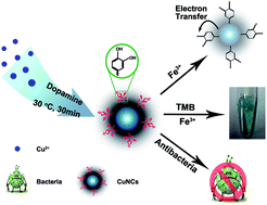
A simple one-pot synthesis method for small copper nanocrystals (CuNCs) was developed by employing dopamine as a reducing and capping reagent. The as-prepared CuNCs exhibited a fluorescence emission at 390 nm, good peroxidase-mimicking catalytic property, and excellent antibacterial activities against Gram-positive Staphylococcus aureus. Based on the fluorescence and peroxidase-mimicking catalytic features, sensing for ferric ions (Fe3+) was made because Fe3+ ions have specific interactions with the catechol groups on the surface of CuNCs with the limits of detection of 1.2 μM and 4.2 μM, respectively, which were much lower than the maximum level (5.4 μM) of Fe3+ permitted in drinking water by the US Environmental Protection Agency. For the antibacterial activities, a minimum inhibitory concentration of 158 μg mL−1 was found which was because of the generation of reactive oxygen species.
Introduction
The ultrasmall nanocrystals bridge the size scales of molecular species and larger nanoparticles or microparticles. They usually reveal elaborate properties, such as size dependent fluorescence, high surface energy, and antibacterial activity, which makes them promising candidates in the fields of sensing, catalytic and biological applications.1–5 Much work has already been done on the fluorescent metal nanoparticles such as gold (Au) and silver.6–10 Meanwhile, reports on the synthesis and properties of fluorescent copper nanocrystals (CuNCs) are emerging because of their low cost, and unique optical and catalytic properties.11–15 Compared to their counterparts, however, fluorescent CuNCs have attracted less attention because of the synthetic difficulties in controlling their ultrafine size and their susceptibility to oxidation upon exposure to air. Recently, Jia et al. developed thiolate ligand CuNCs which have the feature of aggregation induced fluorescence enhancement.16,17 They were used for real applications, where the selected thiolate ligands, just like in the synthesis of Au nanoparticles,18 must play two roles: one was to regulate the CuNC emissions, and the other was to molecularly recognize target species. The thiolate ligand CuNCs are interesting but it has been difficult to find dual-functional thiolate ligands. So, developing an effective method to synthesize brightly luminescent and stable CuNCs with straight and specific interactions with target species is highly desirable, which, if realized, would open up the possibilities of an array of practical applications.
Dopamine (DA), a well-known hormone and neurotransmitter of the catecholamine and phenethylamine families, has received interest because it can act as biocompatible surface stabilizing ligand and reducing agent.19,20 Furthermore, the most interesting property of DA is that it can form versatile biopolymers with many active functional groups such as amines and catechols, which can further interact with other substrates. Recently, DA has been used as reducing agent to synthesise copper nanoparticles (CuNPs).21 However, the use of DA as a capping/reducing reagent for the synthesis of luminescent CuNCs has not been reported.
Meanwhile, copper species are known to exhibit antibacterial activity against a wide variety of bacterial strains.22 Esteban-Cubillo et al. have demonstrated the bactericidal properties of CuNPs prepared in a matrix of sepiolite.23 Mallick et al. reported the good antimicrobial activity for the iodine stabilized CuNPs with chitosan.24 However, the mechanism behind this phenomenon still remains debatable. So it is important to study the antimicrobial activities of prepared CuNCs and address the issue of the species involved in the mechanism of action.
To achieve the goals listed previously, remarkable progress has been made to prepare Cu nanomaterials by using different chemical modifications and with the help of a functional capping reagent with unique and tunable properties.25–32 However, reduced sensitivity by specific interactions between target species limits their extensive multi-applications. So, there is still plenty of opportunity for improving the performances of CuNPs. In this research, a one-pot synthesis strategy was proposed, to prepare small DA derived multifunctional CuNCs. The CuNCs prepared displayed several advantages over the CuNPs.21 Firstly, they showed bright fluorescence and a good peroxidase-mimicking catalytic property. Secondly, by taking advantage of the capping agent, and specific interactions between the catechol groups of DA on the surface of the CuNCs and ferric ions (Fe3+), dual sensing models (the fluorescent and catalytic-based sensing system) were applied for the sensitive and selective detection of Fe3+ ions. The CuNCs also showed good antibacterial ability, allowing the effective killing of Staphylococcus aureus at a low concentration. The mechanism of the antibacterial ability of the CuNCs was also investigated. Thus, this study presents a multifunctional nanomaterial which is an efficient agent for sensing, catalysis and antibacterial applications.
Experimental section
Chemicals and materials
Copper sulfate (CuSO4·5H2O, 99%) was purchased from the Sinopharm Chemical Reagent Co., Ltd. (Shanghai, China). Dopamine hydrochloride and dichlorodihydrofluorescein diacetate (DCFH-DA) were purchased from Sigma-Aldrich (Steinheim, Germany). Standard stock solutions of Fe3+ ions were prepared using iron(III) chloride in deionized water. Different concentrations of Fe3+ ions were obtained by diluting standard stock solutions. All other reagents were of analytical reagent grade and used as received. All experiments were carried out in aqueous Britton–Robinson (BR) buffer (0.2 M, pH 4.0) unless stated otherwise. Deionized water (18.2 MΩ cm; Millipore Co., USA) was used. Other metal salts used in this work included silver nitrate (AgNO3), calcium chloride (CaCl2), cadmium chloride (CdCl2), cobalt chloride (CoCl2), chromium nitrate (Cr(NO3)3), copper chloride (CuCl2), iron(II) chloride (FeCl2), mercury(II) chloride (HgCl2), magnesium sulfate (MgSO4), sodium chloride (NaCl), nickel chloride (NiCl2), lead(II) nitrate (Pb(NO3)2) and zinc chloride (ZnCl2).
Apparatus
Transmission electron microscopy (TEM) and energy-dispersive X-ray spectroscopy (EDS) measurements were obtained using a Tecnai G2 F20 S-TWIN microscope (FEI, USA). Atomic force microscopy (AFM) images were captured using a Dimension Icon ScanAsyst atomic force microscope (Bruker). The X-ray photoelectron spectroscopy (XPS) analysis was conducted using an ESCALAB 250 X-ray photoelectron spectrometer (ThermoScientific, USA). The samples for XPS were made by the deposition of a nanocrystal suspension in water on a silicon substrate. Fourier transform infrared (FT-IR) spectrometry was carried out using a FTIR-8400S spectrophotometer (Shimadzu, Japan). The hydrodynamic diameter and zeta potential of the CuNCs were measured using dynamic light scattering and electrophoretic light scattering (ELS) on a ZEN 3600 Zetasizer (Malvern Instruments). Ultraviolet-visible-near infrared (UV-vis-NIR) absorption spectra were obtained using a UV-3600 spectrophotometer (Hitachi). Steady-state fluorescence spectra were measured with an F-2500 fluorescence spectrophotometer (Hitachi, Japan) with the nanoparticles dispersed in reagents. Fluorescence lifetimes were measured using an FL-TCSPC fluorescence spectrophotometer (Horiba Jobin Yvon Inc., France) using a NanoLED laser light source at the respective excitation wavelength of the dyes. The data were fitted using a double exponential decay model.
Preparation of CuNCs
The ultrasmall CuNCs were prepared by combining aqueous CuSO4·5H2O with DA hydrochloride solution. A typical preparation was composed of 1 mL of CuSO4 (0.1 M), 1 mL of DA (0.1 M), and 3 mL of deionized water by volume (DA
:
Cu2+ = 1). The solutions were combined, mixed well, and allowed to react with constant magnetic stirring at 30 °C. The solution became yellow over the course of 30 min of reaction at ambient temperature. Then the mixture was purified through a 10 kDa dialysis membrane for 24 hours with distilled water.
Fluorescence detection of Fe3+ ions
The detection of Fe3+ ions was performed in BR buffer (0.2 M, pH 4.0). In a typical assay, a CuNC (100 μL, 870 μg mL−1) dispersion was added into the BR buffer (200 μL, pH 4.0), followed by the addition of different concentrations of Fe3+ ions (0, 5, 10, 20, 40, 80, 100, 150, 200, 300, 400, 500, 600, 700, 800, 1000 μM) and water to 500 μL. The fluorescence spectra were recorded after the mixture had reacted for 30 min. The selectivity for Fe3+ was confirmed by adding other metal ion stock solutions (silver (Ag+), calcium (Ca2+), cadmium (Cd2+), cobalt (Co2+), chromium (Cr3+), copper (Cu2+), iron (Fe2+), mercury (Hg2+), potassium (K+), magnesium (Mg2+), sodium (Na+), nickel (Ni2+), lead (Pb+) and zinc (Zn2+)) instead of Fe3+ in a similar way.
Peroxidase mimicking based detection of Fe3+ ions
The detection of the Fe3+ ion was performed in BR buffer (0.2 M, pH 4.0) solution. In a typical assay, CuNCs (100 μL, 870 μg mL−1) and 3,3′,5,5′-tetramethylbenzidine (TMB; 100 μL, 1 mM) dispersion were added into the BR buffer (200 μL, pH 4.0), followed by the addition of different concentrations of Fe3+ ions (0, 6, 10, 16, 20, 25, 30, 40, 50, 80, 100, 160, 200 μM) and water to 500 μL. The UV-vis spectra (652 nm) were recorded after the mixture had reacted for 30 min. The selectivity for Fe3+ was confirmed by adding other metal ion stock solutions (Ag+, Ca2+, Cd2+, Co2+, Cr3+, Cu2+, Fe2+, Hg2+, K+, Mg2+, Na+, Ni2+, Pb+, Zn2+) instead of Fe3+ in a similar way. All the experiments were performed at room temperature.
In vitro antibacterial activity assay
In this study, S. aureus was used as a model bacterium to evaluate the antibacterial activity of the CuNCs adsorbed on to an agar surface. For the qualitative analysis, the inhibition zone of the S. aureus cultured on Luria–Bertani agar plates was assessed.32 Briefly, S. aureus suspension was first spread on the agar plates, and the test samples (100 μL) were added on to the agar plates. The bacteria were then incubated at 37 °C for 24 h, and the inhibition zone was visually inspected for each sample on the plates. The outer and inner diameters and the calculated diameter difference were measured.
Results and discussion
Materials characterization
CuNCs were prepared by a convenient one-step synthesis method. The morphology of the CuNCs was characterized using TEM and AFM. The TEM image showed that the CuNCs were well dispersed and that the average diameter was about 8 nm (Fig. 1A and B). HRTEM analysis was performed to study the nanostructures of CuNCs at the lattice plane level (Fig. 1A inset). Fringes with an inter-planar spacing of 0.20 nm were identified, which was attributed to the (111) diffraction plane of face-centered Cu (JCPDS 89-2838). The AFM image (Fig. 1C and D) showed that the height of the CuNCs was about 2 nm with a uniform distribution. Peaks of Cu were recorded in Fig. 1E, which confirmed the presence of CuNCs by EDS elemental analysis.
 |
| Fig. 1 Structure characterization of CuNCs. (A) TEM image of the small CuNCs (inset: high-resolution TEM (HRTEM) image). (B) Size distributions of the small CuNCs obtained by counting 100 particles. (C) AFM image of the small CuNCs. (D) The height of the CuNCs along the line in image (C). (E) EDS of the particles shown in image (A). | |
XPS analysis (Fig. 2A) was carried out to determine the oxidation state of Cu in the samples. An XPS spectrum showed that samples were composed of all the expected elements: C, O, N, and Cu. Two intense peaks around 951.9 and 932.3 eV were assigned to Cu 2p1/2 and Cu 2p3/2, which were attributed to Cu(0) or Cu(I), and these results were consistent with those presented in a previous report.33 Although it was hard to distinguish it from the Cu(I) from Cu(0) because of a very small difference in their binding energy values (about 0.1 eV), the small shoulder near 942 eV implied the presence of Cu(II) states.16
 |
| Fig. 2 (A) XPS of the CuNCs (upper) and the high-resolution spectra of Cu 2p1/2 and Cu 2p3/2 (lower); (B) FT-IR spectra of free DA molecule and CuNCs. | |
FT-IR was used to gain an understanding of the functional groups involved in the nanoparticles. Representative spectra of the CuNCs and DA are shown in Fig. 2B. The general features of CuNCs were common to those of DA, suggesting that the similar functional groups and structures were likely to be responsible for the formation and DA capping of the nanoparticles prepared. The absorption bands at 900–1300 cm−1 were attributed to the benzene ring and phenol and this hinted at the existence of DA. The broad bands observed near 3400 cm−1 were likely to be because of the –O–H stretch from the hydroxyl groups and the stretching (see Scheme 1) vibration of N–H bonds. The 1601 cm−1 band is because of the overlap of C
C resonance vibrations in the aromatic ring and the band at 1519 cm−1 can be ascribed to N–H scissoring vibrations.34,35
 |
| Scheme 1 A schematic illustration of the synthesis of DA-derived CuNCs and their multifunctional properties. (Upper) Schematic illustration of the fluorescence response of the DA-derived CuNCs to Fe3+ ions. (Middle) Schematic representation of the peroxidase-mimicking catalytic colour reaction of TMB with DA-derived CuNCs and Fe3+ ions. (Lower) Schematic illustration of using dopamine-derived CuNCs for antibacterial applications. | |
As shown in Fig. 3, the absorption spectra with a monotonically exponentially increasing spectrum toward shorter wavelengths indicated the formation of small CuNCs.36 Upon excitation at 320 nm, the CuNCs in aqueous solution displayed one emission peak at 390 nm. When dissolved in aqueous solution, the quantum yield of the CuNCs was 9.6% (ESI Fig. S1†) when compared to quinine sulfate as the reference. And the photoluminescence (PL) lifetime of the emission at 390 nm was 1.03 ns (ESI Fig. S2 and Table S1†), which was in the scope of PL decays of many fluorescent zero value metal NCs and attributed to emission from singlet excited states.37 Furthermore, the fluorescence intensity of CuNCs clearly varied with the increase of the solution pH and the maximal emission can be observed at pH 4 (ESI Fig. S3†). This pH dependent PL behavior might result from the pH sensing groups in DA on the surface of the CuNCs. The fluorescence intensity of the CuNCs in BR buffer (pH = 4) was relatively stable for 24 h when it was kept at room temperature (ESI Fig. S4†). This was verified by testing the zeta potential values (ESI Fig. S5†).
 |
| Fig. 3 UV-vis absorption of DA (black line) and CuNCs (red line); fluorescence excitation (blue line, emission at 390 nm) and emission (dark cyan line, excitation at 320 nm) spectra of the CuNCs. | |
Dual sensing models for Fe3+ detection
Fe3+ are well-known to be highly toxic to the environment.38,39 The detection of such ions is thus highly desirable. Fe3+ ions can oxidize the catechol groups generating the related quinone molecule (ESI Scheme S1†), which is a known, potent electron-acceptor in biological and abiotic systems.21,22 It can act as an electron-acceptor quenching the fluorescence of the CuNCs and can thus be used for highly specific detection of Fe3+ ions (Scheme 1, top).23 Thus, we investigated the capability of the prepared fluorescent CuNCs reduced by DA for Fe3+ ion sensing. As shown in Fig. 4A–C, it was clear that the fluorescence of the probes was gradually quenched by the increasing concentration of the Fe3+ ions and a linear detection range from 5 μM to 300 μM was obtained by fitting the plots of relative intensity versus concentration, where the limit of detection (LOD, 1.2 μM) at the signal-to-noise ratio of 3 was lower than the maximum level of Fe3+ ions (5.4 μM) permitted in drinking water by the US Environmental Protection Agency.40 The response of fluorescent CuNCs to Fe3+ ions was much higher than that to other metal ions, and this was attributed to the specific coordination interaction between Fe3+ ions and catechol groups on the surface of the CuNCs, which implied an excellent selectivity towards the Fe3+ ions.
 |
| Fig. 4 Dual sensing platform for Fe3+ detection. (A) Fluorescence spectra of obtained CuNCs (174 μg mL−1) in the presence of Fe3+ ions with different concentrations (0, 5, 10, 20, 40, 80, 100, 150, 200, 300, 400, 500, 600, 700, 800, 1000 μM). (B) Calibration curve for Fe3+ ion detection using fluorescence intensities (F0/F). (C) Selective relative fluorescent response of CuNCs (174 μg mL−1) to specific metal irons (150 μM). (D) UV-vis absorption of the oxidation product of TMB with increasing Fe3+ concentrations (0, 6, 10, 16, 20, 25, 30, 40, 50, 80, 100, 160, 200 μM) with CuNCs (174 μg mL−1). (E) Calibration curve for Fe3+ ion detection by absorbance using the peroxidase-like activity of CuNCs. (F) The absorbance at 652 nm of TMB (0.2 mM) with the addition of the specific metal irons (50 μM). | |
The catalytic activity such as the enzyme mimetic property of small NCs have become popular for bio-signal amplification, mainly because of their large surface area and high surface energy.3,5 The commonly used target detection involves a chromogenic substrate such as TMB, catalysed in the presence of hydrogen peroxide (H2O2) to form the coloured product. In the following, the enzyme mimicking activity of CuNCs was evaluated using the catalysis of the peroxidase substrate TMB (Scheme 1, middle). The CuNCs could catalyze the oxidation of TMB (ox-TMB) in the presence of H2O2 and produced a deep blue color, with a maximum absorbance at 652 nm. In contrast, CuNCs or H2O2 alone did not produce the significant color change (ESI, Fig. S6†). These results confirmed that the CuNCs exhibited peroxidase mimicking activity toward TMB with H2O2. Fe3+ ions can oxidize the catechol groups to produce the H2O2 (ESI Scheme S1†).21,22 As shown in Fig. 4D, with increasing the concentration of Fe3+ ions, the absorbance at 652 nm for ox-TMB was increased. And a linear detection range from 10 mM to 80 mM was obtained by fitting the plots of relative intensity versus concentration with the detection limit as 4.2 mM (Fig. 4E). Meanwhile, a gradient change of blue color for these solutions can be seen by naked eyes (ESI, Fig. S7†). The selectivity towards Fe3+ ions in the presence of other metal ions was also investigated, and the results were shown in Fig. 4F. It was clear that only Fe3+ ions resulted in a significant absorbance enhancement (at 652 nm) of ox-TMB.
 |
| Fig. 5 Growth inhibition of S. aureus bacteria on agar plates for different samples. (A and B) The antimicrobial activity shown with the decreasing concentration of the CuNCs. Spots 1–9 represent 791, 712, 633, 554, 475, 475, 317, 158, 79 μg mL−1 of the CuNCs. (C) Separate experiments for evaluating the antimicrobial activity with 792 μg mL−1 CuNCs (spot 10), 554 μg mL−1 CuNCs (spot 11), 9.86 mM H2O2 (spot 12), 0.02 M CuSO4 (spot 13), 0.02 M DA (spot 14). | |
To test the practicality of the proposed method, it was applied to the analysis of the aqueous samples spiked with Fe3+ ions (Table 1). The standard addition method was employed to eliminate any matrix effects for synthetic samples prepared with tap water and lake water. The low relative standard deviations (RSDs), ranging from 0.5% to 6.0%, confirmed the accuracy of the two sensing models, thus, the CuNC probes met the test requirements for environmental analysis.
Table 1 Results of the determination of Fe3+ in spiked tap water and lake water samples
|
Spiked amount (μM) |
Measured amount (recovery, μM) |
RSD (%) |
Determination of Fe3+ using fluorescent sensing system. Determination of Fe3+ using a catalytic-based sensing system. |
Tap water |
10 |
9.90 (99%)a |
9.79 (97.9%)b |
2.8a |
5.3b |
40 |
40.2 (100.5%)a |
39.5 (98.7%)b |
1.5a |
3.5b |
80 |
80.8 (101.0%)a |
82.2 (102.8%)b |
0.5a |
5.6b |
Lake water |
10 |
10.2 (102.0%)a |
10.8 (108%)b |
2.5a |
5.8b |
40 |
40.8 (102.0%)a |
40.8 (104.5%)b |
1.0a |
3.3b |
80 |
82.1 (102.6%)a |
82.8 (103.5%)b |
1.3a |
6.0b |
Antibacterial property of CuNCs
Furthermore, the antimicrobial activity of CuNCs using Gram-positive S. aureus bacterial strains was studied (Scheme 1C). Clear bacterial inhibition rings were observed showing their good antimicrobial activity which was dependent on the concentration and the minimum inhibitory concentration was found to be about 158 μg mL−1 (Fig. 5A and B). In order to study the mechanism of the antimicrobial activity of CuNCs, separate experiments were carried out to evaluate the effect of the reagents involved. As shown in Fig. 5C, no antimicrobial activity was detected for the control solution of CuSO4 and DA, which confirms that the Cu2+ ions are not the main factor for the antimicrobial activity. The positive charge of the CuNCs (20.3 mV) excluded the charge effect. These phenomena may be attributed to some species involved in the mechanism of action.41,42 The CuNCs have been reported to produce reactive oxygen species (ROS) for the CuNCs with surface modification which can be determined via oxidation of DCFH. The equivalent [H2O2] generated by the CuNCs can be calculated and was found to increase with the content of the CuNCs (ESI Fig. S8 and S9†). When only the H2O2 (9.86 mM, equal to [H2O2] generated by 791 μg mL−1) was tested, it showed no S. aureus growth inhibition (spot 12). These phenomenon above revealed that the antibacterial activity of the CuNCs may attribute to the interactions between nanoparticles and the ROS generated. When the oxygen was adsorbed at the surface of the nanoparticles, resulting in a contact potential difference between metal and adsorbed oxygen, a Fenton-like process could occur with copper ions (Cu(I) + H2O2 → Cu(II) + OH− + HO˙) according to the XPS of the Cu(II) release.43 Additional studies are needed to determine the role of ROS in the antibacterial activity of CuNCs because of the complexity of the biological environments.
To confirm that the proposed CuNCs had good multifunctional properties, a literature review of the Cu clusters or nanoparticles used for sensing, catalysis and as an antibacterial reagent was carried out (ESI Tables S2–S4†). As can be observed, most of the given examples show only one or two types of properties, which does not take into account factors, such as specific interactions with the target, and special species involved in the mechanism of action. To further confirm and optimize the good performance of the prepared CuNCs, selectivity experiments were carried out using different CuNCs synthesized using various molar ratios of DA to copper salt (1
:
3, 1
:
2, 1
:
1, 2
:
1, 3
:
1) (ESI Fig. S10–S16†). It can be seen that the CuNCs synthesized (DA
:
Cu2+ = 1
:
1) showed the best sensing ability for Fe3+ ion detection. All the CuNCs showed good antimicrobial activity, which further supported the fact that the synthesized CuNCs were quite promising for analytical and biological applications.
Conclusions
In summary, an easy method for the preparation of highly luminescent and peroxidase-mimicking CuNCs, with the DA as the reducing and capping reagent was presented. By taking advantage of the specific interactions between the catechol groups on the surface of CuNCs and the Fe3+ ions, dual sensing models (the fluorescence and peroxidase-mimicking based sensing system) were applied for the sensitive and selective detection of Fe3+ ions. The antibacterial activity of CuNCs showed a distinctive concentration dependence, and this enhancement effect may be attributed to the ROS generated by the CuNCs after surface modification with DA. In view of the convenient synthesis route and attractive multifunctional properties, the CuNCs synthesized are quite promising for diverse analytical and biological applications. Further studies are needed to determine the role of the ROS in the antibacterial activity of CuNCs because of its complexity in biological environments.
Acknowledgements
This study was financially supported by the National Natural Science Foundation of China (NSFC, Grant no. 21375109) and Chongqing Postdoctoral Science Foundation funded project (Xm2014021).
Notes and references
- Y. Lu and W. Chen, Chem. Soc. Rev., 2012, 41, 3594–3623 RSC.
- H. Qian, M. Zhu, Z. Wu and R. Jin, Acc. Chem. Res., 2012, 45, 1470–1479 CrossRef CAS PubMed.
- J. J. Li, J. J. Zhu and K. Xu, TrAC, Trends Anal. Chem., 2014, 58, 90–98 CrossRef CAS PubMed.
- O. V. Kharissova, B. I. Kharisov, V. Manuel Jimenez-Perez, B. Munoz Flores and U. Ortiz Mendez, RSC Adv., 2013, 3, 22648–22682 RSC.
- Y. H. Lin, J. S. Ren and X. G. Qu, Adv. Mater., 2014, 26, 4200–4217 CrossRef CAS PubMed.
- A. Leifert, Y. Pan-Bartnek, U. Simon and W. Jahnen-Dechent, Nanoscale, 2013, 5, 6224–6242 RSC.
- K. Huang, H. Ma, J. Liu, S. Huo, A. Kumar, T. Wei, X. Zhang, S. Jin, Y. Gan, P. C. Wang, S. He, X. Zhang and X. Liang, ACS Nano, 2012, 6, 4483–4493 CrossRef CAS PubMed.
- D. W. Guo, L. Y. Zhu, Z. H. Huang, H. X. Zhou, Y. Ge, W. J. Ma, J. Wu, X. Y. Zhang, X. F. Zhou, Y. Zhang, Y. Zhao and N. Gu, Biomaterials, 2013, 34, 7884–7894 CrossRef CAS PubMed.
- J. Lan, P. Zhang, T. T. Wang, Y. Chang, S. Q. Lie, Z. L. Wu, Z. D. Liu, Y. F. Li and C. Z. Huang, Analyst, 2014, 139, 3441–3445 RSC.
- K. Zhang, K. Wang, X. Zhu, J. Zhang, L. Xu, B. Huang and M. H. Xie, Chem. Commun., 2014, 50, 180–182 RSC.
- X. F. Jia, J. Li, L. Han, J. T. Ren, X. Yang and E. Wang, ACS Nano, 2012, 6, 3311–3317 CrossRef CAS PubMed.
- L. I. Zhang, J. J. Zhao, M. Duan, H. Zhang, J. H. Jiang and R. Q. Yu, Anal. Chem., 2013, 85, 3797–3801 CrossRef CAS PubMed.
- A. D. Brumbaugh, K. A. Cohen and S. K. S. Angelo, ACS Sustainable Chem. Eng., 2014, 2, 1933–1939 CrossRef CAS.
- B. W. Wang, S. X. Chen, J. Nie and X. Q. Zhu, RSC Adv., 2014, 4, 27381–27388 RSC.
- X. J. Zhao and C. Z. Huang, New J. Chem., 2014, 38, 3673–3677 RSC.
- X. F. Jia, J. Li and E. Wang, Small, 2013, 9, 3873–3879 CrossRef CAS PubMed.
- X. F. Jia, X. Yang, J. Li, D. Y. Li and E. Wang, Chem. Commun., 2014, 50, 237–239 RSC.
- Z. T. Luo, X. Yuan, Y. Yu, Q. B. Zhang, D. T. Leong, J. Y. Lee and J. P. Xie, J. Am. Chem. Soc., 2012, 134, 16662–16670 CrossRef CAS PubMed.
- H. Lee, S. M. Dellatore, W. M. Miller and P. B. Messersmith, Science, 2007, 318, 426–430 CrossRef CAS PubMed.
- H. Lee and P. B. Messersmith, Adv. Mater., 2009, 21, 431–434 CrossRef CAS PubMed.
- J. Xiong, X. D. Wu and Q. J. Xue, J. Colloid Interface Sci., 2013, 390, 41–46 CrossRef CAS PubMed.
- N. Cioffi, L. Torsi, N. Ditaranto, G. Tantillo, L. Ghibelli, L. Sabbatini, T. Bleve-Zacheo, M. D'Alessio, P. Giorgio Zambonin and E. Traversa, Chem. Mater., 2005, 17, 5255–5262 CrossRef CAS.
- A. Esteban-Cubillo, C. Pecharroman, E. Aguilar, J. Santaren and J. S. Moya, J. Mater. Sci., 2006, 41, 5208–5212 CrossRef CAS.
- S. Mallick, S. Sharma, M. Banerjee, S. S. Ghosh, A. Chattopadhyay and A. Paul, ACS Appl. Mater. Interfaces, 2012, 4, 1313–1323 CAS.
- H. Y. Cao, Z. H. Chen, H. Z. Zheng and Y. M. Huang, Biosens. Bioelectron., 2014, 62, 189–195 CrossRef CAS PubMed.
- N. Goswami, A. Giri, M. S. Bootharaju, P. Lourdu Xavier, T. Pradeep and S. Kumar Pal, Anal. Chem., 2011, 83, 9676–9680 CrossRef CAS PubMed.
- N. Vilar-Vidal, J. Rivas and M. A. López-Quintela, ACS Catal., 2012, 2, 1693–1697 CrossRef CAS.
- N. Vilar-Vidal, J. Rivas and M. A. López-Quintela, Small, 2014, 10, 3632–3636 CrossRef CAS PubMed.
- L. Hu, Y. Yuan, L. Zhang, J. Zhao, S. Majeed and G. Xu, Anal. Chim. Acta, 2013, 762, 83–86 CrossRef CAS PubMed.
- L. Rastogi and J. Arunachalam, Colloids Surf., B, 2013, 108, 134–141 CrossRef CAS PubMed.
- B. J. Li, Y. Y. Li, Y. H. Wu and Y. B. Zhao, Mater. Sci. Eng., C, 2014, 35, 205–211 CrossRef CAS PubMed.
- V. J. Boyle, M. E. Fancher and R. W. Ross, Antimicrob. Agents Chemother., 1973, 3, 418–424 CrossRef CAS.
- S. Q. Lie, D. M. Wang, M. X. Gao and C. Z. Huang, Nanoscale, 2014, 6, 10289–10296 RSC.
- R. F. Luo, L. L. Tang, S. Zhong, Z. L. Yang, J. Wang, Y. J. Weng, Q. F. Tu, C. X. Jiang and N. Huang, ACS Appl. Mater. Interfaces, 2013, 5, 1704–1714 CAS.
- S. Hong, Y. S. Na, S. Choi, I. T. Song, W. Y. Kim and H. Lee, Adv. Funct. Mater., 2012, 22, 4711–4717 CrossRef CAS PubMed.
- A. D. Brumbaugh, K. A. Cohen and S. S. Angelo, ACS Sustainable Chem. Eng., 2014, 2, 1933–1939 CrossRef CAS.
- J. Zheng, C. Zhang and R. M. Dickson, Phys. Rev. Lett., 2004, 93, 077402 CrossRef.
- S. W. Zhang, J. X. Li, M. Y. Zeng, J. Z. Xu, X. K. Wang and W. P. Hu, Nanoscale, 2014, 6, 4157–4162 RSC.
- B. Xiong, Y. Chen, Y. W. Shu, B. Shen, H. N. Chan, Y. Z. Chen, J. L. Zhou and K. K. Wu, Chem. Commun., 2014, 50, 13578–13580 RSC.
- S. Mirlohi, A. M. Dietrich and S. E. Duncan, Environ. Sci. Technol., 2011, 45, 6575–6583 CrossRef CAS PubMed.
- M. Shi, H. S. Kwon, Z. M. Peng, A. Elder and H. Yang, ACS Nano, 2012, 6, 2157–2164 CrossRef CAS PubMed.
- C. Chen, I. Ahmed and L. Fruk, Nanoscale, 2013, 5, 11610–11614 RSC.
- L. Macomber, C. Rensing and J. A. Imlay, J. Bacteriol., 2007, 189, 1616–1626 CrossRef CAS PubMed.
Footnote |
† Electronic supplementary information (ESI) available: Experimental details, characterization, and some optimization. See DOI: 10.1039/c5ra06240c |
|
This journal is © The Royal Society of Chemistry 2015 |
Click here to see how this site uses Cookies. View our privacy policy here.