Injectable supramolecular hydrogels fabricated from PEGylated doxorubicin prodrug and α-cyclodextrin for pH-triggered drug delivery

Fei Li a, Jinlin He a, Mingzu Zhang a, Kam Chiu Tam b and Peihong Ni *a
aCollege of Chemistry, Chemical Engineering and Materials Science, Suzhou Key Laboratory of Macromolecular Design and Precision Synthesis, Jiangsu Key Laboratory of Advanced Functional Polymer Design and Application, Soochow University, Suzhou 215123, P. R. China. E-mail: phni@suda.edu.cn; Tel: +86 512 65882047
bDepartment of Chemical Engineering, University of Waterloo, 200 University Avenue West, Waterloo, Ontario N2L 3G1, Canada

Received 7th April 2015 , Accepted 10th June 2015

First published on 10th June 2015


Abstract

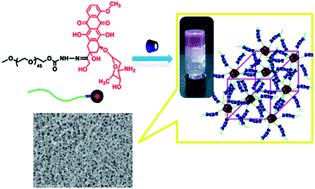
Supramolecular hydrogels, which are held together by noncovalent bonds and show responses to external stimuli, are of great interest in therapeutic delivery and tissue engineering as the injectable depot systems. To obtain a supramolecular hydrogel with multifunctions, such as low cytotoxicity, injectability and stimuli-triggered drug release, we herein report on the synthesis and characterization of a supramolecular hydrogel, which was formed by host–guest interaction between α-cyclodextrin (α-CD) and a PEGylated doxorubicin prodrug linked with an acid-cleavable hydrazone group (mPEG-Hyd-DOX). The polymeric prodrug displayed lower cytotoxicity than the free DOX. The host–guest interaction was demonstrated by X-ray diffraction (XRD) analysis. The structures and morphologies of the supramolecular hydrogels were systematically investigated by differential scanning calorimetry (DSC), scanning electron microscopy (SEM) and thermogravimetric analysis (TGA). The sol–gel transition process was monitored by dynamic and steady rheological analysis. The hydrogels could be degraded in the acidic environment of tumor cells and achieved the controlled delivery of DOX. The results of the pH-responsive property, in vitro cytotoxicity and drug release revealed that the supramolecular hydrogels can be used as a potential injectable matrix for the encapsulation and controlled release of anticancer drugs. This study provides an alternative for the construction of dual- or multi-drug delivery systems.


Introduction

Supramolecular hydrogels are a class of water-swollen polymeric networks with distinct three-dimensional structures and they are usually fabricated by crosslinking with supramolecular interactions.1,2 The concept and achievements of supramolecular hydrogels have drawn immense attentions in recent years and these materials have emerged as promising biomaterials because of their excellent biocompatibility, permeability, and ability to load bioactive agents, such as drugs,3–6 proteins,7,8 genes9 and even living cells.10 The soft and pliable properties of hydrogels could also minimize the mechanical irritation and damage to the surrounding tissue, typically reducing protein adsorption and thus inflammatory response in tissue engineering.11,12 It is worth noting that supramolecular hydrogels, unlike conventional chemical hydrogels, are based on relatively weak non-covalent interactions, such as hydrophobic interaction,13,14 inclusion complexation,15–18 ionic interaction,19–21 metal–ligand coordination,22 biomimetic interaction,23,24 and so on. Among them, the inclusion complexation involving interactions of cyclodextrins (CDs) and various guest molecules have been employed in supramolecular chemistry for many years, and have progressively sparked polymer scientists' attention in this field.2,25–28 For example, Allcock et al. reported an injectable supramolecular hydrogel formed between mPEG modified biodegradable polyphosphazenes and α-CD, and used for protein delivery.29 Harada et al. prepared a transparent supramolecular hydrogel that was quickly formed upon mixing β-CD-modified poly(acrylic acid) (PAA) with ferrocene-modified PAA, and it can be used as the redox-responsive self-healing material.30 Jiang et al. obtained a photoreversible pseudopolyrotaxane hydrogel by simply utilizing the competition of three host–guest interactions among trans-Azo-C1-N+/α-CD, PEG/α-CD, and cis-Azo-C1-N+/α-CD.31 These supramolecular hydrogels are easy to be formed and modulated under mild conditions, and they can carry various drugs for applications in the controlled release and tissue engineering.

Over the past years, rapid growth of drug delivery systems have been achieved, such as nanoparticles,32 polymeric micelles,33–35 hyperbranched polymers,36 dendrimers,37,38 liposomes,39 polymersomes40 and polymeric prodrugs.41,42 In particular, polymeric prodrugs have been considered as one of the most promising manners to improve the efficacy of drug treatment. Typically, the drug molecules are conjugated to the hydrophilic polymers such as poly(ethylene glycol), acrylic polymers, or polysaccharides,41,43,44 and generally possess some advantages including the increased water solubility, lower systemic toxicity, and prolonged circulation time.45 To further strengthen the therapeutic efficacies and pharmaceutical properties of polymeric prodrugs, much more efforts have been done to the hydrogel matrix for the encapsulation and delivery of drugs. For instance, Zhang et al. prepared a hydrogel formed by PEGylated indomethacin with α-CD for the controlled and sustained drug release.46 Shi et al. reported a multifunctional supramolecular hydrogel based on host–guest inclusion for the codelivery of two different anticancer drugs.47

Doxorubicin (DOX) has been demonstrated to possess broad spectrum antitumor activity against various cancers including breast,48 ovarian49 and gastric cancers,50 as well as the acute lymphoblastic–leukemia, alone or in combination with other agents.51 To date, many kinds of DOX-based polymeric prodrug have been proposed,41,45,52–55 however, the supramolecular hydrogels constructed from these prodrugs are rarely investigated. Herein, considering the acidic environment in tumor tissues,56,57 we first synthesized a polymeric prodrug composed of mPEG and DOX linked with acid-labile hydrazone group (designated as mPEG-Hyd-DOX), as shown in Scheme 1. Subsequently, a series of injectable prodrug-encapsulated supramolecular hydrogels were prepared via the inclusion complexation interaction of mPEG-Hyd-DOX and α-CD. The sol–gel transition, mechanical strength and shear-thinning behavior were monitored by dynamic and steady rheological analysis. In addition, the in vitro cytotoxicity and pH-responsive drug release were also studied for this prodrug loaded in the supramolecular hydrogel matrix.


image file: c5ra06156c-s1.tif
Scheme 1 Synthesis of mPEG45-Hyd-DOX prodrug.

Experimental

Materials

Poly(ethylene glycol) monomethyl ether (mPEG45-OH, [M with combining macron]n ≈ 2000 g mol−1, PDI = 1.06, Sigma-Aldrich), 4-nitrophenyl chloroformate (NPC, 97%, Alfa Aesar), doxorubicin hydrochloride (DOX·HCl, 99%, Beijing Zhongshuo Pharmaceutical Technology Development), hydrazine monohydrate (85%, Sinopharm Chemical Reagent), trifluoroacetic acid (TFA, 99%, Acros) and α-cyclodextrin (α-CD, G.R., TCI) were used as received. Triethylamine (TEA, A.R.) and N,N-dimethylformamide (DMF, A.R.) were purchased from Sinopharm Chemical Reagent, separately dried over anhydrous MgSO4 for 24 h, and then distilled under reduced pressure before use. Toluene (A.R.) and dichloromethane (CH2Cl2, A.R.) were obtained from Sinopharm Chemical Reagent, and individually dried over CaH2 for 24 h at room temperature, then followed by distillation just before use. Milli-Q water (18.2 MΩ cm−1) was generated using a water purification system (Simplicity UV, Millipore). All the other reagents and solvents were of analytical grade, obtained from Sinopharm Chemical Reagent and used as received unless otherwise mentioned. All cell culture related reagents were purchased from Invitrogen/Life Technologies.

Instruments

Nuclear magnetic resonance (NMR) spectra were performed on a 400 MHz Bruker NMR spectrometer (INOVA-400, Varian) at 25 °C with CDCl3 or DMSO-d6 as the solvent and TMS as the internal reference. FT-IR spectra were recorded on a Nicolet 6700 spectrometer using the KBr disk method. The interior structures of the hydrogels were observed by scanning electron microscopy (SEM) using a Quanta 200 FEG electron microscope operating at 15 kV. The mixture of prodrug solution and α-CD solution was added on the surface of a silicon SEM specimen holder, let stand for 5 min, quickly frozen in liquid nitrogen and further freeze-dried in a freeze drier at −40 °C for 3 days until all the solvent was sublimed. The freeze-dried hydrogel was sputter coated with gold before observation. XRD patterns were recorded on a Rigaku D/max 2500 X-ray powder diffractometer using Cu Kα (1.54 Å) radiation (50 kV, 250 mA). All samples were scanned from 2θ = 5° to 45° at a speed of 5° min−1. DSC was carried out on a DSC TA-60WS thermal analysis system (Shimadzu, Japan). Samples were first heated from −40 °C to 100 °C at a heating rate of 10 °C min−1 under a nitrogen atmosphere, followed by cooling to −40 °C after stopping at 100 °C for 3 min, and finally heating to 180 °C at 10 °C min−1. Thermogravimetric analysis (TGA) was examined on a Pyris 1 TGA (Perkin-Elmer, USA) with a heating rate of 20 °C min−1 from 80 to 500 °C under a nitrogen atmosphere. HPLC (Agilent 1260 infinity series) uses a C18 reversed phase column (4.6 × 150 mm, 5 mm) at 30 °C and a UV detector conducted at 254 nm. The mobile phase consisted of acetonitrile (HPLC grade, Sinopharm Chemical Reagent)-water (Milli-Q water) (50/50, v/v) with a flow rate of 1.0 mL min−1. The data were analyzed by Chem-Station software. The fluorescence spectra were recorded on a spectrofluorometer (FLS920, Edinburgh). The excitation was carried out at 480 nm and the emission spectra were recorded at 560 nm with the slit width for both excitation and emission set at 1 nm.

Synthesis of mPEG45-Hyd-DOX prodrug

Synthesis of mPEG45-NPC. The mPEG45-OH (2.0 g, 1.0 mmol) was dried by azeotropic distillation with toluene just before use. Under a dry nitrogen atmosphere, the purified mPEG45-OH was dissolved in 10 mL of anhydrous CH2Cl2, and the solution was added dropwise over 30 min into a flask containing NPC (0.8 g, 4.0 mmol) and anhydrous CH2Cl2 (50 mL) at 0 °C, which was then vigorously stirred at 25 °C for 24 h. The mixture was diluted with 60 mL of CH2Cl2 and washed three times with 10 mL of 5% NH4Cl solution. The combined organic phase was dried over anhydrous Na2SO4 for 4 h, concentrated and precipitated into cold diethyl ether twice. The product was collected by filtration and dried under vacuum for 24 h to give mPEG45-NPC with a yield of 90%. 1H NMR (400 MHZ, CDCl3, 25 °C, ppm, Fig. S1(B)): δ 3.38–3.40 (peak a, 3H), δ 3.44–3.84 (peak b, 180H), δ 4.43–4.48 (peak c, 2H), δ 7.38–7.43 (peak d, 2H), δ 8.27–8.32 (peak e, 2H).
Synthesis of mPEG45-Hyd. The hydrazine monohydrate (0.294 g, 5.0 mmol) was dissolved in 50 mL of anhydrous CH2Cl2 in a flask, and mPEG-NPC (1.08 g, 0.5 mmol) dissolved in 20 mL of anhydrous CH2Cl2 was added dropwise over 30 min into the flask at 0 °C overnight under a dry nitrogen atmosphere. The mixture was diluted with 60 mL of CH2Cl2 and washed three times with 10 mL of saturated NaCl solution. The combined organic phase was dried over anhydrous Na2SO4 for 4 h, concentrated and precipitated into cold diethyl ether twice. The product was collected by filtration and dried under vacuum for 24 h to give mPEG45-Hyd with a yield of 72%. 1H NMR (400 MHz, CDCl3, 25 °C, ppm, Fig. S1(C)): δ 3.38–3.40 (peak a, 3H), δ 3.44–3.84 (peak b, 180H), δ 4.22–4.29 (peak c, 2H).
Synthesis of mPEG45-Hyd-DOX. The mPEG45-Hyd (0.12 g, 0.06 mmol) and DOX·HCl (51.3 mg, 0.09 mmol) were dissolved in 10 mL of anhydrous DMF, and the reaction was processed in the presence of one drop of TFA at 60 °C for 48 h. Afterwards, the mixture was purified by dialysis (MWCO 1000) against a mixed solvent of methanol/Milli-Q water (from 2/1 to 1/1, v/v) for 48 h. The red powder mPEG45-Hyd-DOX was finally obtained after lyophilization with a yield of 67%. 1H NMR (400 MHz, CDCl3, 25 °C, ppm, Fig. S1(D)): δ 1.28–1.30 (peak d, 3H), δ 5.35–5.42 (peak e, 2H), δ 7.59–8.56 (peaks f, g, h, 3H) attributed to the protons of aryl groups of DOX.

Rheological analysis of supramolecular hydrogels

To investigate the gelation kinetics of aqueous mPEG45-Hyd-DOX/α-CD system, time sweep tests were performed on a constant oscillatory frequency (1.0 rad s−1) by a RS 6000 rheometer (Thermo Haake) with parallel plate geometry (20 mm diameter, 0.1 mm gap) at 25 °C. In this case, all testing samples were placed on the plate immediately after mixing, let standing for 1 min, and then started the measurement. The viscoelastic parameters were measured as a function of time within the linear viscoelastic region previously determined by a strain scan. Frequency sweep tests were conducted with the help of the same instrument and the hydrogel sample was allowed to consolidate for 24 h before the analysis. The frequency applied to hydrogel sample increased from 0.1 to 100 rad s−1. In addition, steady rate sweep tests were carried out to investigate the shear thinning property of the resultant hydrogels. In this case, the hydrogel samples were also allowed to consolidate for 24 h before the measurements.

In vitro loading and release of DOX

The DOX-loaded supramolecular hydrogel was prepared by an in situ forming method. Briefly, 45 mg of α-CD and 10 mg of mPEG45-Hyd-DOX were individually dissolved in 0.3 and 0.2 mL of Milli-Q water, and the two solutions were mixed thoroughly by stirring for 60 s. Then the mixed solution was divided into 6 pieces in a 1 mL cuvette and stood for 24 h to yield the hydrogel. The cuvette was placed in a test tube with 20 mL of phosphate buffer (PB) (pH 7.4 or pH 5.0) and incubated in a shaking water bath at 37 °C. At the desired time intervals, 5 mL of the released medium was withdrawn for fluorescence analysis and 5 mL of corresponding fresh buffer was added to keep a constant volume. The solution was measured by fluorescence spectroscopy with excitation at 480 nm and emission at 560 nm, and the slit width was set at 1 nm. All the loading and release experiments were carried out in dark.

Cell viability test

HeLa cells were purchased from the American Type Culture Collection. The cells were cultured in DMEM medium containing 10% heat-inactivated fetal bovine serum and 1% penicillin/streptomycin solution at 37 °C under a 5% CO2 atmosphere and were used in their growth state. The culture media were replaced every three days.

The anticancer activity of the supramolecular hydrogel was evaluated by the methyl tetrazolium (MTT) assay using free DOX and mPEG45-Hyd-DOX as the controls. A total of 30 mg of freeze-dried hydrogel sample (containing 1.5 mg DOX) was put into 3 mL of Milli-Q water in a 10 mL centrifuge tube, and then placed in a shaker incubator (37 °C) for two days. After that, the media were filtered with a 0.22 μm of sterile filter into a sterilized container and stored in a refrigerator at 4 °C before use. In addition, in order to investigate the anticancer activity of the released DOX from the hydrogels, HeLa cells were incubated with the released samples separately withdrawn from hydrogels at different time points in pH 5.0 or 7.4 PB solutions. HeLa cells were seeded onto a 96-well plate at a density of about 5 × 10³ cells per well for 12 h. The sample solutions with different concentrations were then added to the wells and cultured for another 48 h. Afterwards, 25 mL of MTT stock solution (5 mg mL−1 in PBS) were added to each well. After incubation for another 4 h, the DMEM medium was removed and the produced purple formazan was dissolved by adding 150 mL of DMSO. The optical density (OD) at 570 nm of each well was measured on a microplate reader (Bio-Rad 680). The absorbance values were normalized to the wells in which cells were not treated with samples. The cell viability was calculated by the equation: ODsample/ODcontrol × 100%, in which ODsample and ODcontrol are the absorbance values of the testing well (in the presence of samples) and the control well (in the absence of samples), respectively. Data are presented as average values with standard deviations.

Results and discussion

Synthesis and characterization of polymeric prodrug

As shown in Scheme 1, the pH-sensitive prodrug mPEG45-Hyd-DOX was synthesized via the following three steps: (i) synthesis of mPEG45-NPC; (ii) synthesis of mPEG45-Hyd; and (iii) preparation of mPEG45-Hyd-DOX. Firstly, the hydroxyl group of mPEG45-OH was activated with 4-nitrophenyl chloroformate to generate mPEG45-NPC. In the second step, the mPEG45-NPC was further reacted with hydrazine monohydrate to give mPEG45-Hyd. The resultant products in each step were characterized by 1H NMR as show in Fig. S1. The peaks at δ 3.44–3.84 and δ 3.39 ppm in Fig. S1(A) were attributed to the protons of the main chain of PEG and the terminal methyl group of PEG, respectively. After modification, comparing with mPEG45-OH, the 1H NMR spectrum of mPEG45-NPC in Fig. S1(B) exhibits new peaks at δ 4.45 ppm (peak c), δ 7.40 ppm (peak d) and δ 8.30 ppm (peak e), demonstrating the successful modification. In addition, the integral value ratio of peaks a, c, d and e (1.56[thin space (1/6-em)]:[thin space (1/6-em)]1.05[thin space (1/6-em)]:[thin space (1/6-em)]0.96[thin space (1/6-em)]:[thin space (1/6-em)]1.00) implies that most of the hydroxyl group of mPEG45-OH was activated. From the 1H NMR spectrum of mPEG45-Hyd shown in Fig. S1(C), the disappearance of the signals at δ 7.40 and 8.30 ppm, as well as the chemical shift from the original signal at δ 4.45 ppm in Fig. S1(B) to δ 4.26 ppm confirmed the successful synthesis of mPEG45-Hyd. FT-IR measurements were also used to characterize the polymers and the results are shown in Fig. S2. Finally, mPEG45-Hyd-DOX was synthesized by the reaction of the hydrazine bond of mPEG45-Hyd with the carbonyl group of DOX, and the obtained mPEG45-Hyd-DOX was confirmed by 1H NMR and HPLC. The successful conjugation of mPEG45-Hyd with DOX was proven by the peaks at δ 7.56–8.59 and 1.27 ppm in the 1H NMR spectrum of mPEG45-Hyd-DOX, as shown in Fig. S1(D). Meanwhile, HPLC showed that the retention time of free DOX was 1.11 min, while that of PEG45-Hyd-DOX was 4.02 min under the same condition, as shown in Fig. S3(A) and S3(B). On the other hand, the hydrazone linker was hydrolized and gradually cleaved at low pH condition. Incubation of PEG45-Hyd-DOX at 1 M HCl for 1 h led to the complete degradation of the hydrazone bonds, resulting in the elution peaks in Fig. S3(C), corresponding to that of free DOX. The DOX content of the polymeric prodrug was about 29 wt%, according to the fluorescence spectroscopy with excitation at 480 nm and emission at 560 nm.

Preparation of supramolecular hydrogel

Supramolecular hydrogel was prepared by directly mixing the α-CD solution with prodrug solution as shown in Scheme 2. The polymeric prodrug mPEG45-Hyd-DOX could be dissolved in water and formed supramolecular hydrogel when α-CD was introduced. This gelation procedure could occur under mild conditions without high temperature and the use of chemical cross-linker, which was only dependent on the amounts of mPEG45-Hyd-DOX and α-CD. The formation of supramolecular hydrogel was based on the host–guest inclusion complexation between α-CD and PEG chain.3 It is well-known that α-CD could form crystalline inclusion complexes with low-molecular-weight PEG in aqueous solution. Here the inclusion complexes formed by α-CD and PEG chain of mPEG45-Hyd-DOX may be thought to aggregate into the microcrystals, which acted as physical crosslinkers and then induced the formation of supramolecular hydrogel networks.
image file: c5ra06156c-s2.tif
Scheme 2 The digital photographs of the sol–gel transition of mPEG45-Hyd-DOX prodrug and α-CD solutions.

Time-sweep rheological analyses were performed to monitor the hydrogel network evolution and evaluate the viscoelastic behavior. Three fixed concentrations of mPEG45-Hyd-DOX aqueous solutions (1.0, 2.0 and 3.0 wt%) and three concentrations of α-CD aqueous solutions (7.0, 9.0 and 11.0 wt%) were used for the study. To investigate the effects of amounts of mPEG45-Hyd-DOX and α-CD on the gelation, time-sweep measurements were carried out, in which the storage modulus (G′) and loss modulus (G′′) were monitored as a function of time. As shown in Fig. 1 and Fig. 2, during the gelation procedure, both moduli increased and the G′ values became larger than the G′′ values, indicating that the systems became more elastic and formed a series of physically cross-linked networks. It is well known that a crossover point (G′ = G′′) of G′ and G′′ curves was detected for a gelation process (from G′ < G′′ to G′ > G′′). The corresponding time of the crossover from a viscous behavior to an elastic response could be regarded as the gelation time. In this work, all the crossover points were observed and the gelation time was very short, indicating that this class of supramolecular hydrogel possesses fast gelation. From Fig. 1(A)–(C), the gelation time was found to decrease with the increase of α-CD amounts. When the concentration of α-CD increased gradually from 7.0, 9.0 to 11.0 wt%, namely, 35, 45 and 55 mg of α-CD alone in 0.5 mL of H2O, while the amount of mPEG45-Hyd-DOX was fixed at 10 mg in 0.5 mL of H2O, the gelation time for the three systems decreased respectively from 167, 108 and finally to 39 s. In contrast, when the α-CD amount was fixed at 45 mg in 0.5 mL of H2O, while the amount of mPEG45-Hyd-DOX was gradually increased from 1.0, 2.0 to 3.0 wt%, that is, 5, 10 and 15 mg alone in 0.5 mL of H2O, the corresponding gelation time decreased from 190, 108 to 101 s, respectively, as shown in Fig. 2(A)–(C). These results indicate that the enhanced amounts of α-CD and mPEG45-Hyd-DOX prodrug would accelerate the gelation time, and the inclusion complexation between α-CD and mPEG45-Hyd-DOX induced the formation of supramolecular hydrogel, which could fix the anticancer drug DOX into the networks.


image file: c5ra06156c-f1.tif
Fig. 1 Effects of the amounts of α-CD on the gelation time of the gelled mPEG45-Hyd-DOX/α-CD systems. The insets show the representative magnification of the crossover of the storage modulus (G′) and loss modulus (G′′) curves. Test conditions: frequency, 1.0 rad s−1; strain, 1%; temperature, 25 °C.

image file: c5ra06156c-f2.tif
Fig. 2 Effects of the amounts of mPEG45-Hyd-DOX on the gelation time of the gelled mPEG45-Hyd-DOX/α-CD systems. The insets show the representative magnification of the crossover of the storage modulus (G′) and loss modulus (G′′) curves. Test conditions: frequency, 1.0 rad s−1; strain, 1%; temperature, 25 °C.

For the resultant supramolecular hydrogels, their storage modulus (G′) and shear-dependent viscosity (η) were also investigated with respect to the effects of the amounts of α-CD and mPEG45-Hyd-DOX. The G′ values increased with the increase of α-CD or mPEG45-Hyd-DOX amount at a fixed frequency or shear rate, as shown in Fig. 3. At a frequency of 1.0 rad s−1, for example, the G′ value increased from 10.4 to 31.6 kPa when the amount of α-CD increased from 7.0 to 9.0 wt%; while the G′ value increased from 6.8 to 20.9 kPa when the amount of mPEG45-Hyd-DOX increased from 1.0 to 3.0 wt%. In addition, all the G′ values showed a weak dependence on frequency, which confirmed that the hydrogels were well cross-linked with insignificant sol fraction. It is widely reported that shear-thinning property is required and important for the injectable drug delivery systems, which have recently drawn great attentions due to the minimization of surgical implantation.46Fig. 4 shows the steady shear viscosity as a function of shear rate for the in situ forming supramolecular hydrogels under various amounts of mPEG45-Hyd-DOX and α-CD. One can find that the viscosity of the hydrogels decreased greatly when it was sheared, which was independent of the amounts of mPEG45-Hyd-DOX or α-CD. This property indicates that the supramolecular hydrogels may be injected into body through a hypodermic needle.


image file: c5ra06156c-f3.tif
Fig. 3 Effects of the amounts of mPEG45-Hyd-DOX and α-CD on the elastic modulus (G′) of the gelled mPEG45-Hyd-DOX/α-CD systems. Test conditions: strain, 1%; temperature, 25 °C.

image file: c5ra06156c-f4.tif
Fig. 4 Effects of the amounts of (A) α-CD and (B) mPEG45-Hyd-DOX on the shear-dependent viscosity of the gelled mPEG45-Hyd-DOX/α-CD systems. Test conditions: strain, 1%; temperature, 25 °C.

Properties of freeze-dried hydrogels

In biomedical applications, such as tissue engineering and drug delivery, the morphological structure of hydrogels is an important factor to be taken into consideration. The morphology of the hydrogels was analyzed by the scanning electron microscopy (SEM) because high water content of the swollen hydrogels will likely lead to highly macroporous scaffolds upon lyophilization. As shown in Fig. 5(A)–(D), the freeze-dried hydrogels clearly exhibited the presence of the typical porous structures. These porous structures are indispensable to allow for tissue growth and diffusion of drugs and nutrients. Therefore, a higher percentage of pore fractions and smaller pore size were obtained by increasing the α-CD amount from 35, 45 to 55 mg, which might be due to the increase of network density.
image file: c5ra06156c-f5.tif
Fig. 5 SEM images of the freeze-dried hydrogel: (A) 10 mg mPEG45-Hyd-DOX and 35 mg α-CD/0.5 mL H2O, scale bar = 100 μm; (B) 10 mg mPEG45-Hyd-DOX and 45 mg α-CD/0.5 mL H2O, scale bar = 100 μm; (C) and (D) 10 mg mPEG45-Hyd-DOX and 55 mg α-CD/0.5 mL H2O with scale bars of 100 μm and 50 μm, respectively.

To confirm the inclusion complexation, we measured XRD patterns of the freeze-dried hydrogel formed from 10 mg of mPEG45-Hyd-DOX and 45 mg of α-CD. As shown in Fig. 6, the freeze-dried hydrogel was observed to have two characteristic diffraction peaks at 2θ = 19.73° and 22.38°, which were not observed in the separate patterns of mPEG45-Hyd-DOX or α-CD. α-CD was characteristic of multiple diffraction peaks corresponding to its crystalline formation, and mPEG45-Hyd-DOX showed two obvious diffraction peaks at 2θ = 19.20° and 23.39°. After the PEG chain was threaded into the α-CD inner core, the different crystallines would be formed. Two new peaks in freeze-dried hydrogel could represent the channel-type crystalline structure of mPEG45-Hyd-DOX/α-CD complex.58


image file: c5ra06156c-f6.tif
Fig. 6 XRD patterns of α-CD, mPEG45-Hyd-DOX and the freeze-dried hydrogel from 10 mg mPEG45-Hyd-DOX and 45 mg α-CD/0.5 mL H2O.

When PEG chain threads into the inner core of α-CD, it will bring different thermal properties to the inclusion complex. From Fig. 7(A), a distinct exothermic peak at 17.2 °C, that is the crystallization temperature (Tc), was observed for mPEG45-Hyd-DOX in the cooling run, corresponding to the crystallization temperature of PEG chain crystalline. However, the freeze-dried hydrogel did not show any obvious peak, which was similar with that of α-CD and implied that PEG chain was included into the inner core of α-CD so as to impede the crystallization of PEG chain. As the same in Fig. 7(B), a strong melting peak was detected for mPEG45-Hyd-DOX at around 53 °C in the second heating run, resulting from the melting of PEG chain. However, there was no observed melting peak corresponding to the melting of mPEG, because the PEG chain in the freeze-dried hydrogel was hided into the inner core of α-CD with the disappearance of any melting behavior. The similar phenomenon has also been reported previously.29 In addition, TGA analysis was also performed to study the supramolecular hydrogel. As shown in Fig. 8, TGA curves revealed that α-CD had a higher decomposition temperature (Td = 315 °C) after threading onto PEG chain than α-CD (Td = 300 °C), implying that the PEG chain included inside the α-CD molecules improve the thermal stability of the α-CD. Meanwhile, the left mPEG45-Hyd-DOX in the freeze-dried hydrogel showed the same decomposition temperature at 377 °C like that of mPEG45-Hyd-DOX when the α-CD was mostly decomposed.


image file: c5ra06156c-f7.tif
Fig. 7 DSC curves of (A) the cooling run and (B) the second heating run of α-CD, mPEG45-Hyd-DOX and the freeze-dried hydrogel.

image file: c5ra06156c-f8.tif
Fig. 8 TGA curves of α-CD, mPEG45-Hyd-DOX and the freeze-dried hydrogel from 10 mg of mPEG45-Hyd-DOX and 45 mg of α-CD/0.5 mL H2O.

In vitro drug release and cytotoxicity

To investigate the effect of pH values on drug release, the in vitro release experiments of DOX were performed in the two different buffer solutions of pH 5.0 and 7.4, respectively. The supramolecular hydrogels were formed by mixing 10 mg of mPEG45-Hyd-DOX and 45 mg of α-CD in pH 7.4 and pH 5.0 conditions at 37 °C. Fig. 9 shows the cumulative release curves of DOX. In the case of pH 5.0, there was a controlled and sustained release behavior without initial burst release. Obviously, the DOX was released much faster at pH 5.0 than that at pH 7.4, indicating that the pH-sensitive hydrazone linker was cleaved faster in the acidic environment, which could accelerate the disintegration of the supramolecular hydrogel and release the native DOX. It is anticipated that other therapeutic agents could be loaded into the hydrogel matrix by simple premixing and released at the tumor environment.
image file: c5ra06156c-f9.tif
Fig. 9 In vitro release curves of the supramolecular hydrogel in pH 5.0 and pH 7.4 PB solutions, respectively.

Finally, the in vitro cytotoxicity of free DOX, mPEG45-Hyd-DOX prodrug and the supramolecular hydrogel formed from 10 mg of mPEG45-Hyd-DOX and 45 mg of α-CD/0.5 mL of H2O were tested by MTT assay against HeLa cells. All the samples were kept at an equivalent DOX concentration. Fig. 10 shows that the cell viability of HeLa cells incubated with free DOX was extremely low, but for the mPEG45-Hyd-DOX prodrug with equal DOX concentration, the cell viability was still 60% after 48 h at a high concentration of 100 mg L−1. PEG does not only improve the water solubility of free DOX, but also decrease its cytotoxicity. Similar results about the polymeric prodrugs have also been reported elsewhere.42,54 Interestingly, the cell viability of hydrogel was higher than that of mPEG45-Hyd-DOX prodrug at the equal concentrations, which could be attributed to the favorable biocompatibility of α-CD. In addition, we have investigated the anticancer activity of the released DOX separately withdrawn from the hydrogels at different time points in pH 5.0 or 7.4 PB solutions. As shown in Fig. 11, the samples released at first 48 h showed minimal cytotoxicity in pH 7.4 PB solution, while the samples released at the same time in pH 5.0 PB solution showed much higher cytotoxicity. In the mean time, both of the released samples showed increased cytotoxicities with the prolongation of release time in 7.4 and 5.0 PB solutions.


image file: c5ra06156c-f10.tif
Fig. 10 Cell viabilities of HeLa cells incubated with free DOX, mPEG45-Hyd-DOX prodrug and hydrogel with different DOX concentrations for 48 h.

image file: c5ra06156c-f11.tif
Fig. 11 Cell viability of HeLa cells incubated with the solutions released at different time points in pH 7.4 and pH 5.0, respectively. All of the samples were incubated with HeLa cells for 48 h.

Conclusions

A series of novel supramolecular hydrogels have been prepared via host–guest inclusion complexation between a polymeric prodrug mPEG45-Hyd-DOX and α-CD. Because the hydrogels contain the acid-cleavable hydrazone bonds, the hydrogels were easy degraded in the tumor cell environment and released the fixed anticancer drug DOX. Meanwhile, the hydrogels possessed the characteristics of supramolecular hydrogels, especially the in situ formation, the porous structure and the shear-thinning property. In addition, the resultant supramolecular hydrogels had the adjustable gelation time and mechanical strength, depending on the amounts of mPEG45-Hyd-DOX prodrug and α-CD. The excess amounts of both prodrug and α-CD could accelerate the gelation process. This study presents a hydrogel carrier based on pH-sensitive polymeric prodrugs to deliver poorly water-soluble drugs, which would find potentials in injectable drug delivery systems.

Acknowledgements

The authors gratefully acknowledge financial supports from the National Natural Science Foundation of China (21374066 and 21074078), the Suzhou Science and Technology Program for Industrial Application Foundation (SYG201429), the Natural Science Foundation of Jiangsu Province (BK20130286), a Project Funded by the Priority Academic Program Development (PAPD) of Jiangsu Higher Education Institutions, and a Soochow-Waterloo University Joint Project for Nanotechnology from Suzhou Industrial Park. We are also thankful to Professor Jian Liu (FUNSOM, Soochow University) for his kind help in the cell-related test.

References

  1. N. M. Sangeetha and U. Maitra, Chem. Soc. Rev., 2005, 34, 821–836 RSC.
  2. X. Ma and Y. L. Zhao, Chem. Rev., 2015 DOI:10.1021/cr500392w.
  3. J. Li, Z. P. Ni and K. W. Leong, J. Biomed. Mater. Res., Part A, 2003, 65, 196–202 CrossRef PubMed.
  4. J. Li, NPG Asia Mater., 2010, 2, 112–118 CrossRef.
  5. H. Zou, W. Guo and W. Z. Yuan, J. Mater. Chem. B, 2013, 1, 6235–6244 RSC.
  6. R. Mateen and T. Hoare, J. Mater. Chem. B, 2014, 2, 5157–5167 RSC.
  7. D. Ma, K. Tu and L. M. Zhang, Biomacromolecules, 2010, 11, 2204–2212 CrossRef CAS PubMed.
  8. H. Hyun, Y. H. Kim, I. B. Song, J. W. Lee, M. S. Kim, G. Khang, K. Park and H. B. Lee, Biomacromolecules, 2007, 8, 1093–1100 CrossRef CAS PubMed.
  9. J. Li and X. J. Loh, Adv. Drug Delivery Rev., 2008, 60, 1000–1017 CrossRef CAS PubMed.
  10. D. Q. Wu, T. Wang, B. Lu, X. D. Xu, S. X. Cheng, X. J. Jiang, X. Z. Zhang and R. X. Zhuo, Langmuir, 2008, 24, 10306–10312 CrossRef CAS PubMed.
  11. K. Y. Lee and D. J. Mooney, Chem. Rev., 2001, 101, 1869–1879 CrossRef CAS PubMed.
  12. N. P. Rhodes, Biomaterials, 2007, 28, 5131–5136 CrossRef CAS PubMed.
  13. L. Yu, G. T. Chang, H. Zhang and J. D. Ding, Int. J. Pharm., 2008, 348, 95–106 CrossRef CAS PubMed.
  14. B. G. Xing, C. W. Yu, K. H. Chow, P. L. Ho, D. G. Fu and B. Xu, J. Am. Chem. Soc., 2002, 124, 14846–14847 CrossRef CAS PubMed.
  15. F. Yuen and K. C. Tam, Soft Matter, 2010, 6, 4613–4630 RSC.
  16. F. van de Manakker, L. M. J. Kroon-Batenburg, T. Vermonden, C. F. van Nostrum and W. E. Hennink, Soft Matter, 2010, 6, 187–194 RSC.
  17. Y. Guan, H. B. Zhao, L. X. Yu, S. C. Chen and Y. Z. Wang, RSC Adv., 2014, 4, 4955–4959 RSC.
  18. J. Yu, W. Ha, J. Chen and Y. P. Shi, RSC Adv., 2014, 4, 58982–58989 RSC.
  19. J. N. Hunt, K. E. Feldman, N. A. Lynd, J. Deek, L. M. Campos, J. M. Spruell, B. M. Hernandez, E. J. Kramer and C. J. Hawker, Adv. Mater., 2011, 23, 2327–2331 CrossRef CAS PubMed.
  20. M. Lemmers, J. Sprakel, I. K. Voets, J. van der Gucht and M. A. Cohen Stuart, Angew. Chem., Int. Ed., 2010, 49, 708–711 CrossRef CAS PubMed.
  21. Y. Xiao, J. L. He, Y. F. Tao, M. Z. Zhang, J. Hu and P. H. Ni, Acta Polym. Sin., 2014, 1, 122–130 Search PubMed.
  22. S. J. Buwalda, P. J. Dijkstra and J. Feijen, J. Polym. Sci., Part A: Polym. Chem., 2012, 50, 1783–1791 CrossRef CAS.
  23. Y. Zhang, H. W. Gu, Z. M. Yang and B. Xu, J. Am. Chem. Soc., 2003, 125, 13680–13681 CrossRef CAS PubMed.
  24. E. A. Appel, J. del Barrio, X. J. Loh and O. A. Scherman, Chem. Soc. Rev., 2012, 41, 6195–6214 RSC.
  25. J. Li, X. Li, Z. H. Zhou, X. P. Ni and K. W. Leong, Macromolecules, 2001, 34, 7236–7237 CrossRef CAS.
  26. G. S. Chen and M. Jiang, Chem. Soc. Rev., 2011, 40, 2254–2266 RSC.
  27. A. Hashidzume and A. Harada, Polym. Chem., 2011, 2, 2146–2154 RSC.
  28. A. Harada and Y. Takashima, Chem. Rec., 2013, 13, 420–431 CrossRef CAS PubMed.
  29. Z. C. Tian, C. Chen and H. R. Allcock, Macromolecules, 2013, 46, 2715–2724 CrossRef CAS.
  30. M. Nakahata, Y. Takashima, H. Yamaguchi and A. Harada, Nat. Commun., 2011, 2, 511–516 CrossRef PubMed.
  31. X. J. Liao, G. S. Chen, X. X. Liu, W. X. Chen, F. Chen and M. Jiang, Angew. Chem., Int. Ed., 2010, 49, 4409–4413 CrossRef CAS PubMed.
  32. K. Cho, X. Wang, S. M. Nie, Z. Chen and D. M. Shin, Clin. Cancer Res., 2008, 14, 1310–1316 CrossRef CAS PubMed.
  33. K. Kataoka, A. Harada and Y. Nagasaki, Adv. Drug Delivery Rev., 2001, 47, 113–131 CrossRef CAS PubMed.
  34. H. R. Wang, J. L. He, M. Z. Zhang, Y. F. Tao, F. Li, K. C. Tam and P. H. Ni, J. Mater. Chem. B, 2013, 1, 6596–6607 RSC.
  35. Y. Zhang, J. L. He, D. L. Cao, M. Z. Zhang and P. H. Ni, Polym. Chem., 2014, 5, 5124–5138 RSC.
  36. D. L. Wang, T. Y. Zhao, X. Y. Zhu, D. Y. Yan and W. X. Wang, Chem. Soc. Rev., 2015, 44, 4023–4071 RSC.
  37. M. Calderón, M. A. Quadir, S. K. Sharma and R. Haag, Adv. Mater., 2010, 22, 190–218 CrossRef PubMed.
  38. C. C. Lee, E. R. Gillies, M. E. Fox, S. J. Guillaudeu, J. M. J. Fréchet, E. E. Dy and F. C. Szoka, Proc. Natl. Acad. Sci. U. S. A., 2006, 103, 16649–16654 CrossRef CAS PubMed.
  39. T. M. Allen and P. R. Cullis, Science, 2004, 303, 1818–1822 CrossRef CAS PubMed.
  40. W. Chen, F. H. Meng, R. Cheng and Z. Y. Zhong, J. Controlled Release, 2010, 142, 40–46 CrossRef CAS PubMed.
  41. P. F. Gou, W. W. Liu, W. W. Mao, J. B. Tang, Y. Q. Shen and M. H. Sui, J. Mater. Chem. B, 2013, 1, 284–292 RSC.
  42. G. Y. Zhang, M. Z. Zhang, J. L. He and P. H. Ni, Polym. Chem., 2013, 4, 4515–4525 RSC.
  43. Y. D. Gu, Y. N. Zhong, F. H. Meng, R. Cheng, C. Deng and Z. Y. Zhong, Biomacromolecules, 2013, 14, 2772–2780 CrossRef CAS PubMed.
  44. M. J. Zou, H. Okamoto, G. Cheng, X. H. Hao, J. Sun, F. D. Cui and K. Danjo, Eur. J. Pharm. Biopharm., 2005, 59, 155–160 CrossRef CAS PubMed.
  45. V. Delplace, P. Couvreur and J. Nicolas, Polym. Chem., 2014, 5, 1529–1544 RSC.
  46. D. Ma and L. M. Zhang, Biomacromolecules, 2011, 12, 3124–3130 CrossRef CAS PubMed.
  47. W. Ha, J. Yu, X. Y. Song, Z. J. Zhang, Y. Q. Liu and Y. P. Shi, J. Mater. Chem. B, 2013, 1, 5532–5538 RSC.
  48. S. M. Swain, F. S. Whaley, M. C. Gerber, M. S. Ewer, J. R. Bianchine and R. A. Gams, J. Clin. Oncol., 1997, 15, 1333–1340 CAS.
  49. R. P. A'Hern and M. E. Gore, J. Clin. Oncol., 1995, 13, 726–732 Search PubMed.
  50. A. M. Murad, F. F. Santiago, A. Petroianu, P. R. S. Rocha, M. A. G. Rodrigues and M. Rausch, Cancer, 1993, 72, 37–41 CrossRef CAS PubMed.
  51. C. Carvalho, R. X. Santos, S. Cardoso, S. Correia, P. J. Oliveira, M. S. Santos and P. I. Moreira, Curr. Med. Chem., 2009, 16, 3267–3285 CrossRef CAS PubMed.
  52. X. L. Hu, S. Liu, Y. B. Huang, X. S. Chen and X. B. Jing, Biomacromolecules, 2010, 11, 2094–2102 CrossRef CAS PubMed.
  53. J. W. Cui, Y. Yan, Y. J. Wang and F. Caruso, Adv. Funct. Mater., 2012, 22, 4718–4723 CrossRef CAS.
  54. X. J. Chen, S. S. Parelkar, E. Henchey, S. Schneider and T. Emrick, Bioconjugate Chem., 2012, 23, 1753–1763 CrossRef CAS PubMed.
  55. X. Guo, C. L. Shi, J. Wang, S. B. Di and S. B Zhou, Biomaterials, 2013, 34, 4544–4554 CrossRef CAS PubMed.
  56. E. S. Lee, Z. G. Gao and Y. H. Bae, J. Controlled Release, 2008, 132, 164–170 CrossRef CAS PubMed.
  57. Y. Huang, Z. H. Tang, X. F. Zhang, H. Y. Yu, H. Sun, X. Pang and X. S. Chen, Biomacromolecules, 2013, 14, 2023–2032 CrossRef CAS PubMed.
  58. W. Ha, J. Yu, X. Y. Song, J. Chen and Y. P. Shi, ACS Appl. Mater. Interfaces, 2014, 6, 10623–10630 CAS.

Footnote

Electronic supplementary information (ESI) available. The results of 1H NMR, FT-IR and HPLC measurements. See DOI: 10.1039/c5ra06156c

This journal is © The Royal Society of Chemistry 2015
Click here to see how this site uses Cookies. View our privacy policy here.