Enhanced visible light photocatalytic activity of a floating photocatalyst based on B–N-codoped TiO2 grafted on expanded perlite

Xin Wang, Wei Wang, Xuejiang Wang*, Jing Zhang, Zaoli Gu, Lijie Zhou and Jianfu Zhao
College of Environmental Science and Engineering, State Key Laboratory of Pollution Control and Resource Reuse, Tongji University, Shanghai 200092, China. E-mail: wangxj@tongji.edu.cn; Tel: +86 021 65984268

Received 5th April 2015 , Accepted 1st May 2015

First published on 1st May 2015


Abstract

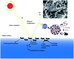
Floating photocatalysts of boron–nitrogen codoped TiO2 grafted on expanded perlite (B–N-TiO2/EP) were prepared by a facile sol–gel method. The catalysts were characterized by thermogravimetric differential thermal analysis (TG-DTA), X-ray diffraction (XRD), N2 adsorption–desorption (BET), scanning electron microscopy (SEM), Fourier transform infrared spectroscopy (FT-IR), X-ray photoelectron spectroscopy (XPS) and UV-vis diffuse reflectance spectroscopy (UV-vis-DRS). The results showed that by modifying the boron doping content in B–N-TiO2/EP, we could effectively obtain photocatalysts with a high BET surface area and porosity. Increasing the boron doping contents would inhibit the transformation of anatase TiO2 to the rutile phase. Compared with N-TiO2/EP, B–N-TiO2/EP exhibits an evident red-shift of the absorption band edge and the absorption intensity of the visible region increases obviously. The enhanced RhB photodegradation rate of B0.57–N-TiO2/EP could reach 94% after 3 h of visible light irradiation. Moreover, the floating photocatalyst could be easily separated and reused, showing great potential for practical applications in environmental cleanup and solar energy conversion.


1. Introduction

Titanium dioxide (TiO2) has been considered as the most promising photocatalyst for the oxidation of organic contaminants in wastewater.1 However, conventional TiO2 photocatalysts can only be activated under UV-light irradiation due to their wide band gap (e.g. Eg ≈ 3.2 eV for anatase),2,3 resulting in a low photo-electronic transition efficiency since UV radiation accounts for only 5% of the total solar spectrum compared to the visible region (∼45%).4–7 Thus, the possibility to extend the photoresponse capacity of TiO2 to the visible region and utilize a higher portion of the solar spectrum has become a topic of great interest within the scientific community. To unravel this problem, one of the most common and promising approaches is doping with impurities. In the approaches dealing with doping impurities, metals (Fe, Cr, Ni, Co or Ag)8–10 or non-metals (N, F, S, C or B)11–14 were utilized as dopants to narrow the band gap of TiO2 materials and decrease the required activation energy. However, the use of metal doped TiO2 for water treatment can lead to possible toxicity due to leakage of the metal ions into the final water, diminishing its quality.15 Nonmetal doping TiO2 has been treated as an effective means to expand the light response range. Among non-metal-doped TiO2 materials, co-doped TiO2 usually shows higher photocatalytic activity in visible range because of the merits benefited from each dopant. As the co-doped elements, B–N co-doping is considered as a much effective way.16 In et al.17 reported that B–N co-doped TiO2 exhibited enhanced photocatalytic activity under UV and visible lights irradiation, which is probably attributed to the synergistic effect between boron and nitrogen, resulting the narrowing of the band gap. Gopal et al.18 reported that B could fill an oxygen vacancy in the form of B- in B-TiO2 and N could serve as a paramagnetic probe for the geometric and electronic structure of other dopants in the lattice as diamagnetic species.

Although the modified TiO2 semiconductor is the most used for high photocatalytic activity and stability, non-toxicity and inexpensiveness, the interest is much more focused nowadays on the synthesis of new photocatalysts to overcome its limitations in application. Some of the important limitations are (1) modified TiO2 are always in the form of powder, it always sinks or suspends into the solution which decreased the using rate of light; (2) TiO2 powder is difficult to recycle, easy to agglomerate, and causes a problem of separation from the solution.19 To overcome the limitations, much attention has been paid to develop supported TiO2 catalysts. In this respect, different types of support for TiO2 have been tested including activated carbon,20 clay,21 silica22 and zeolite et al.23 But these supporting substrates would still sink to the bottom of solution without mechanical agitation. Recently, some studies24,25 developed a new concept of “floating photocatalysts”, which is the TiO2 photocatalyst synthesized on the surface of a floatable substrate (Fig. 1). The floatable photocatalysts are specially interesting for solar remediation of non-stirred and non-oxygenated reservoirs since the process maximizes the: (1) illumination/light utilization (due to their floatable ability), (2) oxygenation of the photocatalyst by the proximity with air/water interface. The optimization of illumination and oxygenation would result in higher rates of radical formation and oxidation efficiencies.26


image file: c5ra06056g-f1.tif
Fig. 1 Schematic representation of a floating photocatalyst.

In this paper, expanded perlite (EP) particles are applied as carrier to support B–N codoped TiO2 powder due to its floatable feature, porous structure, and physical mechanics properties. Sol–gel method was employed to synthesize B–N-TiO2/EP composite. The as-synthesized B–N-TiO2/EP composite was characterized by scanning electron microscope (SEM), thermogravimetric-differential thermal analysis (TG-DTA), X-ray diffraction (XRD), Fourier transform infrared (FT-IR) spectroscopy, X-ray photoelectron spectroscopy (XPS), UV-vis diffuse reflectance spectroscopy (DRS), nitrogen adsorption analyses for Brunauer–Emmett–Teller (BET) specific surface area and mesoporous size distribution. The photocatalytic activity of this floatable photocatalyst was investigated under visible light using Rhodamine B as the pollutant model.

2. Experimental section

2.1 Materials

Polyethylene glycol (PEG-400, GR) was obtained from Sigma-Aldrich Co. LLC. (USA). All other reagents (AR) were purchased from Sinopharm Chemical Reagent Beijing Co., Ltd. (China) and used as received without further purification.

The EP, provided by Xinyang Zhongke Mining Industry Co. Ltd. (Henan, China) with a particle size of 1–2 mm, was used to prepare the composites. Before usage, the original pearlite was activated by nitric acid and thoroughly washed in boiling deionized water until circum-neutral pH of the supernatant, then ultrasonicated for 0.5 h, oven dried in thin layers at 105 °C for 24 h and kept in desiccator for use.

2.2 Preparation of B–N-codoped TiO2/EP composite

A sol–gel preparation of B–N-codoped TiO2/EP was performed as follows. 18 mL tetrabutyl titanate and H3BO3 (0.36, 0.72, 1.8, 3.6 g corresponding to 0.11, 0.23, 0.57, 1.14 of B/Ti molar ratios) were dissolved completely into 50 mL anhydrous ethanol with stirring. Whilst still stirring, a solution containing 1.5 mL HCl (12 mol L−1) and 10 mL PEG-400 (1 wt%) were added drop by drop into the above solution to obtain solution A. Simultaneously, the solution was strongly stirred to sol. 3 g pretreated EP was added into the sol and stirred to uniformity. Following this, 3.6 g urea was dissolved into 4.5 mL deionized water with stirring to obtain solution B. Then solution B was added to solution A while stirring to get the mixture. The resultant mixture was dispersed with ultrasonic for 1 h until a white gel was obtain. The gel was aged at room temperature for 24 h then dried at 105 °C for 12 h to gain the xerogel. The resultant xerogel was crushed to obtain fine powder and further calcined at 550 °C for 2 h to obtain the catalyst. In addition, N-TiO2/EP was prepared according to the above procedure in the absence of H3BO3 for comparison.

2.3 Characterization of B–N-codoped TiO2/EP composite

The TG-DTA was analyzed by Shimadzu TGA-50. For crystal structure analysis of the prepared samples, X-ray diffraction (XRD) analysis was carried out on a Bruker D8 ADVANCE (German) X-ray diffractometer with Cu Kα radiation (40 kV, 40 mA) with a 0.01° step and 2.5 s step time over the range 10°< 2θ < 90°. Nitrogen adsorption–desorption isotherms were used to determine BET surface area and pore size distribution (Micrometritics, ASAP 2020). The morphology of the synthesized materials were observed initially using scanning electron microscopy (SEM, Hitachi S4700) with the working distance of 5–12 mm and an accelerating voltage of 20 keV. The IR spectrum was recorded as KBr pellets at room temperature on a Fourier transform infrared (FTIR) spectroscopy (Nicolet Instrument Corporation, USA). For the surface properties and elemental composition, XPS measurements were conducted by Thermo-VG Scientific ESCALAB 250 XPS system with Al Kα X-ray radiation. The binding energies were corrected by C 1s level at 284.6 eV as a reference to reduce the relative surface charging effect. For the characterization of the light absorption features and band-gap determinations, diffuse reflectance spectra (DRS) of the particles were measured in the range 200–800 nm on a UV-vis-NIR scanning spectrophotometer (Shimadzu UV-2550, Japan) equipped with an integral sphere using BaSO4 as a reference. The photocatalyst powder was placed in the sample holder on an integrated sphere for the reflectance measurements.

2.4 Photocatalytic evaluation with Rhodamine B under visible light

The photocatalytic activities of B–N-TiO2/EP composites were evaluated by decomposing Rhodamine B (RhB) dye under visible light irradiation at room temperature. A floating-bed photoreactor (Fig. 2) including a floating-bed of as-synthesized photocatalyst was applied in this work. Xe lamp (XE-JY500, 500 W) with an UV cut off filter (1 M sodium nitrite solution, λ > 400 nm) was used as visible light source and irradiated from the top. Aeration and recirculation mechanisms would promote a better mass diffusion and cause the supports change their position and face the light. For the photocatalytic experiments, the concentration of RhB solution was 2.5 mg L−1 and the obtained photocatalyst was added at the ratio of 2 g L−1 (surface load of catalyst is 0.018 g cm−2). Prior to irradiation, the mixture was magnetically stirred for 30 min in the dark to reach an adsorption–desorption equilibrium. At predetermined time of 30 min intervals (30–180 min) after 30 min pre-adsorption, the samples were taken out and filtered using 0.45 μm membrane filter for analysis.
image file: c5ra06056g-f2.tif
Fig. 2 Schematic representation of photocatalytic reaction system.

3. Results and discussion

3.1 Characteristics of B–N-TiO2/EP composite

The analysis of TG-DTA is performed to investigate the decomposition behavior of the precursor powders due to heat treatment in N2 (Fig. 3). The TG curve of TiO2/EP particles shows three weight loss stages. As shown, the first event occurred in the region of 50–150 °C, which probably involved in desorption of physically adsorbed water and ethanol on the surface of catalysts,27 leading the major weight loss of 2%. The second and third events occurred in the ranges of 150–350 °C and 350–500 °C, respectively, were recognized as fast weight loss stage and slow weight loss stage. While DTA curve showed the exothermic peak at ∼210 °C for TiO2/EP samples. As discussed previously, this peak may be due to the decomposition of the organic components and then leads to the formation of TiO2 film and evolution of H2O and CO2 gas.28 The differential thermal of B–N-TiO2/EP precursor in the range of 300 to 700 °C was less than that of N-TiO2/EP precursor.
image file: c5ra06056g-f3.tif
Fig. 3 The TGA of (A) N-TiO2/EP and (B) B–N-TiO2/EP.

The XRD patterns of the as-synthesized B–N-TiO2/EP samples were presented in Fig. 4. Comparing the XRD pattern of B–N-TiO2/EP with different boron doping contents, there is no rutile peak in the sample of B1.14–N-TiO2/EP, whereas rutile appears in the samples of B0.11–N-TiO2/EP, B0.23–N-TiO2/EP and B0.57–N-TiO2/EP. It is important to note that the increasing amount of boron dopant inhibits the transformation of anatase TiO2 to rutile phase, which agrees with other literatures.29,30 Even though, in this research the crystal phases of B–N-TiO2/EP consisted of predominantly anatase. Besides the diffraction peak of anatase TiO2, one weak peak at around 30.6° was observed for B1.14–N-TiO2/EP, attributed to the formation of B2O3 on the surface of TiO2.


image file: c5ra06056g-f4.tif
Fig. 4 XRD patterns for B0.11–N-TiO2/EP (a) B0.23–N-TiO2/EP (b) B0.57–N-TiO2/EP (c) B1.14–N-TiO2/EP (d). A is stand for anatase and B is stand for rutile.

The nitrogen adsorption–desorption isotherms of B–N-TiO2/EP are shown in Fig. 5. The N2 sorption isotherm of the samples are consistent with type IV of hysteresis loops, which is a typical mesoporous structure of the material according to the IUPAC classification.29,31 The mesopores allow light to scatter inside their pore channel, thus enhancing the harvesting of light.32 Accordingly, the specific surface area, pore size and total pore volume of B–N-TiO2/EP are presented in Table 1. Comparing the BET surface area of raw EP and B–N-TiO2/EP, it is noted that B–N-TiO2 grafted on EP is able to increase the BET surface area though controlling boron doping amount. When B/Ti ratio increased from 0.11 to 0.57, the BET surface area increased from 17.189 to 33.649 m2 g−1. However, the BET surface area decreased when continued to increase boron doping amount. Considering the BET surface of the sample, the mesopores are believed to be formed by the agglomeration and connection of adjacent nanoparticles in the sample.33 This network nanostructure offers more efficient transportation for reactant molecules to the active sites, which are expected to enhance the photocatalytic activity.34


image file: c5ra06056g-f5.tif
Fig. 5 N2 adsorption–desorption isotherms of B–N-TiO2/EP.
Table 1 The characterization results of different samples
Sample Specific surface area (m2 g−1) Pore size (nm) Total pore volume (cm3 g−1)
EP 5.174 8.528 0.016
B0.11–N-TiO2/EP 17.189 15.898 0.074
B0.23–N-TiO2/EP 28.597 27.314 0.082
B0.57–N-TiO2/EP 33.649 29.553 0.089
B1.14–N-TiO2/EP 13.049 12.503 0.051


The morphologies of EP and B–N-TiO2/EP are observed by SEM (Fig. 6). From Fig. 6, it can be seen the presence of TiO2 particles locates on the surface of EP, and the particle size is in the range of nanometer scale. For B–N-TiO2/EP composites, it can be observed TiO2 agglomerated in higher amounts with most of the EP particle surface. The elemental concentrations of EP and B0.57–N-TiO2/EP are showed in Table 2, the presence of B and N and the high atomic percentage of Ti and O also revealed that the codoping of B–N and the TiO2 coating. The content of TiO2 on the surface of EP is about 8.81 wt% according to the weight variation in EP. These particles are strongly attached to the surface of the composite and present spongy-like surface, which could increase adsorption sites. The results are in agreement with BET analysis.


image file: c5ra06056g-f6.tif
Fig. 6 The SEM of B–N-codoped TiO2/EP samples (A) EP (B) B0.11–N-TiO2/EP (C) B0.23–N-TiO2/EP (D) B0.57–N-TiO2/EP (E) B1.14–N-TiO2/EP.
Table 2 Elemental concentrations of EP and B0.57–N-TiO2/EP
Elements Atomic, %
C Si Ti O N B
EP 25.62% 19.41% 50.47%
B0.57–N-TiO2/EP 25.44% 11.08% 6.80% 48.78% 0.53% 1.35%


The FT-IR spectra of B–N-TiO2/EP samples are provided in Fig. 7. The band at 3342 cm−1 is assigned to the O–H bending modes of adsorbed water molecules, which play an important role in the photocatalytic activity because these groups can inhibit the recombination of photogeneration charges and interact with photogenerated holes to product reactive oxygen species.35 The weak band at 2748 cm−1 is assigned to the stretching of C–H bonds, which should be attributed to the organic precursor from the sol. At lower calcination temperatures, more substance including C–H bonds remained in the surface of the carrier. The band below 1000 cm−1 corresponds to the titania crystal lattice vibration.36,37


image file: c5ra06056g-f7.tif
Fig. 7 FT-IR spectra of B–N-TiO2/EP.

In order to further explore the nature of doped elements, the sample of B0.57–N-TiO2/EP was analyzed by XPS (Fig. 8). Fig. 8(A) shows the high-resolution XPS spectra for B 1s. The weak B 1s peak at around 193.0 eV suggests the formation of B2O3 phase coupled on the surface of TiO2. The B 1s peak at about 191.4 eV is ascribed to the B 1s state of doped B3+ ions in interstitial mode, as the binding energy locate between that of B 1s in boron doped TiO2 (190.6 eV) and B 1s in B2O3 (193.2 eV). The N 1s XPS spectra shows two constituent peaks at 399.3 and 401.2 eV, without the peak at 396–397 eV definitely assigned to substitutional nitrogen.


image file: c5ra06056g-f8.tif
Fig. 8 XPS spectra of B0.57–N-TiO2/EP (A) B 1s (B) N 1s.

The UV-vis diffuse reflectance spectra of B–N-TiO2/EP samples and reference materials are presented in Fig. 9(a). The reference samples (EP blank and TiO2/EP) have no significant absorbance in the visible light region (λ > 420 nm). However, the absorption edges of all doped TiO2/EP samples are shifted to a lower energy region and the elements doped into TiO2 lattice are responsible for the red-shift absorption band of these samples. Comparing N-TiO2/EP and B–N-TiO2/EP, it is found that the amount of boron doped into N-TiO2/EP affected the optical absorption of the sample in visible regions. Either too much or too less doping amount would inhibit the absorption of visible light. The samples of B0.23–N-TiO2/EP and B0.57–N-TiO2/EP showed the stronger photo-absorption in the visible light region than N-TiO2/EP, which could imply promise for higher visible light photocatalytic activity. The energy band gap was estimated by plotting (Ahν)2 dependence on photon energy (), assuming the indirect band gap.38 The value of indirect band gap for each sample is determined by the linear extrapolation in high slope of the corresponding curve and is provided in Fig. 9(b). The band gap energies are estimated to be 3.14, 3.02, 3.05 and 3.07 eV for B0.11–N-TiO2/EP, B0.23–N-TiO2/EP, B0.57–N-TiO2/EP and B1.14–N-TiO2/EP, respectively. The narrowed band gap arises from the Ti–N–B–O structure formed by B–N co-doped atoms.29


image file: c5ra06056g-f9.tif
Fig. 9 UV-vis absorbance (a) and band gaps (b) of synthetic samples.

3.2 Photocatalytic activity of B–N-TiO2/EP composite under visible light

The photocatalytic degradation of RhB on the as-synthesized B–N-TiO2/EP is evaluated under visible light irradiation and displayed in Fig. 10(a). It is clearly shown that RhB is hardly diminished under visible light irradiation with EP only. When boron content is at a lower level (B/Ti = 0.11 and B/Ti = 0.23), B–N-TiO2/EP exhibits a poorer performance on photodegradation of RhB than N-TiO2/EP. However, the photocatalytic activities of B–N-TiO2/EP increased gradually with an increase in boron content. When B/N ratio is 0.57, the sample of B0.57–N-TiO2/EP exhibits the best performance on photodegradation of RhB (94%) after 3 h irradiation. The high photocatalytic activity of B0.57–N-TiO2/EP is attributed to its high specific surface (33.649 m2 g−1) and the formation of Ti–N–B–O structure. The high specific area is responsible for providing strong adsorption ability toward target molecules and thus the generation of photoinduced electron–hole pairs of active sites while the Ti–N–B–O structure effectively narrowed the band gap and then easily generated electron–hole pairs. But the excess amount of B–N species would also lead to the recombination of electrons and holes.
image file: c5ra06056g-f10.tif
Fig. 10 The photodegradation for RhB on B–N-TiO2/EP under visible light irradiation (a) and the pseudo-first-order kinetics (b).

Fig. 10(b) shows the kinetic studies of the photodegradation of RhB over B–N-TiO2/EP with different boron contents. It is observed that the photocatalytic reactions of RhB obey the pseudo-first-order kinetics according to the Langmuir–Hinshelwood model and may be expressed as:39

image file: c5ra06056g-t1.tif
where k is the observed rate constant, C0 is the equilibrium concentration of RhB and C is the concentration at time t. Furthermore, according to the kinetics model, the k and R2 of different B–N-TiO2/EP samples and N-TiO2/EP are calculated and summarized in Table 3. The results clearly demonstrates that the optimum boron doping content is the sample of B0.57–N-TiO2/EP with a k value of 0.84960 h−1, which is 1.5 times larger than of N-TiO2/EP.

Table 3 Photocatalytic kinetic parameters of RhB solution by different B–N-TiO2/EP samples
Photocatalyst k/h−1 R2
N-TiO2/EP 0.53984 0.98986
B0.11–N-TiO2/EP 0.43065 0.96626
B0.23–N-TiO2/EP 0.45242 0.97825
B0.57–N-TiO2/EP 0.84960 0.97585
B1.14–N-TiO2/EP 0.70927 0.95365


Several factors may account for the high photocatalytic activity of the as-synthesized floating B–N-TiO2/EP photocatalysts. First, B–N-TiO2/EP has a favorable band structure that ensures it efficiently utilizes the visible light, as well as has strong redox ability during photocatalytic degradation. Besides the band-to-band transition, the photogenerated electrons could transferred from the energy level of interstitial N in the TiO2 crystal lattice to conduction band of TiO2. Moreover, surface B2O3 species allowed the transfer of the photogenerated electrons from conduction band TiO2 to conduction band of B2O3 to contribute to the degradation of RhB. Second, it is generally accepted that the catalytic process mainly on the adsorption of reactant molecules to the catalytic surface.40 The as-synthesized B–N-TiO2/EP photocatalysts' higher specific surface area with mesoporous feature allows more efficient transport for the reactant molecules to access its active sites, hence enhancing the photocatalytic efficiency. Thirdly, high photocatalytic efficiency is related to the floating feature of EP, which provided efficient conversion pathways for visible light.26,41

4. Conclusions

In summary, floating N–B-TiO2/EP photocatalysts with different doping contents of boron were successfully prepared by using a facile sol–gel method and confirmed by TG-DTA, XRD, BET, SEM, FT-IR, XPS and UV-vis-DRS measurements. The B–N-TiO2/EP has a specific surface area of 13–34 m2 g−1, which implies a typical mesopore structure. The presence of boron and nitrogen species have been inferred to play a key role in extending the photoactivity to visible light region, effectively narrowing the band gap, and inhibiting the transformation of anatase TiO2 to rutile phase. The investigation of photocatalytic ability showed that the B–N-TiO2/EP activity was greatly influenced by boron doping content. At an optimal B/Ti molar ratio of 0.57, the RhB degradation reached 94% within 3 h visible light irradiation, which indicated to be very promising photocatalysts and could be employed to remediate contaminated waters.

Acknowledgements

This work was supported by National Natural Science Foundation of China (no. 21277097, 51179127).

Notes and references

  1. A. Amorisco, I. Losito, T. Carbonara, F. Palmisano and P. Zambonin, Rapid Commun. Mass Spectrom., 2006, 20, 1569–1576 CrossRef CAS PubMed.
  2. L. Lin, Y. Chai, Y. Yang, X. Wang, D. He, Q. Tang and S. Ghoshroy, Int. J. Hydrogen Energy, 2013, 38, 2634–2640 CrossRef CAS PubMed.
  3. X. Chen, H. Cai, Q. Tang, Y. Yang and B. He, J. Mater. Sci., 2014, 49, 3371–3378 CrossRef CAS PubMed.
  4. H. Cai, X. Chen, Q. Li, B. He and Q. Tang, Appl. Surf. Sci., 2013, 284, 837–842 CrossRef CAS PubMed.
  5. C. Z. Wen, H. B. Jiang, S. Z. Qiao, H. G. Yang and G. Q. M. Lu, J. Mater. Chem., 2011, 21, 7052–7061 RSC.
  6. Q. Xiang, J. Yu, W. Wang and M. Jaroniec, Chem. Commun., 2011, 47, 6906–6908 RSC.
  7. J. Yu, Q. Xiang and M. Zhou, Appl. Catal., B, 2009, 90, 595–602 CrossRef CAS PubMed.
  8. J. Choi, H. Park and M. R. Hoffmann, J. Phys. Chem. C, 2009, 114, 783–792 Search PubMed.
  9. J. Gong, W. Pu, C. Yang and J. Zhang, Electrochim. Acta, 2012, 68, 178–183 CrossRef CAS PubMed.
  10. L. Lin, Y. Yang, L. Men, X. Wang, D. He, Y. Chai, B. Zhao, S. Ghoshroy and Q. Tang, Nanoscale, 2013, 5, 588–593 RSC.
  11. M. Pelaez, A. A. de la Cruz, E. Stathatos, P. Falaras and D. D. Dionysiou, Catal. Today, 2009, 144, 19–25 CrossRef CAS PubMed.
  12. K. Gopalakrishnan, H. M. Joshi, P. Kumar, L. Panchakarla and C. Rao, Chem. Phys. Lett., 2011, 511, 304–308 CrossRef CAS PubMed.
  13. C. Yu, L. Wei, X. Li, J. Chen, Q. Fan and J. C. Yu, J. Mater. Sci. Eng. B, 2013, 178, 344–348 CrossRef CAS PubMed.
  14. C. Yu and J. Yu, Catal. Lett., 2009, 129, 462–470 CrossRef CAS.
  15. M. Pelaez, P. Falaras, V. Likodimos, A. G. Kontos, A. A. De la Cruz, K. O'shea and D. D. Dionysiou, Appl. Catal., B, 2010, 99, 378–387 CrossRef CAS PubMed.
  16. X. Zhou, F. Peng, H. Wang, H. Yu and J. Yang, J. Solid State Chem., 2011, 184, 134–140 CrossRef CAS PubMed.
  17. S. In, A. Orlov, R. Berg, F. García, S. Pedrosa-Jimenez, M. S. Tikhov, D. S. Wright and R. M. Lambert, J. Am. Chem. Soc., 2007, 129, 13790–13791 CrossRef CAS PubMed.
  18. N. O. Gopal, H.-H. Lo and S.-C. Ke, J. Am. Chem. Soc., 2008, 130, 2760–2761 CrossRef CAS PubMed.
  19. X. Wang, Z. Wu, Y. Wang, W. Wang, X. Wang, Y. Bu and J. Zhao, J. Hazard. Mater., 2013, 262, 16–24 CrossRef CAS PubMed.
  20. Y. Liu, S. Yang, J. Hong and C. Sun, J. Hazard. Mater., 2007, 142, 208–215 CrossRef CAS PubMed.
  21. V. Belessi, D. Lambropoulou, I. Konstantinou, A. Katsoulidis, P. Pomonis, D. Petridis and T. Albanis, Appl. Catal., B, 2007, 73, 292–299 CrossRef CAS PubMed.
  22. X.-T. Wang, S.-H. Zhong and X.-F. Xiao, J. Mol. Catal. A: Chem., 2005, 229, 87–93 CrossRef CAS PubMed.
  23. C. Wang, H. Shi and Y. Li, Appl. Surf. Sci., 2012, 258, 4328–4333 CrossRef CAS PubMed.
  24. L. C. R. Machado, C. B. Torchia and R. M. Lago, Catal. Commun., 2006, 7, 538–541 CrossRef CAS PubMed.
  25. F. Magalhães and R. M. Lago, Solar Energy, 2009, 83, 1521–1526 CrossRef PubMed.
  26. F. Magalhães, F. C. Moura and R. M. Lago, Desalination, 2011, 276, 266–271 CrossRef PubMed.
  27. T. Wang, G. Yang, J. Liu, B. Yang, S. Ding, Z. Yan and T. Xiao, Appl. Surf. Sci., 2014, 311, 314–323 CrossRef CAS PubMed.
  28. P. Huo, Y. Yan, S. Li, H. Li and W. Huang, Desalination, 2010, 256, 196–200 CrossRef CAS PubMed.
  29. K. Zhang, X. Wang, T. He, X. Guo and Y. Feng, Powder Technol., 2014, 253, 608–613 CrossRef CAS PubMed.
  30. J. Yuan, E. Wang, Y. Chen, W. Yang, J. Yao and Y. Cao, Appl. Surf. Sci., 2011, 257, 7335–7342 CrossRef CAS PubMed.
  31. G. Zhang, Y. C. Zhang, M. Nadagouda, C. Han, K. O'Shea, S. M. El-Sheikh, A. A. Ismail and D. D. Dionysiou, Appl. Catal., B, 2014, 144, 614–621 CrossRef CAS PubMed.
  32. D.-G. Huang, S.-J. Liao, J.-M. Liu, Z. Dang and L. Petrik, J. Photochem. Photobiol., A, 2006, 184, 282–288 CrossRef CAS PubMed.
  33. G. Tian, H. Fu, L. Jing, B. Xin and K. Pan, J. Phys. Chem. C, 2008, 112, 3083–3089 CAS.
  34. D. M. Antonelli and J. Y. Ying, Angew. Chem., Int. Ed. Engl., 1995, 34, 2014–2017 CrossRef CAS PubMed.
  35. G. Liu, C. Han, M. Pelaez, D. Zhu, S. Liao, V. Likodimos, A. G. Kontos, P. Falaras and D. D. Dionysiou, J. Mol. Catal. A: Chem., 2013, 372, 58–65 CrossRef CAS PubMed.
  36. F. Wei, L. Ni and P. Cui, J. Hazard. Mater., 2008, 156, 135–140 CrossRef CAS PubMed.
  37. P. R. Palaniappan and K. S. Pramod, Spectrochim. Acta, Part A, 2011, 79, 206–212 CrossRef CAS PubMed.
  38. M. Shao, J. Han, M. Wei, D. G. Evans and X. Duan, Chem. Eng. J., 2011, 168, 519–524 CrossRef CAS PubMed.
  39. J.-C. Sin, S.-M. Lam, I. Satoshi, K.-T. Lee and A. R. Mohamed, Appl. Catal., B, 2014, 148–149, 258–268 CrossRef CAS PubMed.
  40. D. Ma, S. Huang, W. Chen, S. Hu, F. Shi and K. Fan, J. Phys. Chem. C, 2009, 113, 4369–4374 CAS.
  41. A. T. Bell, Science, 2003, 299, 1688–1691 CrossRef CAS PubMed.

This journal is © The Royal Society of Chemistry 2015
Click here to see how this site uses Cookies. View our privacy policy here.