
Development of a topical adapalene-solid lipid nanoparticle loaded gel with enhanced efficacy and improved skin tolerability
Harshad Harde,
Ashish Kumar Agrawal,
Mahesh Katariya,
Dnyaneshwar Kale and
Sanyog Jain*
Centre for Pharmaceutical Nanotechnology, Department of Pharmaceutics, National Institute of Pharmaceutical Education and Research (NIPER), S.A.S Nagar, Mohali-160062, Punjab, India. E-mail: sanyogjain@niper.ac.in; sanyogjain@rediffmail.com; Fax: +91-172-22914692; Tel: +91-172-2292055
First published on 8th May 2015
Abstract
The present investigation substantiates the efficacy of adapalene loaded solid lipid nanoparticles (Ada-SLNs) in ameliorating the skin irritation potential of adapalene owing to its altered skin distribution. The Ada-SLNs were prepared by a hot homogenization technique and optimized using Box–Behnken design. Stable Ada-SLNs with a 102 ± 5 nm particle size and >85% entrapment efficiency were prepared, and thence formulated as a dispensable Cabopol gel with optimal viscosity (24.57 ± 0.27 Pa s) and high spreadability (12.39 ± 2.62 cm2). In vitro dermatokinetics revealed increased dermal bioavailability by 4.69 and 3.19 fold for 0.1% w/w Ada-SLNs gel (∼0.48 μg cm−2) and 0.1% w/w Ada-SLNs (∼0.37 μg cm−2) respectively in comparison to its free counterpart 0.1% Adiff gel (∼0.12 μg cm−2), while it was comparable (p > 0.005) to clinically superior 0.3% Adiff gel (∼0.41 μg cm−2). No detectable amount of adapalene permeated through the skin in the receptor compartment. Confocal microscopy of cryosectioned skin illustrated a significant appreciation in follicular localization of fluorophore labelled SLNs followed by its diffusion in surrounding dermis. Skin irritation studies using reconstructed human epidermis (EpiSkin) and transepidermal water loss (TEWL) revealed better skin tolerability of Ada-SLNs over Adiff gel even after higher dermal drug distribution. This visual and histological observation further clarified the enhanced anti-acne potential of Ada-SLNs gel in comparison to Adiff gel. The present SLNs can be a promising carrier for follicular delivery of adapalene along with a minimized irritation effect.
1. Introduction
Acne vulgaris is a most prevalent chronic skin disorder of the pilosebaceous unit. The severity of the disease can be perceived by statistical data which clearly reveals that nearly 70–80% of young adults and adolescents are affected by acne.1,2 Acne therapy can be challenging owing to the unpredictability of the response and the need for a long-term treatment. It may also cause serious physical and emotional scarring as well as significantly affect the quality of life, if not treated properly.3Topical retinoids are the first-line treatment used either alone or in combination with other antimicrobials.4 Among, androgen induced hyperseborrhea, hyperkeratinisation, narrowing of hair the follicle opening by hypercornification, and medication induced exacerbation are ideally healed by retinoids only.2 Adapalene is a third generation topical retinoid which helps in treating the mild to moderate acne by modulating the skin differentiation and production of oil. It is also helpful in other skin conditions such as keratosis pilaris. However, current conventional topical therapy for acne suffers lot of criticism owing to poor patient compliance, unpredictable skin pharmacokinetic, and severe topical side effects. Conventional adapalene formulations such as Differin and Adiff gel may cause severe adverse events such as burning, skin peeling, erythema, dryness, and itching which may further lessen the applicability of adapalene.5
The success rate of acne treatment can be improved by designing a formulation with dual mechanistic of controlled release with targeting potential to pilosebaceous unit.6,7 The targeting to the hair follicles can provide a long term storage up to 10 days in comparison to stratum corneum (SC) which provides the short term reservoir due to physiological process of desquamation.8 Nanoparticles can be suitable alternative for the site specific topical delivery of numerous drugs including anti-acne, which have already been well-explored by using PLGA microspheres9 and microemulsion.10 Among, solid lipid nanoparticles (SLNs) as topical carrier have been reported to increase drug deposition in the pilosebaceous unit which ultimately reduces the dose frequency and unwanted topical adverse events. Apart, SLNs can be an ideal carrier for dermal delivery due to biocompatibility, entrapment of lipophilic or hydrophilic drugs, and protection of labile compounds.11 The ease of scalability and less stringent regulatory requirements make SLNs an ideal topical delivery carrier over the other colloidal drug carriers.
A strong scientific rationale encouraged us to develop adapalene loaded SLNs (Ada-SLNs) with controlled release profile and enhanced localization in the pilosebaceous unit. In the present work, we have formulated an applicable topical formulation of Ada-SLNs in Carbopol gel matrix using quality by design (QbD) approach. The skin penetration and mechanistic understanding of SLNs permeation was studied by using Franz diffusion cell and confocal laser scanning microscopy (CLSM), respectively. Moreover, irritation potential of the developed formulation was evaluated by using in vitro skin irritation testing on EpiSkin and by measuring the transepidermal water loss (TEWL). Furthermore, efficacy of the developed formulation was tested on testosterone induced acne animal model.
2. Material and method
2.1. Materials
Adapalene, testosterone (TS), coumarin-6 (Cou6), stearic acid, cetyl palmitate, tristearin, Brij 78, Pluronic F68, Tween 80, Span 20, Sodium Dodecyl Sulphate (SDS), and 3-(4,5-dimethylthiazol-2-yl)-2,5-diphenyltetrazolium bromide (MTT) were purchased from Sigma, USA. Glyceryl monostearate (GMS), Compritol 888 ATO, and Precirol ATO 5 were procured from Gattefosse, India. Glyceryl monooleate (GMO) was obtained from Danisco Co., Denmark. Carbopol 980 NF, Carbopol Ultrez 10 NF and Pemulen TR-1 were received as gift samples from Lubrizol, USA. Ethanol, acetonitrile, tetrahydrofuran (THF) acetone, and ethyl acetate were purchased from Merck, India. Cryomatrix was obtained from Thermo Shandon, USA. Adiff gel (0.1% w/w and 0.3% w/w) from Bionova, India was purchased from local pharmacy. In house ultrapure water was used for all experimentation.2.2. Selection of solid lipid
For methodical screening of the solid lipids, 5 mg of adapalene was mixed with the incremental quantity of molten solid lipids at 80 °C viz. steric acid, trimyristin, GMO, GMS, Compritol 888 ATO, Precirol ATO 5 until the drug gets completely solubilized and a clear lipid melt containing adapalene was obtained.122.3. Preparation of adapalene loaded solid lipid nanoparticles (Ada-SLNs)
The Ada-SLNs were prepared by well-established hot melt homogenization method.13–15 Briefly, a hot melt of 1% w/w adapalene in 1000 mg GMS was prepared at 80 °C, which was further added to 20 mL hot aqueous surfactant solution (0.5–1.5% w/v). The resultant pre-emulsion was homogenized at 15![[thin space (1/6-em)]](https://www.rsc.org/images/entities/char_2009.gif) 000–25000 rpm for 10 min using high shear homogenizer (T25, UltraTurrex, IKA works Inc., Wilmington, USA) to obtain hot o/w nanoemulsion, which was then cooled at 4 °C to obtain the Ada-SLNs. The final optimized parameters for Ada-SLNs were selected on the basis of particle size, polydispersity index (PDI), and %entrapment efficiency (%EE).
000–25000 rpm for 10 min using high shear homogenizer (T25, UltraTurrex, IKA works Inc., Wilmington, USA) to obtain hot o/w nanoemulsion, which was then cooled at 4 °C to obtain the Ada-SLNs. The final optimized parameters for Ada-SLNs were selected on the basis of particle size, polydispersity index (PDI), and %entrapment efficiency (%EE).
2.4. Selection of surfactant
The preliminary screening of surfactants at arbitrarily fixed concentration of 1% w/v was carried out with different categories of nonionic surfactants like Brij 78, Pluronic F68, Tween 80, and Span 20 by keeping the other parameters like homogenization speed, lipid concentration and the drug content constant.2.5. Optimization of surfactant concentration and homogenization parameter
Response surface optimization was applied to determine the critical parameters for the preparation of Ada-SLNs using 32 factorial designs. The effect of the two independent variables namely surfactant (Pluronic F68) concentration (X1) and homogenization speed (X2) on the dependent variables viz. particle size (PS) (Y1), entrapment efficiency (%EE) (Y2) and PDI (Y3) was checked. Three surfactant concentrations (0.8%, 1.0% and 1.2%) and three homogenization speeds (15000, 20000, and 25000 rpm) were investigated to determine the optimum range. Total nine experiments were designed with the six centre points and each factor was tested at 3 designated levels −1, 0 and +1 (Table 1). Experiments were run in random order to increase the predictability of the model. Response surface methodology (RSM) plots were generated to identify the effect of significant variables using SAS-JMP statistical software. “Actual by predicted plot”, “Sorted estimate parameter”, and ANOVA was applied to determine the significance and magnitude of interaction between independent and response variables. The regression model was used to generate the contour plots for analysing interactions of the independent variables.16,17
| Run | Pattern | Surfactant concentration (%w/v) | Homogenization speed (RPM) | Size (nm) | PDI | %EE |
|---|---|---|---|---|---|---|
| 1 | −− | 0.5 | 15000 |
146 ± 6 | 0.259 ± 0.011 | 92.8 ± 1.6 |
| 2 | a0 | 0.5 | 20000 |
117 ± 6 | 0.229 ± 0.014 | 88.8 ± 2.0 |
| 3 | −+ | 0.5 | 25000 |
159 ± 5 | 0.261 ± 0.012 | 80.0 ± 1.0 |
| 4 | 0a | 1 | 15000 |
139 ± 6 | 0.244 ± 0.009 | 89.2 ± 1.2 |
| 5 | 0 | 1 | 20000 |
102 ± 2 | 0.204 ± 0.010 | 85.3 ± 2.0 |
| 6 | 0A | 1 | 25000 |
144 ± 5 | 0.257 ± 0.007 | 74.1 ± 1.5 |
| 7 | +− | 1.5 | 15000 |
126 ± 6 | 0.222 ± 0.011 | 78.4 ± 2.1 |
| 8 | A0 | 1.5 | 20000 |
97 ± 5 | 0.208 ± 0.014 | 77.7 ± 1.3 |
| 9 | ++ | 1.5 | 25000 |
123 ± 6 | 0.247 ± 0.008 | 70.7 ± 1.2 |
2.6. Preparation of Ada-SLNs gel
Different gelling agents from Carbopol family such as Carbopol 980 NF, Carbopol Ultrez 10 NF and Pemulen TR-1 (all approved by FDA for human use) were screened depending on their gel formation ability, consistency and rheological behaviour for topical use. The weighed quantity of gelling agent (0.5, 1, and 1.5% w/w) was mixed and dispersed in SLNs dispersion using overhead stirrer at 1000 rpm for 6 h. After complete hydration, mixture of preservatives namely methyl paraben (0.2% w/v) and propyl paraben (0.02% w/v) in propylene glycol (5% w/v), and sodium metabisulphite (0.02% w/v) was added with continuous stirring and the pH was adjusted to ∼6.0 by drop wise addition of 10% w/v triethanolamine solution.182.7. Characterization of formulations
2.7.1.1. Particle size and zeta potential. The particle size, PDI and zeta potential of Ada-SLNs were determined by the dynamic light scattering (DLS) using zeta sizer (Nano ZS, Malvern Instruments, UK). Both particle size and zeta potential were recorded after 1
:10 dilution with distilled water; 2 min of equilibration time followed by 20 runs. Ada-SLNs were diluted with distilled water before particle size and PDI determination.
2.7.1.2. Scanning electron microscopy (SEM). The shape and morphology of Ada-SLNs was determined by SEM. Briefly, Ada-SLNs were deposited on a glass cover slip which was previously adhered to carbon tape attached to the metallic stub, followed by air drying. Finally, it was surface coated using gold sputter and visualized under SEM (S-3400N, Hitachi, Japan).19
2.7.1.3. Drug loading and entrapment efficiency (%EE). Entrapment efficiency is the percentage of adapalene incorporated in SLNs, which was determined by direct quantification of encapsulated adapalene in SLNs.20 A 200 μL of the Ada-SLNs dispersion was added to 800 μL NaCl solution (25% w/w) in microcentrifuge tube and vortexed. The resultant suspension was centrifuged at 42
000g for 30 min (High speed centrifuge, 3K30, Sigma, USA). The pellet was dissolved in THF and the solution was analyzed by validated HPLC method using Shimadzu LC system (Kyoto, Japan) with SPD-M20A PDA detector and C18-RP column. The chromatographic analysis was performed isocratically at 25 °C using acetonitrile–phosphate buffer (60:40 v/v; pH 2.5) as the mobile phase. The flow rate was 1.0 mL min−1, and the PDA detector was set at 321 nm. The injection volume was 20 μL for all solutions.21
2.7.2.1. Drug content. For the determination of drug content, 100 mg Ada-SLNs gel was dissolved in THF and filtered through 0.22 μm membrane filters. Amount of adapalene in the filtrate was determined by validated HPLC method.
2.7.2.2. Viscosity and rheological measurements. The viscosity of different Ada-SLNs gels formulated using different grades of Carbopol was measured using programmable cone and plate rheometer (Bohlin C-VOR, Malvern Instruments Ltd., Worcestershire, UK) at 25 ± 1 °C. Briefly, 100 mg of the gel sample was placed on the sample holder and spindle (plate with 20 mm diameter and 4° cone angle) was lowered and allowed to equilibrate for 5 min. The spindle was rotated at shear rate of 10/s−1 and corresponding viscosity (Pa s) was noted. Based on the single point viscosity, desired grade of Carbopol was selected.
Rheological properties of Ada-SLNs gel were studied by continuous shear investigations, which were performed in order to evaluate the shear rate [1/s] as a function of shear stress [Pa]. The study was done at shear stress of 80 to 250 to 80 Pa in 60 steps with 10 s equilibration time at each point and the resulting shear rate was measured at 25 °C.22
2.7.2.3. Spreadability. The spreadability of the Ada-SLNs gel was checked by reported method.12 A 500 mg of gel sample was positioned within the circle of 1 cm diameter pre-marked glass plate over which second glass plate was placed. The 500 g weight was applied on upper glass plate for 5 min and increase in diameter was noted.
2.8. Storage stability
The physicochemical stability, of the Ada-SLNs and Ada-SLNs gel, was assessed at different storage conditions viz. 2–8 °C, 25 °C/60% RH (relative humidity) and 40 °C/75% RH after 1 and 3 month. Any change in the particle size, PDI and %EE was evaluated for Ada-SLNs while assay, viscosity and spreadability were monitored for Ada-SLNs gel.2.9. In vitro release
In vitro release of adapalene from Ada-SLNs and Ada-SLNs gel was determined by dialysis bag method using cellulose acetate membrane (molecular weight cut-off 12000 Da).23 The dialysis bags, containing 1 mL formulations, were immersed in the vials containing 20 mL release medium [mixture of phosphate buffer (pH 6.0) and THF (60:40 v/v)]24 and placed in shaker bath at 80 rpm at 37 °C. At each time interval, 1 mL of aliquot was withdrawn from receptor compartment and replaced with fresh medium to maintain the sink conditions. The samples were analyzed by using validated HPLC method.
2.10. In vitro skin permeation and penetration study
The study protocols for all animal studies were duly approved by the Institutional Animal Ethics Committee of National Institute of Pharmaceutical Education and Research (NIPER), SAS Nagar, India. All animal studies were performed according to Committee for the Purpose of Control and Supervision of Experiments on Animals (CPCSEA) guideline, India.In vitro skin permeation and penetration of Ada-SLNs, Ada-SLNs gel and Addif gel was evaluated using pig ear skin. The fresh pig ears were obtained from a local slaughter house. The hairs on the skin were removed by Sterling-2 animal hair clipper (Wahl, USA) carefully to avoid any damage to the skin. Then adhering tissue and subcutaneous fat was completely removed using isopropyl alcohol followed by washing with PBS (pH 7.4). The average thickness of the skin was ∼0.48 mm and average hair follicles density was 58/cm−2.
The skin samples were mounted on modified Franz diffusion cells (surface area of 0.64 cm2 and a receptor volume of 5 mL) in such a way that the dermal side of the skin was exposed to the receptor medium and SC remained in contact with the donor compartment. The receptor medium consisted of phosphate buffer (pH 6.0) and THF (60:40 v/v) solution. A 0.1 g formulation was applied in the donor compartment enabling a gel film to cover the entire skin surface. The temperature was maintained at 37.0 ± 1.0 °C and sampling was done at 1, 4, 6, 8, 12 and 24 h. At each time point, 250 μL aliquot was withdrawn from receptor compartment and replaced with equivalent volume of fresh medium to maintain the sink conditions in the receptor compartment.25 The concentration of the adapalene in receptor fluid was analyzed using validated HPLC method.
The in vitro skin penetration was determined by tape stripping technique.26–28 The skin samples were unclipped from Franz diffusion cell after 4, 12, and 24 h and the exposed surface was washed thrice with phosphate buffer saline (PBS, pH 7.4). The 19 mm Scotch cellophane tape (3M, USA) was used for tape stripping. The first strip was discarded as it mainly contained superficially adhered formulations. For the removal of SC layer, 10 strips were detached in such a way that the whole area of the tape was utilized.29 The skin (represents adapalene adsorbed in dermis and part of epidermis) and tape strips (represents SC and part of epidermis) were chopped into small pieces and soaked in THF at 120 rpm for 24 h in order to extract the adapalene. The samples were then sonicated using bath sonicator for 1 h for the complete extraction. The extracted sample was centrifuged at 10000g for 10 min (High speed centrifuge, 3K30, Sigma, USA) and supernatant was analyzed by validated HPLC method.
The “drug amount retained on skin” was also determined after completion of study. The formulation was wiped out from the skin and dissolved in 100 mL THF followed by bath sonication for 1 h at 40 °C. The extracted adapalene was determined by validated HPLC method. Then mass balanced was done after quantification of adapalene in formulation retained on skin, stripped tape, incubated skin and media in the receptor compartment (Table 6). The amount reached in receptor compartment is the index of transdermal delivery, while amount in SC, epidermis and dermis is the index of topical delivery.
2.11. Mechanistic understanding of skin distribution using confocal laser scanning microscopy (CLSM)
Coumarin-6 (Cou6) was selected as model tracer for the visualization of transcutaneous pathway followed by SLNs after topical application.18 Briefly, Cou6 loaded SLNs (Cou6-SLNs) were also prepared by hot melt homogenization method. A hot melt of 0.05% w/w coumarin-6 in 1 g GMS was prepared at 80 °C and added to 20 mL 1% w/v hot aqueous Pluronic F68 solution. The resultant pre-emulsion was homogenized at 20000 rpm for 10 min at 80 °C using high shear homogenizer (T25, UltraTurrex, IKA works Inc., Wilmington, USA) to obtain hot o/w nanoemulsion, which was then cooled at 4 °C to obtain the Cou6-SLNs. The Cou6-SLNs gel was then prepared by mixing 1% w/v Carbopol Ultrez 10 with Cou6-SLNs dispersion at 1000 rpm for 3 h followed by addition of methyl paraben (0.2% w/v), propyl paraben (0.02% w/v) in propylene glycol (5% w/v), and sodium metabisulphite (0.02% w/v). The spontaneous Cou6-SLNs gel was formed at ∼6.0 pH following drop wise addition of 10% w/v triethanolamine solution. Similarly, free Cou6 gel was prepared by mixing 1% w/v Carbopol Ultrez 10 to Cou6 solution (0.05% w/v). Methyl paraben (0.2% w/v), propyl paraben (0.02% w/v) in propylene glycol (5% w/v), and sodium metabisulphite (0.02% w/v) was also added. The Cou6 gel was formed at ∼6.0 pH and used as a control.
The pig ear skin samples were mounted on modified Franz diffusion cells. The receptor medium consisted of phosphate buffer (pH 6.0) and THF (60:40 v/v) solution. A 0.1 g Cou6-SLNs and Cou6 gel were applied in the donor compartment and temperature was maintained at 37.0 ± 1.0 °C. At the end of exposure period (24 h), skin was unclipped and washed with phosphate buffer saline (PBS; pH 7.4). The excised skin was embedded in cryomatrix and cryo-sectioned using cryostat microtome (Thermo scientific instruments, Microme, Germany). The skin sections of 40 μm were mounted on the glass slides and visualized using confocal laser scanning microscopy (CLSM; Olympus FluoView 1000, Tokyo, Japan).28,30 To study the SLNs transport pathways, the localization of Cou6-SLNs gel in hair follicles was studied using cyanoacrylate stripping method.31 Cou6-SLNs gel (0.1 g) was applied over 4 cm2 area of live Sprague Dawley (SD) rat using applicator. After 4 h of application, hair was extracted using thin film of cyanoacrylate spreaded over the surface of glass slide. The glass slide was then pressed against the skin. The extracted SC along with hair follicles on the glass slide as well as depilated skin was visualized under CLSM to understand transcellular and paracellular pathways of skin penetration followed by SLNs.
2.12. Skin irritation
Test substances, [group I: Ada-SLNs gel; group II: 0.1% Adiff gel; group III: 0.3% Adiff gel; group IV: PBS (negative control); and group V: Sodium Dodecyl Sulphate (SDS 5%) (positive control)], were topically applied on EpiSkin for 42 min (following the protocol supplied with the product) at room temperature, followed by rinsing with PBS and dried. Epidermis were then transferred to fresh medium and incubated at 37 °C for 42 h. Cell viability was assessed by incubating the tissues for 3 h with 0.3 mL MTT (0.1% w/v) solution. The formazan crystals were dissolved in DMSO for 2 h at RT and quantified colorimetrically at 570 nm. For each treated tissue, the cell viability was expressed as the percentage of the mean negative control tissues. The test substance was classified as irritant if cell viability <50% was observed.33
2.13. Anti-acne potential
Male Swiss albino mice were divided into four groups, each group consisting of 5 animals. A 2% ethanolic solution of testosterone (TS) was applied to dorsal side of each animal per day till the completion of study. Acne was induced after three weeks, subsequently treatment using different adapalene formulation was started. Group I did not receive any treatment (positive control), while group II–IV received 0.1% Adiff gel, 0.3% Adiff gel and 0.1% Ada-SLNs gel. Different formulations (0.1 g per 4 cm2) were applied once-a-day for four consecutive weeks. Application site was not covered with cotton slab, as it may increases the hydration and thus penetration of drugs through skin. Skin was observed for the presence of papules and/or other visible change(s). The anti-acne potential of various formulations was calculated as a function of reduction in the papule density per 4 cm2 area at the end of treatment.35,36After completion of the study, the animals were sacrificed and the treated skin area was harvested. Each specimen was fixed in 10% formalin; embedded in paraffin and sectioned transversely. Skin samples obtained were appropriately processed and stained with haematoxylin and eosin. The processed skin specimens were examined microscopically for the changes in sebaceous gland number and size.
2.14. Statistical analysis
The optimization of multiple explanatory variables was accomplished using SAS-JMP statistical software (Version 10.0.0, SAS Institute Inc.). DDsolver, an excel addin,37 was used for comparison of different release profiles. The statistical analysis, of in vitro and in vivo results, was performed using SigmaStat (version 3.5, Systat Software Inc.) utilizing one-way ANOVA followed by Tukey's pairwise multiple comparison procedures. Significance was evaluated at p-value of 0.05.3. Results and discussion
3.1. Solubility of adapalene in lipids
Lipids are basic units of SLNs which strictly affect the drug loading, entrapment efficiency and particle size. Loading capacity of the drug in the SLNs is mainly controlled and dependent on solubility of drug in the lipid melt.11 Lipid selection for the preparation of Ada-SLNs was carried out using preliminary solubility studies of adapalene in lipid melt (Fig. 1A). Adapalene showed highest solubility in GMS in comparison with other lipids. The high solubilizing potential coupled with biocompatibility as well as acceptability of GMS for topical, peroral and parenteral route12 has favoured its selection for further study.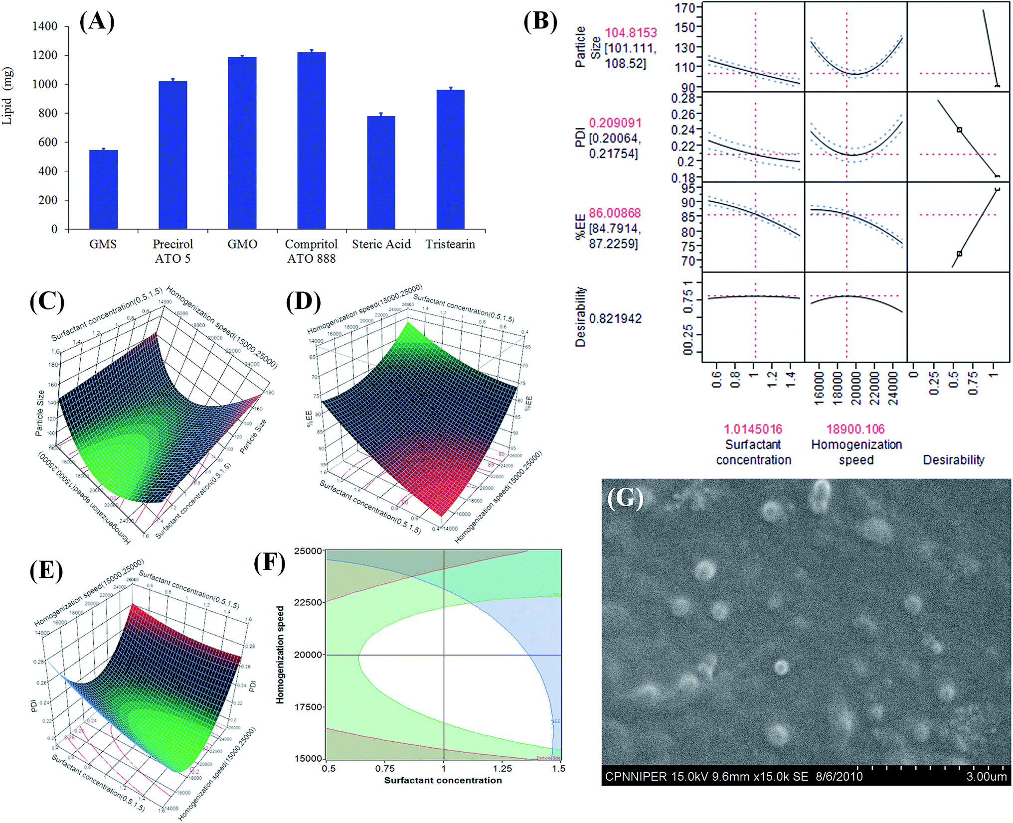 | ||
| Fig. 1 Graph (A) represents amount of solid lipid required to solubilise 5 mg of adapalene. Prediction profiler (B) representing the effect of independent variable on responses along with desirability. Contour plot representing the effect of homogenisation speed and surfactant concentration on responses such as (C) particle size, (D) %EE, and (E) PDI. Design space (F) representing the effect of independent factors on responses. Photograph (G) displays shape and morphology of Ada-SLNs using scanning electron microscopy. | ||
3.2. Selection of surfactant
Surfactants play pivotal role in the stabilization of SLNs by preventing the coalescence of highly unstable surfaces. They act as skin permeation enhancer either by skin lipid extraction or by skin lipid bilayer fluidization.38 In order to reversibly alter the SC barrier function to drug permeation different categories of nonionic surfactants like Brij 78, Pluronic F68, Tween 80 and Span 20 were screened because of their non-toxic nature and permeation enhancement ability. Table 2 indicates the superiority of Pluronic F68 in stabilizing Ada-SLNs in terms of size, PDI and %EE over the other surfactants.| Surfactant | Size (nm) | PDI | ZP (mV) | %EE |
|---|---|---|---|---|
| a ND: not determined due to aggregation of particles; values are represented as mean ± SD (n = 6). | ||||
| Pluronic F68 | 98 ± 6 | 0.184 ± 0.007 | −12.4 ± 0.82 | 86.78 ± 1.75 |
| Tween 80 | 162 ± 14 | 0.328 ± 0.012 | −7.75 ± 1.50 | 82.53 ± 4.36 |
| Brij 78 | 208 ± 20 | 0.274 ± 0.004 | −17.5 ± 0.92 | 85.35 ± 0.78 |
| Span 20 | ND | ND | ND | ND |
3.3. Development and optimization of Ada-SLNs using QbD approach
International Conference on Harmonization Q8 guideline on pharmaceutical development, deals with QbD concept, stating that quality should not be tested into the products, but should be built in.39 Herein, Ada-SLNs were prepared and optimized by QbD using surfactant concentration and homogenization speed as independent variables. ANOVA results confirmed the adequacy of the quadratic model (p < 0.001) for all the responses.The Box–Behnken design (SAS-JMP statistical software, SAS Institute Inc.) of Ada-SLNs suggested that both Pluronic F68 concentration and homogenization speed were predominant factors that notably affected (p < 0.0001) all the responses. The “sorted parameter estimate” confirmed the importance of explanatory variables, while “actual vs. predicted plot” demonstrated that independent variables significantly (p < 0.0001) altered the particle size, PDI and %EE of Ada-SLNs with acceptable coefficient of determination (data not shown). Fig. 1B depicts that the particle size was reduced upon increasing the surfactant concentration (0.5 to 1.5% w/v), which revealed that concentration of surfactant should be sufficient enough to cover the lipid globules and thus to stabilize the SLNs (smaller particle size).11,40 Similarly, particle size was decreased with increase in homogenization speed (15000 to 20000 rpm), however further increase in homogenization speed (>21000 rpm) had negative impact on responses. It suggested that the increase in energy might have led to increase in collision between newly formed globules and thus resulted in the aggregation to form larger SLNs.40 Similarly, both independent variables had detrimental effect on PDI at extreme levels of the model (Fig. 1E). Centre points of the design space produced desirable PDI which may be attributed to formation of desirable formulation with expected characteristics. Surfactant concentration and homogenization speed were also critical parameter to obtain the desired quality attributes of SLNs (Fig. 1C–E). Very high surfactant concentration and homogenization speed had deleterious impact on encapsulation efficiency which might be attributed to the solubilization of adapalene in aqueous phase.
000–17000 rpm for homogenization speed. Same design space was used to validate the model and it was observed that the model was accurate as well as precise within desired design space. The predicted and experimental values for Y1 and Y2 were precise and comparable (Table 3). The values were in very close agreement with model and established the reliability of the optimization procedure.
| Surfactant concentration (%w/v) | Homogenization speed (RPM) | Actual by predicted | Particle size (nm) | %EE | PDI |
|---|---|---|---|---|---|
| 1 | 20000 |
Predicted | 104 | 85.03 | 0.209 |
| Actual | 102 ± 5 | 86.61 ± 2.10 | 0.207 ± 0.009 | ||
| Prediction error | 4.17 | 2.47 | 2.87 | ||
| 0.9 | 18000 |
Predicted | 111 | 88.02 | 0.215 |
| Actual | 113 ± 5 | 88.95 ± 1.61 | 0.213 ± 0.012 | ||
| Prediction error | 3.9 | 1.72 | 3.72 | ||
| 0.8 | 21000 |
Predicted | 111 | 85.69 | 0.22 |
| Actual | 111 ± 4 | 85.97 ± 2.21 | 0.222 ± 0.008 | ||
| Prediction error | 2.4 | 1.86 | 2.58 |
SEM analysis further revealed that Ada-SLNs were almost spherical in shape with particle size below 200 nm which was in good correlation with the dynamic light scattering (Fig. 1G).
3.4. Preparation and characterization of Ada-SLNs gel
Maximum ‘slip’ and ‘drag’ as well as high viscosity were important for comfortable application of the formulation on inflamed region. Therefore, the viscosity and spreadability of Ada-SLNs loaded gel was checked to obtain an elegant pharmaceutical composition with optimum consistency, spreadability and ease of application. The comparative viscosity and spreadability as a function of increased surface area after the application of constant weight for constant period are summarized in Table 4. The adapalene content of Ada-SLNs gel was found to be 97.20 ± 4.20% w/v, while pH was found to be 6.8 which are in acceptable limits (Table 5).| Carbopol grade | Conc. (%w/v) | Viscosity (Pa s) | Spreadability (cm2) |
|---|---|---|---|
| a Values reported as mean ± SD (n = 6). | |||
| 980 NF | 0.5 | 6.32 ± 0.165 | 8.39 ± 1.36 |
| 1 | 14.26 ± 0.425 | 7.83 ± 2.52 | |
| 1.5 | 18.02 ± 0.348 | 5.28 ± 1.94 | |
| Ultrez 10 | 0.5 | 12.62 ± 0.206 | 14.24 ± 0.84 |
| 1 | 24.72 ± 0.275 | 12.39 ± 2.62 | |
| 1.5 | 34.52 ± 0.407 | 9.95 ± 2.45 | |
| Pemulen TR-1 | 0.5 | 70.37 ± 0.127 | 10.05 ± 3.72 |
| 1 | 12.45 ± 0.231 | 8.24 ± 2.85 | |
| 1.5 | 17.53 ± 0.362 | 6.23 ± 1.46 | |
| (a) Critical quality attributes of Ada-SLNs | |||||||||
|---|---|---|---|---|---|---|---|---|---|
| Storage conditions | 2–8 °C | 25 °C/60% RH | 40 °C/75% RH | ||||||
| Particle size (nm) | PDI | %EE | Particle size (nm) | PDI | %EE | Particle size (nm) | PDI | %EE | |
| a Values reported as mean ± SD (n = 6). | |||||||||
| Initial | 102 ± 5 | 0.207 ± 0.009 | 86.61 ± 2.10 | 102 ± 5 | 0.207 ± 0.009 | 86.61 ± 2.10 | 102 ± 5 | 0.207 ± 0.009 | 86.61 ± 2.10 |
| 1 month | 98 ± 8 | 0.185 ± 0.014 | 85.92 ± 1.54 | 105 ± 10 | 0.212 ± 0.021 | 86.18 ± 1.38 | Aggregation | ||
| 3 month | 105 ± 11 | 0.215 ± 0.018 | 86.58 ± 2.05 | 101 ± 14 | 0.218 ± 0.012 | 86.72 ± 3.15 | Aggregation | ||
| (b) Characteristics of Ada-SLNs gel | |||||||||
|---|---|---|---|---|---|---|---|---|---|
| Storage conditions | Assay Ada | Viscosity (Pa s) | Spreadability (cm2) | Assay Ada | Viscosity (Pa s) | Spreadability (cm2) | Assay Ada | Viscosity (Pa s) | Spreadability (cm2) |
| Initial | 100 | 24.72 ± 0.27 | 12.39 ± 2.62 | 100 | 24.72 ± 0.27 | 12.39 ± 2.62 | 100 | 24.72 ± 0.27 | 12.39 ± 2.62 |
| 1 month | 98.73 ± 2.15 | 26.06 ± 1.64 | 10.28 ± 1.41 | 99.46 ± 0.84 | 24.62 ± 1.32 | 12.38 ± 1.54 | 99.46 ± 0.84 | 23.74 ± 1.25 | 12.84 ± 1.33 |
| 3 month | 99.82 ± 2.57 | 27.85 ± 2.14 | 9.80 ± 1.82 | 98.32 ± 2.04 | 24.58 ± 1.74 | 12.34 ± 1.05 | 98.32 ± 2.04 | 22.83 ± 2.10 | 12.86 ± 1.37 |
All grades of Carbopol produced stable gel without inducing agglomeration of SLNs at all tested concentrations. However, Carbopol Ultrez 10 was selected as preferred gelling agent on the basis of high viscosity and high spreadability (Table 4). Carbopol Ultrez 10 might have formed severe interconnected network of polymer chains in presence of water producing high viscosity at low concentrations. Additionally, ease of dispersibility in comparison to conventional gelling agents (e.g. acacia, hydroxyl propyl methyl cellulose, cellulose, etc.) and low thermosensitivity in aqueous system makes Carbopol Ultrez 10 a suitable gelling agent.41
The change in thixotropic behavior after addition of SLNs to Carbopol gel was also studied (Fig. 2A). Rheological curves clearly depicted the increase in viscosity of Carbopol gel upon addition of Ada-SLNs (Fig. 2B). Additionally, increase in thixotropic area from 839 mm2 to 1934 mm2 was observed, which may be attributed to hindrance offered by Ada-SLNs during recovery of gel ultrastructure by forming intermolecular hydrogen bonds.
 | ||
| Fig. 2 Rheology representing (A) thixotropic behaviour and (B) viscosity of Ada-SLNs loaded Carbopol Ultrez 10 gel and Carbopol Ultrez 10 gel. In vitro release representing (C) %cumulative release of adapalene from Free-Ada, Ada-SLNs and Ada-SLNs gel and (D) mathematical modelling representing best suitable release mechanism through Ada-SLNs and Ada-SLNs gel. | ||
3.5. Storage stability
Storage stability signified that Ada-SLNs can be stored at room temperature without any notable (p > 0.05), albeit prone to aggregation at 40 °C/75% (Table 5a). Aggregation at 40 °C/75% could be attributed to the temperature dependent thermodynamic instability of lipids. Wang et al. also suggested that slow oxidation and crystal structure changes in lipids at higher temperature might be attributed to relatively high instability.42 The results were in line with previous reports in which thick gel formation was observed at 40 °C.43,44Ada content in Ada-SLNs gel was not significantly altered upon storage up to three months (Table 5b). However, the viscosity and spreadability were notably deviated at lower (2–8 °C) and higher temperature conditions (40 °C/75%). Cool condition might have increased the slip-drag forces between Carbopol matrix and water molecules resulting in high viscosity and poor spreadability. On contrary, hot condition might have disturbed the Carbopol matrix which decreased the viscosity and lower the spreadability. Therefore, Ada-SLNs bulk and finished Ada-SLNs gel is recommended to be stored at room temperature.
3.6. In vitro release
The amount of adapalene released from Free-Ada suspension demonstrated ∼98% release in 8 h, while ∼38% and ∼30% adapalene was released from Ada-SLNs and Ada-SLNs gel, respectively in 48 h (Fig. 2C). Univariate ANOVA revealed a significant difference (P < 0.001) in the release profiles of Free-Ada, Ada-SLNs and Ada-SLNs gel. Mathematical modelling using DD solver suggested that Free-Ada followed zero order release kinetics, while the Ada-SLNs and Ada-SLNs gel demonstrated Higuchi type matrix release behaviour (Fig. 2D). It suggested that diffusion dependent release of adapalene via erosion of solid matrix of lipids or gel might be the primary mechanism of drug release. The Ada-SLNs followed characteristic initial burst followed by sustained release which may be ascribed to adapalene adhered to the surface of the lipid and adapalene encapsulated in solid matrix of lipid, respectively. Carbopol gel matrix provided an additional barrier, therefore further retarded the drug release from Ada-SLNs gel as compared to Ada-SLNs dispersion. Viscous nature of the Carbopol might have constituted thicker stagnant layer around SLNs matrix which initially restricted the drug transport, but its subsequent dispersion in release media might have facilitated the drug release at later time point. DDsolver also confirmed the significant difference in release profile of free adapalene dispersion (0.1% w/v adapalene dispersed in 0.05% w/v methyl cellulose solution) and Ada-SLNs or Ada-SLNs gel on the basis of similarity factor [f2 ∈ (50,100)], difference factor [f1 ∈ (0,15)] and Rescigno index. A sustained release of adapalene from Ada-SLNs not only retained the drug within skin layers for prolonged period, but also reduced the possibility of concentration dependent irritation caused by adapalene.3.7. In vitro skin permeation and penetration study
The in vitro skin permeation and penetration study was carried out to estimate the systemic exposure and dermal bioavailability of Ada-SLNs and Ada-SLNs gel. The cumulative amount of adapalene, permeated and penetrated (expressed as %dose) after 24 h, has been shown in Table 6. For topical drug products, the evaluation of systemic exposure is an important safety criterion. Therefore, the amount of adapalene in the receptor compartment, which represents the absorbed dose, was calculated and found to be below the limit of quantification (LOQ, 10 ng mL−1). This data was well correlated with existing literature information such as non-dectectable (only 0.01%) adapalene of the applied dose has been penetrated through the skin.45,46| Parameters | 0.1% Adiff gel | 0.3% Adiff gel | 0.1% Ada-SLNs dispersion | 0.1% Ada-SLNs gel |
|---|---|---|---|---|
| Assay | 98.17 ± 2.81 | 100.08 ± 1.38 | 98.61 ± 2.46 | 97.20 ± 4.20 |
| pH | 6.5 | 6.5 | 6.8 | 6.8 |
| Total number of samples | 6 | 6 | 6 | 6 |
| Actual dose applied (μg) | 100.8 ± 2.2 | 298.8 ± 3.8 | 97.5 ± 0.9 | 99.3 ± 0.18 |
| Amount retained on skin (μg) | 76.6 ± 0.76 | 271.9 ± 9.5 | 66.6 ± 2.8 | 71.2 ± 0.9 |
| Epidermis + SC (μg) (A) | 1.2 ± 0.15 | 3.8 ± 0.1 | 4.5 ± 0.5 | 3.4 ± 0.35 |
| Dermis (μg) (B) | 0.12 ± 0.1 | 0.41 ± 0.02 | 0.37 ± 0.03 | 0.48 ± 0.06 |
| Absorbed dose (μg) (C) | Below LOQ | Below LOQ | Below LOQ | Below LOQ |
| [A + B + C] total penetrated (μg) | 1.3 ± 0.21 | 4.2 ± 0.12 | 4.8 ± 0.5 | 3.8 s ± 0.4 |
| Mass balance (μg) | 77.9 ± 0.9 | 276.0 ± 9.6 | 71.5 ± 3.4 | 74.9 ± 1.3 |
The tape stripping technique was employed to establish the skin penetration of adapalene formulations in same set of experiments and the results are represented in Table 6. The dermal bioavailability of adapalene was increased by 4.69 and 3.19 fold form Ada-SLNs and 0.1% Ada-SLNs gel, respectively as compared to 0.1% Adiff gel, while it was comparable (p > 0.005) to that of 0.3% Adiff gel. It clearly indicated that 0.1% Ada-SLNs gel was equally efficacious as compared to 0.3% Adiff gel on the basis of dermal bioavailability after application for 24 h.47 Various mechanisms such as pilosebaceous, transcellular and paracellular routes have been proposed to demonstrate the enhanced absorption of the nanoparticles through skin.48 Ada-SLNs might have predominantly followed pilosebaceous and transcellular pathways resulted in high topical and dermal bioavailability.49
3.8. Mechanistic understanding of skin drug distribution
The mechanism of pathway followed by SLNs through skin was studied by CLSM analysis using fluorescence labelled SLNs. High affinity for cell membrane lipids, almost similar molecular weight (<500 Da) and high logP (>5.0) makes ‘Cou6’ a suitable tracer. Adapalene possesses similar physicochemical properties as that of Cou6 and hence was expected to follow similar permeation properties. The distribution pathways followed by Cou6-SLNs gel indicated that SLNs preferentially followed transfollicular route and to some extent transcellular route (Fig. 3B). Loosening of tight junction between keratinocytes owing to skin hydration (occlusive effect of SLNs) and surfactant action of Poloxamer F68 might have facilitated permeation of SLNs via transcellular route.48 Additionally nanometer size of the developed SLNs could assisted into the permeation of SLNs into the deeper layers of skin and accumulation in the hair follicles which further offer a reservoir for topically applied drug products.
 | ||
| Fig. 3 CLSM images of skin sections treated with (A) 24 h control (free Cou6 gel) and (B) 24 h treatment with Cou6-SLNs gel. CLSM image of cyanoacrylate strips after 4 h of application (C) first strip showing hair follicular pathway (D) second strip showing intercellular and transcellular penetration pathway. | ||
To investigate the permeation pathways, the follicular extraction was performed using cyanoacrylate striping. Two simultaneous strips were removed out of which the first strip extracted the majority of hair follicles, while the second strip removed remaining SC. Fluorescence image of first strip showed the green fluorescence majorly localized in hair follicles confirming the follicular permeation pathway (Fig. 3C–D).50 Additionally, limited permeation through transcellular route was also observed which could be attributed to improved skin hydration leading to widening of skin pores or slow diffusion of drug through lipid matrix of SLNs.
3.9. Skin irritation
 | ||
| Fig. 4 Skin irritation study: (A) %cell viability after treatment of RHE according to EpiSkin-42 protocol, the line represents the classification cut-off (50%) and (B) skin barrier properties as a function of Transepidermal Water Loss (TEWL). ‘a’ and ‘b’ refers to the comparison with 0.3% and 0.1% Adiff gel, respectively. ‘***’ and ‘*’ represents the confidence interval of 99.9% and 95.0%, respectively. | ||
The changes in TEWL produced by Ada-SLNs gel was found to be significantly (p < 0.05) lower than 0.1% Adiff gel (Fig. 4B) indicating reduced toxicity caused by SLNs gel, albeit higher dermal distribution. The decrease in TEWL after application of Ada-SLNs gel could be attributed to occlusive effect of SLNs on skin, which also assisted in improving the skin permeation of Ada-SLNs. Further it can additionally help in preventing the skin dryness, one of the side effect of adapalene.5,15
3.10. Anti-acne potential
Fig. 5 displayed dynamic changes in skin after treatment of adapalene formulations. Visual changes in animal skin illustrated that application of TS developed severe acne in animal after 3 weeks, which were notably reduced following treatment with adapalene formulations (Fig. 5). Fig. 5I evidenced significant (p < 0.001) reduction in papule density in the adapalene treated groups in comparison to untreated group (visual observation). The order of mean percentage reductions in papule density was 0.1% Adiff gel (43.8 ± 6.3%) < 0.3% Adiff gel (60.0 ± 7.8) < 0.1% Ada-SLNs gel (86.3 ± 5.7) (Fig. 5(III)). | ||
| Fig. 5 Evaluation of anti-acne potential of Ada loaded formulations in TS induced acne model. Digital photograph (I) and histology (II) of mice skin is represented as (A) control; (B) TS induced acne (untreated), (C) 0.1% Adiff gel, (D) 0.3% Adiff gel, and (E) and 0.1% Ada-SLNs gel. Graph (III) represents anti-acne efficacy of formulation as function of percentage reduction in number of papule (lesion) density per 4 cm2. | ||
The histological evaluation demonstrated sebaceous hyperplasia (both increase in number and size of sebaceous gland) and follicular hyperkeratosis in pilosebaceous unit which are major indication of development of acne (Fig. 5B). On the contrary, adapalene treated animals showed marked reduction of the lesion, sebaceous gland hyperplasia and seborrhea (Fig. 5C–E). Surprisingly, the skin histology of 0.3% Adiff gel and Ada-SLNs gel were as same as normal skin which revealed significant reduction in papule density and acne lesion. Additionally, Ada-SLNs gel treated group did not show any evidence of skin peeling and desquamation, indicating better tolerability and less toxicity in comparison to Adiff gel. Sustained release of adapalene and higher skin penetration were possible explanations for the enhanced therapeutic efficacy and increased tolerability of Ada-SLNs gel vis-a-vis marketed Adiff gel.
4. Conclusions
In present work, Ada-SLNs gel was successfully prepared by strategic hot homogenization technique followed by Carbopol gelation. The developed Ada-SLNs gel exhibited improved dermal delivery of adapalene via pilosebaceous unit with better skin tolerability and enhanced anti-acne potential. Easy scale up, devoid of organic solvents, and patient friendly nature are the few hidden advantages of present technology which can make it a clinical reality. A combinational drug therapy containing adapalene and benzoyl peroxide as major therapeutic agent is being evaluated in our lab for further effective and synergistic treatment of acne. The present formulation strategy can also be a viable alternative for delivery of drugs similar to adapalene “a drug seeking higher concentration at sight of action”.Acknowledgements
Authors acknowledge Director, NIPER for providing necessary infrastructure facilities. We are also grateful to Dr Kusum Joshi, Dr B. D. Radotra and Dr Pankaj from Post Graduate Institute of Medical Education and Research, Chandigarh for their help. Technical assistance provided by Mr Rahul Mahajan in SEM analysis is also duly acknowledged.References
- A. Krautheim and H. P. Gollnick, Clin. Dermatol., 2004, 22, 398–407 CrossRef PubMed.
- W. D. James, N. Engl. J. Med., 2005, 352, 1463–1472 CrossRef CAS PubMed.
- E. Mallon, J. Newton, A. Klassen, S. Stewart-Brown, T. Ryan and A. Finlay, Br. J. Dermatol., 1999, 140, 672–676 CrossRef CAS.
- M. Kawashima, S. Harada, C. Loesche and Y. Miyachi, J. Dermatol. Sci., 2008, 49, 241–248 CrossRef CAS PubMed.
- W. Cunliffe, R. Caputo, B. Dreno, L. Förström, M. Heenen, C. Orfanos, Y. Privat, A. R. Aguilar, J. Meynadier and M. Alirezai, J. Am. Acad. Dermatol., 1997, 36, S126–S134 CrossRef CAS.
- G. M. E. Maghraby, A. C. Williams and B. W. Barry, J. Pharm. Pharmacol., 2006, 58, 415–429 CrossRef PubMed.
- M. Foldvari, I. Badea, S. Wettig, D. Baboolal, P. Kumar, A. L. Creagh and C. A. Haynes, Mol. Pharmaceutics, 2010, 7, 751–762 CrossRef CAS PubMed.
- F. Knorr, J. Lademann, A. Patzelt, W. Sterry, U. Blume-Peytavi and A. Vogt, Eur. J. Pharm. Biopharm., 2009, 71, 173–180 CrossRef CAS PubMed.
- A. Rolland, N. Wagner, A. Chatelus, B. Shroot and H. Schaefer, Pharm. Res., 1993, 10, 1738–1744 CrossRef CAS.
- G. Bhatia, Y. Zhou and A. K. Banga, J. Pharm. Sci., 2013, 102, 2622–2631 CrossRef CAS PubMed.
- H. Harde, M. Das and S. Jain, Expert Opin. Drug Delivery, 2011, 8, 1407–1424 CrossRef CAS PubMed.
- K. A. Shah, A. A. Date, M. D. Joshi and V. B. Patravale, Int. J. Pharm., 2007, 345, 163–171 CrossRef CAS PubMed.
- M. R. Aji Alex, A. J. Chacko, S. Jose and E. B. Souto, Eur. J. Pharm. Sci., 2011, 42, 11–18 CrossRef CAS PubMed.
- Z. Mei, X. Li, Q. Wu, S. Hu and X. Yang, Pharm. Res., 2005, 51, 345–351 CrossRef CAS PubMed.
- S. Jain, M. A. Mistry and N. K. Swarnakar, Drug Delivery Transl. Res., 2011, 1, 395–406 CrossRef CAS PubMed.
- H. Harde, A. K. Agrawal and S. Jain, Nanomedicine, 2014, 1–19, DOI:10.2217/nnm.2213.2225.
- H. Harde, A. K. Agrawal and S. Jain, J. Biomed. Nanotechnol., 2014, 11, 363–381 CrossRef PubMed.
- R. Sonawane, H. Harde, M. Katariya, S. Agrawal and S. Jain, Expert Opin. Drug Delivery, 2014, 11, 1833–1847 CrossRef CAS PubMed.
- A. K. Agrawal, H. Harde, K. Thanki and S. Jain, Biomacromolecules, 2014, 15, 350–360 CrossRef CAS PubMed.
- R. K. Subedi, K. W. Kang and H.-K. Choi, Eur. J. Pharm. Sci., 2009, 37, 508–513 CrossRef CAS PubMed.
- L. A. Martins, L. Z. Meneghini, C. A. Junqueira, D. C. Ceni and A. M. Bergold, J. Chromatogr. Sci., 2011, 49, 796–800 CAS.
- P. Batheja, L. Sheihet, J. Kohn, A. J. Singer and B. Michniak-Kohn, J. Controlled Release, 2011, 149, 159–167 CrossRef CAS PubMed.
- V. Venkateswarlu and K. Manjunath, J. Controlled Release, 2004, 95, 627–638 CrossRef CAS PubMed.
- J. Y. Fang, C. L. Fang, C. H. Liu and Y. H. Su, Eur. J. Pharm. Biopharm., 2008, 70, 633–640 CrossRef CAS PubMed.
- C. Herkenne, I. Alberti, A. Naik, Y. N. Kalia, F.-X. Mathy, V. Préat and R. H. Guy, Pharm. Res., 2008, 25, 87–103 CrossRef CAS PubMed.
- B. N'Dri-Stempfer, W. Navidi, R. Guy and A. Bunge, Pharm. Res., 2009, 26, 316–328 CrossRef PubMed.
- W. Navidi, A. Hutchinson, B. N'Dri-Stempfer and A. Bunge, J. Pharmacokinet. Pharmacodyn., 2008, 35, 337–348 CrossRef PubMed.
- X. Wu, P. Griffin, G. J. Price and R. H. Guy, Mol. Pharmaceutics, 2009, 6, 1449–1456 CrossRef CAS PubMed.
- E. Touitou, V. M. Meidan and E. Horwitz, J. Controlled Release, 1998, 56, 7–21 CrossRef CAS.
- S. Jain, A. Mittal and A. K. Jain, Curr. Nanosci., 2011, 7, 524–530 CrossRef CAS.
- G. E. Pierard, C. Pierard-Franchimont, J. Arrese Estrada, P. Delvoye, C. Henry, M. Damseaux, T. Le, J. F. Hermans, B. Letot and P. Melotte, et al., J. Cutaneous Pathol., 1989, 16, 180–182 CrossRef CAS PubMed.
- F. Netzlaff, C.-M. Lehr, P. Wertz and U. Schaefer, Eur. J. Pharm. Biopharm., 2005, 60, 167–178 CrossRef CAS PubMed.
- N. Alépée, C. Tornier, C. Robert, C. Amsellem, M.-H. Roux, O. Doucet, J. Pachot, M. Méloni and A. de Brugerolle de Fraissinette, Toxicol. In Vitro, 2010, 24, 257–266 CrossRef PubMed.
- V. Jenning, A. Gysler, M. Schäfer-Korting and S. H. Gohla, Eur. J. Pharm. Biopharm., 2000, 49, 211–218 CrossRef CAS.
- K. Raza, B. Singh, S. Singla, S. Wadhwa, B. Garg, S. Chhibber and O. P. Katare, Mol. Pharmaceutics, 2013, 10, 1958–1963 CrossRef CAS PubMed.
- D. V. M. F. Neumann and W. Elger, J. Invest. Dermatol., 1966, 46, 561–572 Search PubMed.
- Y. Zhang, M. Huo, J. Zhou, A. Zou, W. Li, C. Yao and S. Xie, AAPS J., 2010, 12, 263–271 CrossRef CAS PubMed.
- G. Cevc, G. Blume, A. Schatzlein, D. Gebauer and A. Paul, Adv. Drug Delivery Rev., 1996, 18, 349–378 CrossRef CAS.
- R. A. Lionberger, S. L. Lee, L. Lee, A. Raw and X. Y. Lawrence, AAPS J., 2008, 10, 268–276 CrossRef CAS PubMed.
- W. Mehnert and K. Mäder, Adv. Drug Delivery Rev., 2001, 47, 165–196 CrossRef CAS.
- M. Fresno Contreras, A. Ramírez Diéguez and M. Jiménez Soriano, Il Farmaco, 2001, 56, 437–441 CrossRef CAS.
- Y. Wang, L. Zhu, Z. Dong, S. Xie, X. Chen, M. Lu, X. Wang, X. Li and W. Zhou, Colloids Surf., B, 2012, 98, 105–111 CrossRef CAS PubMed.
- R. Shegokar, K. Singh and R. Müller, Int. J. Pharm., 2011, 416, 461–470 CrossRef CAS PubMed.
- A. Radomska, R. Dobrucki and R. Müller, Pharmazie, 1999, 54, 903–909 CAS.
- J. Allec, A. Chatelus and N. Wagner, J. Am. Acad. Dermatol., 1997, 36, S119–S125 CrossRef CAS.
- E. G. de Jalon, M. J. Blanco-Prieto, P. Ygartua and S. Santoyo, Int. J. Pharm., 2001, 226, 181–184 CrossRef CAS.
- D. Thiboutot, D. M. Pariser, N. Egan, J. Flores, J. H. Herndon Jr, N. B. Kanof, S. E. Kempers, S. Maddin, Y. P. Poulin and D. C. Wilson, J. Am. Acad. Dermatol., 2006, 54, 242–250 CrossRef PubMed.
- P. Desai, R. R. Patlolla and M. Singh, Mol. Membr. Biol., 2010, 27, 247–259 CrossRef CAS PubMed.
- A. K. Jain, A. Jain, N. K. Garg, A. Agarwal, A. Jain, S. A. Jain, R. K. Tyagi, R. K. Jain, H. Agrawal and G. P. Agrawal, Colloids Surf., B, 2014, 121, 222–229 CrossRef CAS PubMed.
- X. Wu, G. J. Price and R. H. Guy, Mol. Pharmaceutics, 2009, 6, 1441–1448 CrossRef CAS PubMed.
- S. Mukherjee, A. Date, V. Patravale, H. C. Korting, A. Roeder and G. Weindl, Clin. Interventions Aging, 2006, 1, 327–348 CrossRef CAS.
- G. A. Castro, A. L. L. Coelho, C. A. Oliveira, G. A. Mahecha, R. L. Oréfice and L. A. Ferreira, Int. J. Pharm., 2009, 381, 77–83 CrossRef CAS PubMed.
- B. Sotoodian and H. I. Maibach, Clin. Dermatol., 2012, 30, 301–310 CrossRef PubMed.
| This journal is © The Royal Society of Chemistry 2015 |