DOI:
10.1039/C5RA05834A
(Paper)
RSC Adv., 2015,
5, 56210-56218
Au@ZnO hybrid nanostructures: correlation between morphology and optical response†
Received
1st April 2015
, Accepted 18th June 2015
First published on 18th June 2015
Abstract
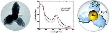
Au@ZnO hybrid nanostructures (HNs) have been synthesized in aqueous media by implementing a simple chemical methodology. These HNs consist of 40 nm mean size almost spherical Au cores, over which the heterogeneous formation of ZnO is observed. By increasing the concentration of ZnO precursors in the system, it is possible to change the morphology of the material formed over the Au core from branched to shell like structures, which, in turn, significantly modifies the extinction properties of the naked Au core nanoparticles. This effect has been rationalized by means of electrodynamics simulations based on two different approaches: the Mie theory for coated spheres and the Discrete Dipole Approximation (DDA). The changes measured in the extinction spectra as the amount of ZnO formed around the Au cores increases are properly described by both methodologies, while the good correlation between experimental and theoretical spectra suggest that the ZnO material includes a significant amount of water. Furthermore, based on the Mie theory results, a graphical method was implemented which allows us to predict the main morphological parameters of the Au@ZnO HNs. In addition, the combination of optical measurements, morphological characterization and DDA modeling allowed us to estimate that the water content of the shell surrounding the metallic core is 65%. The methodology presented in this work provides a useful tool to characterize the structural properties of HNs and can be straightforwardly generalized to other systems.
Introduction
Hybrid nanostructures (HNs) composed of noble metal and semiconductor materials are currently materials of great interest as this particular kind of system may exhibit novel and unique physical chemistry properties, achieving potential applications in diverse areas such as biological detection, catalysis and solar energy conversion.1–6 Regarded separately, the optical properties of noble metal (NPs) and semiconductor (QDs) nanoparticles are characterized by the elusive localized surface plasmon resonances (LSPR) and excitons, respectively. In both cases, the excitation wavelength required to produce such excitations strongly depends on the NP nature, size, shape and local environment.7–9 Consequently, synthetic methodologies capable to produce NPs and QDs with narrows size and shape distributions are necessary in order to rationally employ the optical properties of nanosized matter in a given device.10–14 Nonetheless, when considering the optical properties of HNs, it may be different from that observed for NPs and QDs separately as a consequence of the interaction between the respective HNs building blocks. Moreover, the interaction between the constitutive parts of a given HN can be tuned by controlling the three-dimensional arrangement, size and shape of the HN building blocks. Energy transfer processes may take place if QDs and NPs are located adjacent within a given HN and Förster resonance energy transfer as well as nanometal surface energy transfer mechanisms had been employed to characterize these processes.15–19 For instance, Viste et al. showed the possibility of tuning under control the emission efficiency of HNs for a large range of Au NP sizes and demonstrated that the modification factor of emission of the hybrid nanosources compared to the bare QDs strongly depends on the Au NP size.20 In addition, interfacial charge transfer processes, which are relevant in photocatalytic reactions and light energy conversion, may also occur if QDs and NPs are close enough.21–23 In this regard, it has been demonstrated that the photo-generated electron transfer and the resulting catalytic activity of ZnO–Au composites can be controlled by the size of the mediating Au NP.24
Several chemical strategies have been developed and implemented to prepare HNs, resulting in a wide variety of different structures with distinct morphology and structural parameters, which, in turn, allow the study of optical phenomena that might result from the interaction between NPs and QDs. Indeed, general growth strategies has been reported for the synthesis of water dispersible Au@X (X = ZnS, AgS, CuS, PbS, CdS, CdSe, CdTe) core shell HNs, which achieves precise control of the morphologies of the structures.25,26 Au–TiO2 nanocomposites has also been successfully synthesized by several authors.27 In particular, Au@ZnO HNs with various morphologies have been prepared by different methods. Composites composed by large ZnO domains (several hundreds of nm) decorated with Au NPs likely comprise the kind of Au–ZnO HN most described in literature.28 Nonetheless, Haldar et al. have implemented a simple method to prepare water soluble and stable Au@ZnO core shell NHs in the size range below 10 nm.29 More recently, the synthesis of Au@ZnO HNs with a novel hexagonal pyramid-like structure has also been reported.30 However, to the best of our knowledge, reports concerning the preparation of Au@ZnO core shell HNs are rare.
On the other hand, the employment of theoretical approaches to describe the optical properties of HNs is of great importance to achieve a better understanding of the observed phenomena. For instance, an analytical model based on an approximate Mie's theory has been employed to study the optical features of Au@Cu2O core shell HNs prepared by controlling epitaxial growth of Cu2O on as-prepared Au NPs in aqueous solution.31 Wang and co-workers applied the Mie theory for coated spheres to interpret the complexity of extinction spectral line shapes and geometry-dependent optical tunability of the Au@Cu2O HNs.32 More recently, these authors employed the Mie theory for coated spheres along with effective medium theory to achieve a quantitative understanding of the geometry-dependent optical tenability of Au@Cu2O HNs with porous Cu2O shells.33 The same theoretical approach has been employed to reveal the growth mechanism in Ag@ZnO core shell HNs.34 The Mie theory and the four-flux method has been applied to study theoretically the influence of an oxide (SiO2, ZrO2, or TiO2) shell on a metal core (Ag, Au, or Cu) NP.35 Shaviv et al. studied the light absorption properties of CdS–Au and CdSe–Au NHs by comparing experimental results with electrodynamics simulations obtained by applying the DDA method.36 In this case, a qualitative agreement between DDA simulations and experimental data for CdS–Au HNs has been found and it has been stated that DDA simulations provide insights on the gold tip shape and its interface with the semiconductor material. For the CdSe–Au case, a shortcoming of the electrodynamics model was found, as it does not predict properly the optical features, which was ascribed to strong interaction of the metal and semiconductor excitations, that spectrally overlap in the CdSe case. However, as far as we know, the employment of electrodynamics simulations to obtain further insight on the optical properties of Au@ZnO HNs has not been reported so far.
In this work, a simple chemical method has been implemented in order to obtain Au@ZnO HNs. The synthesized HNs consist in 40 nm mean size almost spherical Au NPs cores, over which the heterogeneous formation of ZnO is observed. It was found that the morphology of the semiconductor material deposited over the core particles depends on the experimental conditions. In turn, these morphological changes induce significant modifications on the extinction spectra, which have been rationalized by means of two different electrodynamics approaches: the Mie theory for coated spheres and the DDA method. In both cases an effective medium theory was used to describe the dielectric constant of the shell. The good correlation between experimental and theoretical spectra indicates that the ZnO material that surrounds the Au core NP includes a significant amount of water. By implementing a graphical method to analyze the extinction spectra obtained by means of the exact extended Mie theory, it was possible to perform an accurate estimation of the main morphological parameters of the synthesized HNs. In addition, the DDA method was employed to address particularly the effect of the shape of the shell on the optical response of the HNs. By considering the shape of the surrounding material in a more realistic way within the DDA method, it was possible to estimate its water content, a parameter that could be rather hard to obtain by others approach.
Methodologies
Experimental methods
Materials. Sodium citrate (Na3C6H5O7, Mallinckrodt), chloroauric acid (HAuCl4, Carlo Erba), zinc nitrate (Zn(NO3)2·6H2O, Anedra) and potassium hydroxide (KOH, Cicarelli) were used as received without further purification. Aqueous solutions were prepared with ultrapure water (18.2 mΩ resistivity).
Synthesis of Au NPs. Nearly spherical Au NPs of mean size 40 nm were obtained according to the Turkevich method. Aliquots of 0.1 M sodium citrate aqueous solution (0.25 mL) and of 0.01 M acid chloroauric aqueous solution (2.5 mL) were added to 97.25 mL of boiling water under vigorous stirring. After 2–3 minutes of reaction an intense red wine color was observed indicating the formation of Au NPs. The system was kept at boiling temperature during 5 minutes to complete reaction and then it was cooled at room temperature. Au NPs were purified by performing centrifugation of 13 mL of the aqueous suspension of Au NPs during 10 minutes at 5000 rpm. The resulting precipitate was redispersed in 12 mL of 3.33 mM KOH aqueous solution. The alkaline dispersion of Au NPs was then separated in four aliquots (A, B, C, D) of 3 mL each one.
Synthesis of Au@ZnO HNs. By implementing a very simple chemical methodology HNs dispersed in aqueous media were obtained. To each one of the 3 mL Au NPs alkaline dispersions (aliquots A, B, C and D), 2 mL of zinc nitrate aqueous solution of a certain concentration was added in order to obtain samples with increasing Zn(NO3)2 concentration as displayed in Table 1. Note that 2 mL of water ([Zn(NO3)2] = 0 mM) were added to aliquot A in order to have an unmodified Au NPs sample as a control experiment. After adding the zinc nitrate solution, each one of the 5 mL volume samples A, B, C and D were kept at 80 °C during 15 minutes and, finally cooled and stored at room temperature.
Table 1 Zn(NO3)2 and KOH concentrations employed in each different sample
Sample |
Volume/mL |
[Zn(NO3)2]/mM |
[KOH]/mM |
A |
5 |
0.0 |
2.0 |
B |
5 |
0.1 |
2.0 |
C |
5 |
0.5 |
2.0 |
D |
5 |
1.0 |
2.0 |
Optical and morphological characterization. UV-vis spectroscopy characterization was performed using a Shimazdu UV-1700 PharmaSpec spectrophotometer with a 1 cm quartz cell at room temperature. To determine the mean size (Z-average diameter) and the particle size distribution, photon correlation spectroscopy (PCS) measurements were performed with a DelsaNano C Particle Analyzer Beckman Coulter instrument using a 1 cm quartz cell at room temperature. Transmission electron microscopy (TEM) images were obtained using a JEM-JEOL 1120 EXII instrument under an accelerating voltage of 80 kV. Samples were prepared by seeding a drop (∼20 μL) of the colloidal dispersion to be characterized onto a holey carbon/formvar-coated copper TEM grid (100 mesh).
XRD characterization. The XRD instrument employed was a PANalytical X-Pert Pro using a Cu Kα radiation (λ = 1.5406 Å). The diffractograms were obtained in the range 30–70° from powders of the particles. The respective powders were obtained by depositing droplets containing the nanostructures over glass slides, and evaporating the solvent in a fumehood at room temperature.
Theoretical methods
Mie theory for coated spheres. A complete and detailed description of this method can be found in the book written by Bohren and Huffman.37 This theory constitutes an exact solution to the problem of absorption and scattering of light by an object composed by concentric spheres. We have used the BHCOAT code, which implements such a solution, to simulate the extinction properties of Ag@ZnO core shell NHs.
DDA method. Details about this methodology can be found elsewhere.38–41 Briefly, this approximated method allows the calculation of the optical properties of arbitrarily shaped objects with a high degree of accuracy. In this method, the continuum target, i.e. the nanostructure, is approximated by a cubic array of N polarizable elements (dipoles) at positions ri with dipole polarizabilities αi. Due to the freedom to locate dipoles at any site of the cubic array, it is possible to model the optical properties of arbitrarily shaped objects with domains having different compositions. The dielectric constant of each material forming the target ε(λ) is introduced externally is input, and the method employs the lattice dispersion relation to determine the αi values so that an infinite array with finite lattice spacing mimic an extended solid with dielectric constant ε(λ).42 Furthermore, the number of dipoles used to represent a given particle can be thought as a control parameter of the code. By increasing the dipole number, the accuracy of the calculation is improved as well as the computational time increases. The number of dipoles used in the calculations should be chosen in such a way that convergence has been reached. In this work, we have used the DDSCAT 7.3 code to simulate the optical response of Au@ZnO NHs.43 A lattice spacing (inter dipole distance) of 1.33 nm was used in all simulations whereas the total number of dipoles to represent the target was kept in the range 726
744 ± 63.
Results and discussion
Experimental extinction spectra of Au@ZnO HNs
Fig. 1 shows the extinction spectra of suspensions of Au@ZnO HNs synthesized according to the procedure described above. The extinction spectrum of the unmodified Au NPs colloidal dispersion (black curve), which correspond to [Zn(NO3)2] = 0.0 mM, resembles the typical spectrum expected for this system, exhibiting a peak centered at 528 nm which is attributed to the excitation of the Au NPs LSPR. When a [Zn(NO3)2] of 0.1 mM was used in the synthesis (red curve), the LSPR peak position slightly red shifts 2 nm being centered at 530 nm, while a moderate increase in the extinction intensity for wavelength (λ) values shorter than 380 nm is observed in comparison with the previous case (black curve, [Zn(NO3)2] = 0.0 mM).
 |
| Fig. 1 Extinction spectra of Au@ZnO HNs suspensions obtained for different concentrations of Zn(NO3)2 employed in the synthesis as indicated in the inset. | |
These changes are more important when the [Zn(NO3)2] employed in the experiment is 0.5 mM (green curve). In this case, the LSPR peak increases its intensity and appears centered at 540 nm, while the extinction intensity for λ values shorter than 380 nm is noticeably amplified and exhibit a shoulder at 350 nm. More pronounced changes are observed for the case of HNs synthesized with [Zn(NO3)2] = 1.0 mM (blue curve): the intensity of the LSPR is increased and the peak red-shifts to 550 nm whereas the extinction intensity in the UV region is enlarged almost four times with respect to the spectrum of the naked Au NPs and displays a defined shoulder at 350 nm. The description of the main phenomena suggests that the remarkable enlargement of the extinction intensity in the UV region, particularly for λ values around 350 nm, is consistent with the absorption features of ZnO. On the other hand, the considerable redshifts of the LSPR peak position can be attributed, in principle, to two principal causes: plasmonic coupling between Au NPs or increase of the dielectric constant of the environment where the NPs are located. It is noteworthy that these two phenomena may occur simultaneously.
Morphological characterization of Au@ZnO HNs
Fig. 2 shows representative TEM images of the HNs obtained for Zn(NO3)2 concentrations of (a) 0.0, (b) 0.1, (c) 0.5 and (d) 1.0 mM (samples A, B, C and D respectively). TEM images of the sample containing the unmodified Au NPs cores, i.e. Au NPs obtained by means of the Turkevich method, which were then purified by centrifugation, redispersed in basic media and heated at 80 °C during 15 minutes, are shown in Fig. 2a. It is observed that, in the absence of Zn2+, nearly spherical Au NPs with mean size 40 nm were obtained. Note that the Au NPs size and shape distributions that can be appreciated from Fig. 2a is consistent with the extinction spectrum of the sample shown in Fig. 1 (black curve). More detailed information on the initial particle size distribution used for growing the HNs, was obtained by performing PCS measurements of unmodified Au NPs suspensions. The intensity size distribution obtained is shown in Fig. S1,† which presents a single peak centered at 45 nm, indicating that the hydrodynamic radius and the particle size distribution is consistent with the morphological information obtained by TEM. The TEM images shown in Fig. 2b–d clearly reveal the appearance of a different material closely connected to the Au NPs ascribed as ZnO. In the TEM image shown in Fig. 2b, this material is visualized as protuberances or branches that arise from the Au NPs surface. The number of branches in each Au NP varies between 2 and 6, being 50 nm its mean length. Qualitatively, similar HNs were obtained for [Zn(NO3)2] = 0.5 mM (Fig. 2c). The TEM image corresponding to sample D (Fig. 2d), obtained with [Zn(NO3)2] = 1.0 mM, shows qualitatively similar HNs, but in this case a considerably larger amount of ZnO attached to the Au NPs is observed in comparison with the HNs obtained with lower [Zn(NO3)2] values. Furthermore, the Au NPs appears as immersed into a ZnO matrix that resemble a shell which mean thickness is approximately 60 nm. It can be noted in the respective TEM images that the amount of ZnO that surrounds the Au cores increases proportionally to the Zn2+ concentration used in each experiment. Concomitantly, the width of the ZnO shell remains roughly at a constant value around 60 nm. Besides, irrespective of the [Zn(NO3)2] used, the mean size of the Au NPs is 40 nm and its size and shape distributions are practically the same to that observed in Fig. 2a for the naked Au NPs. This observation suggests that the Au NPs are not modified by adding Zn2+ or by increasing temperature. Furthermore, aggregation of Au NPs, which could lead to plasmonic coupling, and, consequently, to a red-shift of the extinction peaks, is not appreciated in these images.
 |
| Fig. 2 Representative TEM images of the as-prepared Au@ZnO HNs obtained for different concentrations of Zn(NO3)2 employed in the synthesis: (a) 0.0 mM, (b) 0.1 mM, (c) 0.5 mM and (d) 1.0 mM. | |
In general, the TEM analysis indicates that the synthesized HNs display a flower-like morphology and that the degree of coverage of the Au core NPs by ZnO increases with the value of [Zn(NO3)2] employed in the synthesis. Importantly, the TEM images also show that, in all cases, the ZnO formation uniquely takes place on the Au NPs surface. This observation suggests that the Au NPs behave as nucleation centers for the heterogeneous precipitation of ZnO, and provides evidence that the LSPR redshift can be attributed to an increase of the local dielectric constant generated by oxide material that surrounds the Au NPs. Therefore, the morphological characteristics of the HNs revealed by the TEM images analysis are in a quite good qualitative agreement with the interpretation regarding the changes observed in the extinction spectra.
XRD characterization
Fig. 3 shows the XRD patterns of the powders obtained from the unmodified Au NPs and from the Au@ZnO HNs (synthesized with [Zn(NO3)2] = 1.0 mM) denoted as (a) and (b), respectively. For comparison, the standard data for fcc metal Au (JCPDS 4-784) and hexagonal wurtzite ZnO (JCPDS 36-1451) are also shown at the bottom as blue and red bars, respectively.
 |
| Fig. 3 XRD patterns of (a) Au NPs and (b) Au@ZnO HNs. The reference XRD pattern of Au (JCPDS 4-784) and ZnO (JCDPS 36-1451) are also shown in the lower part of the figure as blue and red bars, respectively. The patterns have been arbitrarily shifted in the y-axis. | |
The diffraction pattern of Au NPs shows three peaks at 2θ = 38.2, 44.4 and 64.6, which are assigned to the reflections of the planes (111), (200) and (220) of Au, respectively. On the other hand, the diffraction pattern of Au@ZnO HNs shows peaks at 2θ = 31.7, 34.4, 36.3, 56.7, 62.9 and 68.0 which are assigned to the reflections of the planes (100), (002), (101), (110), (103) and (112) of ZnO, respectively, in addition to the peaks attributed to reflections of the Au phase. The background signal, particularly its increase for 2θ → 30, is attributed to amorphous SiO2 (JCPDS 29-0085) which constitutes the material employed to support the sample during measurements, whereas the additional peaks present in the Au@ZnO HNs pattern at 2θ = 31.7, 34.4 might be assigned to reflections of the planes (102) and (211) of Quartz (JCPDS 33-1161). Therefore, these results confirm the formation of an irregular ZnO shell on the Au cores, and further support the interpretation of the measured changes in the optical properties. Furthermore, the Scherrer equation (see ESI†) was applied to estimate the crystallite size of the respective materials (instrumental broadening was considered negligible and was not included in the analysis). In particular for Au NPs, the mean coherence length perpendicular to the planes (111) gives a value of 26 nm, while for ZnO the mean coherence length perpendicular to the planes (101) gives a value of 24 nm.
Modelling of the extinction spectra of HNs
Probably the simplest model to describe the optical response of the synthesized HNs is graphically schematized in Fig. 4. It consists in two concentric spheres: a Au core of diameter D surrounded by a uniform shell of thickness S dispersed in water.
 |
| Fig. 4 Graphic scheme of the model used in Mie theory for coated spheres simulations to represent the synthesized Au@ZnO HNs. | |
Note that the composition of the shell is heterogeneous as it is composed by water and ZnO. Thus, the simple proposed model aims to capture the most relevant morphological features observed in the TEM images. The extinction spectrum of the proposed concentric spheres model to describe the optical response of the synthesized HNs, can be exactly calculated in the frame of classical electrodynamics by using the Mie theory for coated spheres as formulated by Aden and Kerker (ref. 37, page 181). The dielectric constants of the Au core as well as of the shell are essential quantities in order to perform such simulations. The dielectric constant of the core (Au) was obtained from literature44 whereas the dielectric constant of the shell was obtained by means of effective-medium theories. In this case, the expression reported by Garnett (ref. 37, page 217) has been employed to determine the dielectric constant of the shell εeff:
|
 | (1) |
where
ε stands for the dielectric constant of ZnO (which was obtained from the values reported by Postava
et al.45),
εm (= 1.33) the dielectric constant of water and
f the filling factor defined as the ratio of the volume of the shell occupied by ZnO to the total volume of the shell. The simulated extinction efficiency spectra
Qext of Au@ZnO HNs with fixed geometrical parameters (
D = 40 nm,
S = 50 nm) calculated according to the approach described above are shown in
Fig. 5a. In this set of spectra the filling factor
f is varied from 0 to 0.8. Qualitatively, it can be clearly observed that as the
f values increase from 0 to 0.8, the changes in the extinction spectra are quite similar to those observed in the measured extinction spectra (see
Fig. 1). On the one hand, a large increase of the extinction intensity in the UV region particularly for
λ values around 350 nm is observed, where the shoulder located at around 350 nm appears as a clearly defined peak as the value of
f → 1. This increase of the extinction in the UV region is attributed to the increasing fraction of ZnO that compose the shell of the HNs. On the other hand, a substantial red-shift of the Au NP LSPR from 530 to 590 nm when the
f value increases from 0 to 0.8 is observed. This redshift is attributed to an increase of the dielectric constant of the environment where the Au core is located as the filling factor
f → 1.
Fig. 5b shows the effect of increasing the shell thickness on the simulated extinction spectra of Au@ZnO HNs with fixed diameter and filling factor
f values (
D = 40 nm,
f = 0.3). In this case, qualitatively similar changes to those described in
Fig. 5a are found: a large increase of the extinction intensity in the UV region as well as a noticeable red-shift of the Au NP LSPR. However, the magnitude of the LSPR redshift observed in
Fig. 5b, from 530 to 550 nm when the
S value increases from 0 to 80 nm, is almost a half in comparison to that observed in
Fig. 5a. In addition, note that the LSPR red-shift from 545 to 550 nm when the
S value increases from 20 to 80 nm, for a constant
f = 0.3 value. This fact indicates that the value of the dielectric constant of the environment experienced by the Au core is more sensitive to the filling factor
f value than to the shell thickness.
 |
| Fig. 5 Effects of (a) filling factor f and (b) shell thickness S on the simulated extinction efficiency spectra Qext of Au@ZnO HNs according to the Mie theory for coated spheres. | |
Fig. 6 summarize the main features of the simulated spectra shown in Fig. 5 along with the respective data for other not shown spectra corresponding to HNs with different geometries. Fig. 6a shows the variation of the LSPR spectral position (λres) with the filling factor f for several shell thickness S. For a given thickness, it is observed that λres varies almost linearly with f. In addition, for a given f value, the sensitivity of λres upon a thickness change is inversely proportional to the shell size. For instance, for f = 0.5, λres changes from 530 to 550 nm (Δλres = 20 nm) when S increases from 10 to 20 nm (ΔS = 10 nm). However, λres changes from 550 to 560 nm (Δλres = 10 nm) when S increases from 20 to 30 nm (ΔS = 10 nm). Moreover, λres changes from 563.5 to 565 nm (Δλres = 1.5 nm) when S increases from 50 to 60 nm (ΔS = 10 nm). This trend strongly suggests that the magnitude of the redshift of the LSPR is mostly determined by the properties of the adjacent environment to the Au core NP. Fig. 6b shows the magnitude of the parameter R defined as the ratio between the values of the extinction efficiency Qext valuated at λres to the value of Qext valuated at 350 nm vs. f, for several shell thickness S values. In these set of curves it can be observed that the relation between R and f is largely influenced by the shell thickness. For S = 10 nm, the values of R increase with f, which implies that the extinction at λres is higher with respect to that at 350 nm. On the contrary, the opposite trend is found for S ≥ 30 nm.
 |
| Fig. 6 Variation of the simulated (a) λres and (b) R values with filling factor f for different shell thickness S according to the Mie theory for coated spheres. | |
The spectral information summarized in Fig. 6 can be used as an accurate guide to obtain information about the morphology of the experimentally prepared Au@ZnO HNs. Note that both quantities λres and R are easily accessible experimentally by means of extinction measurements. If considered the sample obtained with [Zn(NO3)2] = 1.0 mM, it is found that its characteristic spectral values are λres = 549 nm and R = 0.58. These parameters can be used along with Fig. 6 to estimate the average shell thickness S and the filling factor f for this sample. It can be noted from Fig. 6a that there are a large number of different combinations between S and f values that present λres = 549 nm, for instance, S = 10 nm and f = 0.5 as well as S = 50 nm and f = 0.3 (the black dot line at λ = 549 nm it is plotted to guide the eye). On the other hand, it can also be observed from Fig. 6b that there are a large number of different combinations between S and f values that present R = 0.58 (see the black dot line at R = 0.58). However, there is a unique set of S and f values that simultaneously match the λres = 549 nm and R = 0.58 values. In the case under consideration, these values are S = 61 nm and f = 0.29. It is possible to perform the same procedure for the experimentally obtained λres and R values corresponding to samples A, B and C, and determine its morphological parameters S and f associated. Fig. 7 show the extinction spectra simulated with the S and f values obtained for each sample according to the procedure described above.
 |
| Fig. 7 Au@ZnO HNs simulated extinction efficiency spectra with the f and S values described in the inset. In all cases the diameter of the Au core NP is 40 nm. | |
It is found that the computational results obtained exhibit a very good qualitative as well as quantitative agreement with the experimentally measured spectra (Fig. 1). Importantly, note that the S and f values obtained for each sample are in close agreement with the morphologies that can be appreciated from the TEM images (Fig. 2). This comparison shows that the optical response of the Au@ZnO NHs chemically synthesized can be accurately described by means of a coated sphere model along with effective medium theory to represent the dielectric constant of the ZnO shell.
To obtain further insight into the optical properties of the HNs we have also performed DDA simulations. This versatile and powerful methodology allows simulate the optical properties of nanostructures with arbitrary composition and geometry, at difference with the Mie theory which is restricted to spherical shaped particles. Therefore we employed it to address in particular the effect of the shape of the material that surrounds the Au core NP on the optical response of the HNs. Specifically, focus has been made in simulate the optical properties of the HNs obtained when a [Zn(NO3)2] = 1.0 mM concentration was used in the experiments (Fig. 1, blue curve). The schemes shown in Fig. 8 illustrates some of the nanostructures computationally generated to calculate its extinction properties, which describe the synthesized HNs in a more approximate way than the core shell structures, and consist in a 40 nm diameter Au core NP located at the intersection between a given number of ellipsoids, whose number range from 2 to 6 (note that Fig. 8 illustrates the HNs composed by 2, 3 and 4 ellipsoids). More specifically, the 40 nm diameter Au core was represented by 14
147 dipoles arranged in a spherical way in all cases. The code applies the lattice dispersion relation to determine the αi values of the 14
147 dipoles so that an infinite array mimic the dielectric constant ε(λ) of Au bulk.44 On the other hand, the surrounding material was described by 712
597 ± 63 dipoles arranged in the volume defined between the external surface of the ellipsoids and the Au core.
 |
| Fig. 8 Schemes of the structures employed in the DDA method to simulate the extinction spectra of the synthesized HNs, which consist in a 40 nm diameter Au core NP at the intersection between (a) 2, (b) 3 and (c) 4 ellipsoids, respectively. | |
In addition, the dimensions of the ellipsoids in each case (see Table 2) were determined in order that the volume occupied by them keeps constant and equal to 1.69 × 10⁶ nm3. Besides, in each case the volume of these nanostructures is contained within a sphere of 80 nm radius. The effective medium theory was used to obtain the dielectric constant of the material surrounding the Au core NP, i.e. the ellipsoids, using a fDDA = 0.35 value. Note that this fDDA (= 0.35) value should not be directly compared with the f (= 0.29) value employed in the Mie theory simulations because, although the ZnO volume is essentially the same in both cases, it is referred to different total volumes: 2.11 × 10⁶ nm3 for the case of the spherical shell in the Mie theory simulations, whereas to 1.69 × 10⁶ nm3 for the case of the ellipsoids in the DDA method. In other words, the total volume is reduced when changing the morphology from a spherical shell to the other one less regular defined by the ellipsoids, therefore the f value increases as stated by its definition. In order to obtain comparable f values, the following argument was employed to calculate the fDDA value in relation to the volume of the spherical shell. In DDA simulations, the total volume occupied by the ellipsoids irrespective of its number (1.69 × 10⁶ nm3) is represented by 712
650 dipoles, and considering the fDDA = 0.35 value, it can be thought that 249
427 dipoles represents the ZnO material.46 On the other hand, the total volume occupied by the spherical shell (2.11 × 10⁶ nm3) is represented by 890
781 dipoles. Thus, the ratio 249
427/890
781 gives a value of 0.28, which is quite similar to the f (= 0.29) value determined according to Mie theory simulations. It is important to note that, in this case, the DDA method applied the lattice relation dispersion to determine the polarizabilties of the dipoles that constitute the surrounding material so that an infinite array mimic the dielectric constant calculated using the effective medium theory given by eqn (1).
Table 2 Dimensions of the nanostructures employed in DDA calculations to simulate the extinction spectra of the synthesized HNs. In all cases the diameter of the core Au NP is 40 nm
Number of ellipsoids |
2 |
3 |
4 |
5 |
6 |
Minor axes/nm |
100 |
100 |
100 |
100 |
100 |
Major axes/nm |
135 |
128 |
119 |
113 |
107 |
Fig. 9 shows the simulated extinction spectra for HNs with 2, 3, 4, 5 and 6 ellipsoids, where it can be appreciated that the main spectral features, i.e. a shoulder and a peak centered at 350 and 550 nm, respectively, clearly resemble the extinction spectrum of the HNs obtained for [Zn(NO3)2] = 1.0 mM (Fig. 1, blue curve). In addition, the DDA simulated spectra are not affected by changing the shape of the material surrounding the Au core NP as this set of spectra significantly overlap. Importantly, the fDDA = 0.35 value employed in the simulations indicates that the precipitated material around the Au core contain a relatively high amount of water (65%), and show a very good agreement with the f = 0.4 value reported previously for Ag@ZnO core shell HNs synthesized by means of a similar methodology.35 Note that within the DDA method a more realistic representation of the morphology of the synthesized HNs can be performed in comparison with the Mie approach, and, consequently, a more approximated value of the parameter f can be obtained.
 |
| Fig. 9 DDA calculated extinction spectra of Au@ZnO HNs composed by a spherical Au core located at the intersection between different numbers of ZnO ellipsoids as indicated in the inset. | |
Conclusions
In this work a very simple chemical methodology has been implemented which allows to obtain Au@ZnO HNs in aqueous media. The HNs obtained consist in almost spherical Au core NPs, whose mean size is 40 nm, surrounded by an irregular ZnO shell. According to TEM images, the ZnO material appears as protuberances that merge from the Au NPs surface. The quantity of ZnO protuberances, which mean length is around 60 nm, vary between 2 and 6. The major effect of increasing the amount of [Zn(NO3)2] employed in the synthesis is to increase the value of the filling factor f of ZnO within the shell, rather than increase the shell thickness. We propose that the synthetic methodology implemented in this work can be easily applied to other Au NPs shape and shell material, broadening the possibilities of synthesizing HNs in aqueous media. In addition, as a consequence of the ZnO shell formation significant changes in the extinction spectra of the Au NPs were observed. These changes in the optical response have been rationalized by means of electrodynamics simulations based on two different approaches: the Mie theory for coated spheres and the discrete dipole approximation. In both cases, the effective medium theory was used to describe the dielectric constant of the ZnO shell. The good correlation between experimental and theoretical spectra indicates that the model employed properly describes the optical response of Au@ZnO HNs. By implementing a graphical method to analyze the extinction spectra simulated with the Mie theory, it was possible to perform an accurate prediction of the main morphological parameters of the synthesized Au@ZnO HNs. In addition, the combination of DDA modeling with optical measurements and morphological information, allows estimate that the water content of the shell surrounding the metallic core is 65%. Lastly, we believe the methodology implemented in this work to study the optical properties of Au@ZnO HNs provides a useful tool to obtain, in addition, relevant structural information, which could also be applied to study other systems composed by different plasmonic and dielectric materials.
Acknowledgements
The authors thank CONICET (PIP 112-201101-00430), FONCYT (PICT-2012-2286) and SECyT (UNC) for financial support. The authors thank Dra. Cecilia Blanco for technical assistance in XRD measurements.
References
- F. Chunga, Z. Zhub, P. Luoa, R. Wua and W. Li, Sens. Actuators, B, 2014, 199, 314–319 CrossRef PubMed.
- Y. Chen, D. Zeng, K. Zhang, A. Lu, L. Wang and D. Peng, Nanoscale, 2014, 6, 874–881 RSC.
- C. Mondal, Y. Pal, M. Ganguly, A. K. Sinha, J. Jana and T. Pal, New J. Chem., 2014, 38, 2999–3005 RSC.
- Y. Quab and X. Duan, Chem. Soc. Rev., 2013, 42, 2568–2580 RSC.
- P. Li, Z. Wei, T. Wu, Q. Peng and Y. Li, J. Am. Chem. Soc., 2011, 133, 5660–5663 CrossRef CAS PubMed.
- X. Feng, G. Hu and J. Hu, Nanoscale, 2011, 3, 2099–2117 RSC.
- L. Zhang, L. Yin, C. Wang, N. Iun, Y. Qi and D. Xiang, J. Phys. Chem. C, 2010, 114, 9651–9658 CAS.
- E. R. Encina and E. A. Coronado, J. Phys. Chem. C, 2010, 114, 3918–3923 CAS.
- E. R. Encina and E. A. Coronado, J. Phys. Chem. C, 2007, 111, 16796–16801 CAS.
- M. Rycenga, C. M. Cobley, J. Zeng, W. Li, C. H. Moran, Q. Zhang, D. Qin and Y. Xia, Chem. Rev., 2011, 111, 3669–3712 CrossRef CAS PubMed.
- M. R. Jones, K. D. Osberg, R. J. Macfarlane, M. R. Langille and C. A. Mirkin, Chem. Rev., 2011, 111, 3736–3827 CrossRef CAS PubMed.
- Z. Zhuang, Q. Peng and Y. Li, Chem. Soc. Rev., 2011, 40, 5492–5513 RSC.
- J. Watt, S. Cheong and R. D. Tilley, Nano Today, 2013, 8, 198–215 CrossRef CAS PubMed.
- M. R. Buck and R. E. Schaak, Angew. Chem., 2013, 52, 6154–6178 CrossRef CAS PubMed.
- Y. Chen, K. Munechika, I. Jen-La Plante, A. M. Munro, A. M. Skrabalak, Y. Xia and D. S. Ginger, Appl. Phys. Lett., 2008, 93, 053106 CrossRef PubMed.
- K. Munechika, Y. Chen, A. F. Tillack, A. P. Kulkarni, I. Jen-La Plante, A. M. Munro and D. S. Ginger, Nano Lett., 2010, 10, 2598–2603 CrossRef CAS PubMed.
- T. Torimoto, H. Horibe, T. Kameyama, K. Okazaki, S. Ikeda, M. Matsumura, A. Ishikawa and H. Ishihara, J. Phys. Chem. Lett., 2011, 2, 2057–2062 CrossRef CAS.
- M. Li, S. K. Cushing, Q. Wang, X. Shi, L. A. Hornak, Z. Hong and N. Wu, J. Phys. Chem. Lett., 2011, 2, 2125–2129 CrossRef CAS.
- H. Naiki, A. Masuhara, S. Masuo, T. Onodera, H. Kasai and H. Oikawa, J. Phys. Chem. C, 2013, 117, 2455–2459 CAS.
- P. Viste, J. Plain, R. Jaffiol, A. Vial, P. M. Adam and P. Royer, ACS Nano, 2010, 4, 759–764 CrossRef CAS PubMed.
- A. Wood, M. Giersig and P. Mulvaney, J. Phys. Chem. B, 2001, 105, 8810–8815 CrossRef CAS.
- V. Subramanian, E. Wolf and P. V. Kamat, J. Phys. Chem. B, 2003, 107, 7479–7485 CrossRef CAS.
- M. Jakob, H. Levanon and P. V. Kamat, Nano Lett., 2003, 3, 353–358 CrossRef CAS.
- J. Lee, H. S. Shim, M. Lee, J. K. Song and D. Lee, J. Phys. Chem. Lett., 2011, 2, 2840–2845 CrossRef CAS.
- Z. Sun, Z. Yang, J. Zhou, M. H. Yeung, W. Ni, H. Wu and J. Wang, Angew. Chem., Int. Ed., 2009, 48, 2881–2885 CrossRef CAS PubMed.
- J. Zhang, J. Tang, K. Lee and M. Ouyang, Science, 2010, 327, 1634–1638 CrossRef CAS PubMed.
- N. Zhang, S. Liu, X. Fu and Y. Xu, J. Phys. Chem. C, 2011, 115, 91369145 Search PubMed.
- Y. Qin, Y. Zhou, J. Li, J. Maa, D. Shi, J. Chen and J. Yang, J. Colloid Interface Sci., 2014, 418, 171–177 CrossRef CAS PubMed.
- K. K. Haldar, T. Sen and A. Patra, J. Phys. Chem. C, 2008, 112, 11650–11656 CAS.
- N. P. Herring, K. A. Zeid, M. B. Mohamed, J. Pinsk and M. S. El-Shall, Langmuir, 2011, 27, 15146–15154 CrossRef CAS PubMed.
- D. Liu, S. Ding, H. Lin, B. Liu, Z. Ye, F. Fan, B. Ren and Z. Tian, J. Phys. Chem. C, 2012, 116, 4477–4483 CAS.
- L. Zhang, D. A. Blom and H. Wang, Chem. Mater., 2011, 23, 4587–4598 CrossRef CAS.
- L. Zhang, H. Jing, G. Boisvert, J. Z. He and H. Wang, ACS Nano, 2012, 6, 3514–3527 CrossRef CAS PubMed.
- E. R. Encina, M. A. Pérez and E. A. Coronado, J. Nanopart. Res., 2013, 15, 1688 CrossRef.
- K. Laaksonen, S. Suomela, S. R. Puisto, N. K. J. Rostedt, T. Ala-Nissila and R. M. Nieminen, J. Opt. Soc. Am. B, 2014, 31, 494–502 CrossRef CAS.
- E. Shaviv, O. Schubert, M. Alves-Santos, G. Goldoni, R. Di Felice, F. Vallée, N. Del Fatti, U. Banin and C. Sönnichsen, ACS Nano, 2011, 5, 4712–4719 CrossRef CAS PubMed.
- C. F. Bohren and D. R. Huffman, Absorption and Scattering of Light by Small Particles, Wiley-Interscience, New York, 1983 Search PubMed.
- K. L. Kelly, E. A. Coronado, L. L. Zhao and G. C. Schatz, J. Phys. Chem. B, 2003, 107, 668–677 CrossRef CAS.
- E. M. Purcell and C. R. Pennypacker, Astrophys. J., 1973, 186, 705–714 CrossRef PubMed.
- B. T. Draine, Astrophys. J., 1988, 333, 848–872 CrossRef CAS PubMed.
- B. T. Draine and P. J. Flatau, J. Opt. Soc. Am., 1994, 11, 1491–1499 CrossRef.
- B. T. Draine and J. Goodman, Astrophys. J., 1993, 405, 685–697 CrossRef PubMed.
- http://code.google.com/p/ddscat/.
- Handbook of Optical Constant of Solids, ed. E. D. Palik, Academic Press, New York, 1985 Search PubMed.
- K. Postava, H. Sueki, M. Aoyama, T. Yamaguchi, C. Ino, Y. Igasaki and M. Horie, J. Appl. Phys., 2000, 87, 7820 CrossRef CAS PubMed.
- According to the inter dipole separation (1.333 nm) the volume corresponding to each dipole is 2.47 nm3.
Footnote |
† Electronic supplementary information (ESI) available. See DOI: 10.1039/c5ra05834a |
|
This journal is © The Royal Society of Chemistry 2015 |
Click here to see how this site uses Cookies. View our privacy policy here.