DOI:
10.1039/C5RA05731K
(Paper)
RSC Adv., 2015,
5, 52817-52823
Morphological study of microwave-assisted facile synthesis of gold nanoflowers/nanoparticles in aqueous medium and their catalytic application for reduction of p-nitrophenol to p-aminophenol
Received
31st March 2015
, Accepted 10th June 2015
First published on 10th June 2015
Abstract
The present work reports a facile, microwave assisted one-pot synthesis of gold (Au) nanostructures with different morphologies. The Au nanoparticles were synthesized using an aqueous solution of AuCl3 and dimethyl sulfoxide (DMSO) as a structure directing agent. DMSO plays a crucial role in the synthesis as it acts as a reducing agent and capping agent. It also controls the crystal morphology of Au nanoparticles. The phase identification, morphology and elemental composition of the synthesized Au nanoparticles were studied using XRD, FEG-SEM, HRTEM and EDS respectively. In addition, we showed the catalytic activity of Au nanoparticles for the reduction of p-nitrophenol to p-aminophenol in the presence of sodium borohydride at room temperature. The catalytic activity was evaluated using UV-Vis spectroscopy. This is a simple, rapid, additive free, economic and greener approach for the synthesis of Au nanoparticles.
Introduction
The development of synthetic protocols for size and shape selective nanostructured materials is a major focus of current research in nanoscience and nanotechnology. There are several general reasons that explain the interest in nanomaterials. Prominent among them are a high surface ratio, extraordinary physicochemical properties, narrow size distribution, high stabilization and good catalytic activity.1 Among the various noble metal nanomaterials, gold (Au) nanoparticles are finding increasing use in electronics, sensors, disease diagnosis, drug delivery, therapeutics, detection of toxic ions, electrochemistry, catalysis and applications in biological and biomedical fields.1,2 Although, the synthesis of spherical Au nanoparticles is a well known, but the development of flower shape Au nano or a microparticles is a challenging task.
Most of the synthetic strategies to obtain gold nanostructures developed using seed mediated growth methods.3 Seed mediated synthesis of Au nanoparticles makes the use of nucleation and growth i.e. pre-formation of metal seeds and then growth of seeds to metallic branches using capping and reducing agents.4 Seed mediated synthesis is an inexpensive synthetic method to generate arrays of noble metal nanoparticles. It includes the use of stabilizing and capping agents like cetyl trimethylammonium bromide (CTAB), polyvinylpyrrolidone (PVP), sodium dodecyl sulfate (SDS), polyethylene glycol (PEG) and a suitable mild reducing agent for nanoparticle development.1 Based on these synthetic strategies, impressive progress has been achieved towards the shape selective synthesis of Au nanoparticles in past few years. The examples include triangular or hexagonal nanoplates,5 nanobelts,6 rhombic dodecahedra and bipyramids,7 nanorods,2,8 nanocages and nanoframes,9 truncated tetrahedral and icosahedral nanoparticles,10 polyhedra,11 nanostars,12 nanowires, nanosheets, nanotubes,13 and nanodendrites14 etc. Synthesis of Au nanoflowers has also been well explored using various methods.15
Microwave assisted synthesis of nanomaterials has gained attention due to several advantages such as volumetric heating, energy efficiency, fast kinetics, homogeneity, selectivity, time economy and easy operation.16 Recently we have reported effective syntheses of nanomaterials and advantages of microwave-assisted method.17 In general, considering all these advantages, microwave assisted syntheses can be regarded as simple, economic and greener methods of nanofabrication. The efficiency of microwave heating is given by the following equation:
where,
P,
c,
E,
f and
ε′′ are microwave power dissipation per unit volume in solvent, radiation velocity, an electric field in the material, radiation frequency and dielectric loss constant respectively. The dielectric loss constant (
ε′′) is a significant parameter that determines the ability of a material to gain heat in the microwave field. In this protocol we have achieved Au nanoflowers using dimethyl sulfoxide (DMSO) as a structure directing agent as well as reducing agent without addition of any other surfactants. The DMSO has high dielectric loss constant of 46.7, high boiling point (189 °C) and low vapor pressure (0.556 mbar or 0.42 mm Hg at 20 °C) which greatly minimizes solvent emission in the atmosphere.
Liu et al.18 used the microwave heating for the preparation of spherical nanometer gold particles, but the protocol has disadvantages such as high temperature, longer heating time duration for synthesis of gold nanoparticles, and the use of sodium citrate as reducing agent. Cui et al.19 have prepared sub-micron tubes of Pd, Pt, Au, and Ag using DMSO to by electrochemical route. However the protocol suffers from some limitations such as use of template (AAO) and longer reaction time (5 h). Seol et al.20 have reported synthesis of Au nanoparticles via microwave technique but this protocol has disadvantages like tedious synthetic procedure, use of non-ambient temperature for the reaction (95 °C) and use of sodium citrate as a reducing agent as well as for pH adjustment of reaction mixture. Guin et al.21 carried out synthesis of iron oxide nanoparticles in DMSO by thermal decomposition. Liu et al.22 have prepared Pd nanoparticles using DMSO as solvent via reduction method. Rodriguez-Gattorno et al.23 have demonstrated the synthesis of silver nanoparticles by spontaneous reduction of silver 2-ethylhexanoate in presence of DMSO. However, most of these protocols have disadvantages like use of high temperatures, longer reaction times (24 h), use of reducing agents (NaBH4) and need of excess chemicals.
Herein, we report a simple, facile, rapid, seedless, surfactant free and template free approach for synthesis of Au nanoflowers as well as nanoparticles via microwave route. We have studied the effect of microwave power in watt and the effect of time of microwave irradiation on the morphology of Au nanoparticles. We could able to achieve the formation of nanoflowers and nanoparticles at different microwave power and time intervals. Microwave energy acts as a driving force to speed up the reaction and DMSO acts as a capping agent as well as reducing agent which controls the crystal morphology. Furthermore we demonstrate the catalytic activity of as prepared Au nanoflowers for the reduction of p-nitrophenol (p-NP) to p-aminophenol (p-AP). The catalytic activity was examined using UV-Vis spectroscopy.
Experimental
Materials
Gold(III) chloride (AuCl3) was purchased from Parekh Industries Pvt. Ltd. Mumbai, India and dimethyl sulfoxide (DMSO) was purchased from S. D. Fine Chemicals Pvt. Ltd. India. All the chemicals were used as received without further purification.
Synthesis of Au nanoparticles
The synthesis of Au nanoparticles was carried out by microwave irradiation in a domestic microwave oven (LG intellowave, operating at 100% power of 800 watt (W) and frequency of 2.45 GHz). In a typical experimental procedure, 5 mL of AuCl3 (1 mM) was mixed with 0.5 mL DMSO in a 100 mL glass beaker and kept in microwave oven for 2–3 minutes at different electrical powers in watts i.e. 360 W, 600 W and 800 W with on/off mode having time interval of 30 s. The reaction progress was monitored by observing change in the color of reaction mixture. A color change from colorless to pale pink indicated the formation of gold nanoparticles. After completion of the reaction, the product was isolated by subjecting the reaction mixture to centrifugation at 10
000 rpm for 10 min. The product obtained as sediment at the bottom of the centrifugation tube was washed with distilled water and absolute ethanol several times.
Method of characterization of Au nanoparticles
The morphology of as prepared Au nanoparticles was examined by field emission gun-scanning electron microscopy (FEG-SEM) analysis using Tescan MIRA 3 model. The energy dispersive X-ray spectrum (EDS) was recorded using Oxford instrument (model 51-ADD0007). Phase identification of Au nanoparticles was carried out using X-ray diffractometer (Shimadzu XRD-6100 using Cu Kα radiations = 1.5405 Å) with scanning rate 2° per min and 2 theta (θ) angle ranging from 30° to 90° at current 30.0 mA and voltage 40.0 kV. The high resolution TEM (HRTEM) analysis was recorded using JEOL JEM-2100F and UV-Vis spectrum was recorded on Shimadzu UV-2450.
Results and discussion
Characterization of Au nanoparticles
The crystal structure of the Au nanoparticles was carried out using X-ray diffractometer (XRD). XRD of Au nanoparticles was carried out on thin glass plate. As shown in Fig. 1, the sharp diffraction peaks of 2θ values at 38.4°, 44.6°, 64.8°, 77.8°, and 81.9° indexed to the (111), (200), (220), (311), and (222) reflection planes confirm the formation of pure nanocrystalline Au nanoparticles with face-centered cubic (fcc) structure (JCPDS card no. 04-0784).14,15b
 |
| Fig. 1 XRD pattern of the Au nanoparticles synthesized using DMSO via microwave irradiation. | |
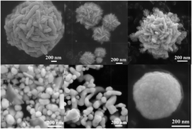
The effect of microwave power and microwave irradiation time on the morphology of the nanoparticles was studied by varying microwave irradiation power as 360 W, 600 W and 800 W for the duration of 2 min and 3 min respectively. The low magnification to high magnification FEG-SEM images (Fig. 2a–c) are the Au nanoparticles synthesized by microwave irradiation at 360 W for 2 min. The FEG-SEM images clearly show formation of spherical microparticles, and the spheres built from smaller, elongated rod-like building blocks. Similarly the FEG-SEM images (Fig. 2d–f) of Au nanoparticles synthesized by microwave irradiation at 360 W for 3 min depict that the formation of spherical hierarchical nanoflowers having size range in between 300 nm to 1 μm takes place.
 |
| Fig. 2 FEG-SEM images (a–c) Au microspheres synthesized at 360 W for 2 min, (d–f) spherical hierarchical Au nanoflowers synthesized at 360 W for 3 min by microwave irradiation. | |
The effect of microwave power on the morphology of Au nanoparticles was further studied by microwave irradiation of the reaction mixture at 600 W for 2 min and 600 W for 3 min. At 600 W for 2 min it was observed that formation of spherical nanoparticles along with rather triangular nanoplates with approximate size of nanoparticles in the range of 100 to 200 nm and the size of triangles in the range of 300 to 500 nm (Fig. 3a–c) takes place. The morphological effect was studied at 600 W for 3 min, the formation of spherical as well as tubular morphology of nanoparticles was observed in FEG-SEM analysis (Fig. 3d–f). On the other hand when the reaction was carried out at 800 W for 2 min and 800 W for 3 min it was observed that well dispersed 3D flower-like morphology of Au nanoparticles emerges in FEG-SEM analysis at 800 W for 2 min (Fig. 4a–c). In addition formation of spherical Au nanoparticles was detected with good dispersion of nanoparticles in FEG-SEM analysis subjected to 800 W microwave powers for 3 min (Fig. 4d–f). Low magnifications to high magnification FEG-SEM micrographs of spherical Au nanoparticles (800 W for 3 min) are shown in Fig. 4d–f. The rapid microwave energy brings drastic change in the morphology. The size and morphology of Au nanoparticles depend on the applied microwave power and time of microwave irradiation. We further tried to check the effect of conventional heating on the morphology of Au nanoparticles and carried out reaction at 80 °C for 1 h using same other reaction parameters. It was observed that formation of random nanoparticles takes place by conventional heating in contrast to the well defined morphologies obtained by microwave irradiation. This indicates that microwave energy plays a crucial role in the formation of Au nanoflowers.
 |
| Fig. 3 FEG-SEM images (a–c) spherical and triangular morphology of Au nanoparticles synthesized at 600 W for 2 min, (d–f) spherical and tubular morphology of Au nanoparticles synthesized at 600 W for 3 min by microwave irradiation. | |
 |
| Fig. 4 FEG-SEM images (a–c) 3D flower-like morphology of Au synthesized at 800 W for 2 min, (d–f) spherical Au nanoparticles synthesized at 800 W for 3 min by microwave irradiation. | |
The Fig. 5 show EDS spectrums of Au nanoparticles synthesized at different microwave powers in watt and different times in minutes such as Fig. 5 (a) 360 W for 2 min (b) 360 W for 3 min (c) 600 W for 2 min (d) 600 W for 3 min (e) 800 W for 2 min (f) 800 W for 3 min. The entire spectrum contains peak for Au and carbon. The carbon peak arises from the carbon tape used for EDS analysis. The EDS analysis indicates the purity of the synthesized nanoparticles. Among all synthesized Au nanoparticles, Au nanoflowers showed good catalytic activity for reduction reaction. Fig. 6a showed the TEM image of Au nanoflowers synthesized at 800 W for 2 min, which gives flower-like morphology. The HRTEM images showed in Fig. 6b and c are part of Au nanoflowers. It shows the interplanar spacing of 0.24 nm and 0.26 nm respectively. The inset image of Fig. 6c is the corresponding fast Fourier transform (FFT) image. A selected area electron diffraction (SAED) pattern of Au nanoflowers shows the Scherrer ring patterns indicate the fcc gold which is nanocrystalline in nature (Fig. 6d). The classical tautomeric equilibrium and then reduction of aqueous AuCl3 in to Au(I) as AuCl2− by chloride transfer under microwave energy can be suggested as a plausible mechanism for the synthesis of Au nanoparticles in DMSO (Fig. 7). Further disproportionation takes place form Au(I) to Au(0) which results in well dispersed and stable nanoparticles.24 In the disproportionation step of Au(I) to Au(0), the growth of Au nanoparticles takes places.
 |
| Fig. 5 EDS spectrum of Au nanoparticles synthesized at (a) 360 W for 2 min (b) 360 W for 3 min (c) 600 W for 2 min (d) 600 W for 3 min (e) 800 W for 2 min (f) 800 W for 3 min by microwave irradiation. | |
 |
| Fig. 6 (a) TEM and (b and c) high resolution TEM images of part of Au nanoflowers synthesized at 800 W for 2 min, inset of (c) is its corresponding fast Fourier transform (FFT) image. (d) Selected area diffraction pattern of Au nanoflowers. | |
 |
| Fig. 7 Plausible reaction mechanisms for the synthesis of Au nanoparticles with DMSO. | |
Catalytic application of Au nanoparticles
It is well-recognized that the size and shape of metal nanoparticles determines the activity and selectivity of the catalyst. The catalytic activity of synthesized Au nanoparticles was checked for the reduction of p-nitrophenol (p-NP) to p-aminophenol (p-AP). The reduction of p-NP to p-AP occurs in presence of metal as catalyst with a reducing agent.25 A general representation of catalytic reduction of p-NP to p-AP using Au nanoparticles is shown in Fig. 8. The p-NP (10 mM) shows absorption peak in the visible region at 320 nm (Fig. 9a), which undergoes a red shift to 400 nm on addition of NaBH4 (0.1 M) due to formation of nitrophenolate anion (Fig. 9b). The color of p-NP changes from light yellow to dark yellow. It can be concluded that the absorption by nitrophenolate anion (p-NP/NaBH4) at 400 nm remains the same even after addition of excess NaBH4 in absence of Au nanoparticles (Fig. 9b), indicates that the reaction cannot proceed without addition of Au nanoparticles. When Au nanoparticles were added to the reaction mixture the reduction reaction completed within 5 min. The reaction was monitored on a UV-Vis spectrophotometer for various time intervals such as 0 min, 1 min, 3 min and 5 min (Fig. 9c). At the end of reduction reaction, after 5 minutes corresponding change in color from pale yellow to colorless was observed which indicates the complete reduction of p-NP to p-AP (Fig. 9d). A rapid catalytic reduction of p-NP to p-AP was observed after the addition of Au (0.394 mg) nanoflowers (synthesized at 800 W for 2 min) as indicated by a gradual decrease in the peak intensity of p-NP at 400 nm and successive increase in the peak intensity of p-AP at 299 nm (Fig. 9c).
 |
| Fig. 8 Reduction of p-nitrophenol to p-aminophenol using Au nanoparticles. | |
 |
| Fig. 9 UV-Visible spectrum of (a) p-nitrophenol (b) nitrophenolate anion (c) reduction of p-nitrophenol to p-aminophenol at different intervals of time (d) change in color before and after the reaction. | |
Recyclability of catalyst decreases the overall cost of process. Recycling study was performed to explore the stability of as synthesized Au nanoflowers (Fig. 10). The response time of Au nanoflowers shows high stability towards reduction reaction. The catalyst exhibiting good catalytic activity and can be recycled up to fifth recycles. In order to check the effect of sodium borohydride on the morphology of Au nanoflowers, the FEG-SEM analysis of recycled Au nanoflowers was done (Fig. 11). The morphology of Au nanoflowers is almost remain same and no aggregation is observed after first recycle (Fig. 11b), whereas the Au nanoflowers get agglomerated after fifth recycle (Fig. 11c).
 |
| Fig. 10 Recyclability study of Au nanoflowers for reduction reaction. | |
 |
| Fig. 11 FEG-SEM images of (a) fresh (b) first recycle (c) fifth recycle of Au nanoflowers. | |
Conclusions
In summary, we demonstrated a simple, rapid and economic microwave approach for the synthesis of various morphologies of Au nanoparticles using DMSO. The DMSO plays dual role in reaction and it act as a reducing agent as well as a capping agent to develop the crystal morphology of Au nanoparticles. Various morphologies of Au nanoparticles such as microspheres with rod like surface (360 W for 2 min), hierarchical flower shape (360 W for 3 min), spherical with triangles (600 W for 2 min), spherical with tubular morphology (600 W for 3 min), nanoflowers (800 W for 2 min), and spherical nanoparticles (800 W for 3 min) were obtained by applying different microwave power in watt and different time duration of microwave irradiation. The applied microwave power and time of microwave irradiation is responsible for the development of specific morphology of Au nanoparticles. The as synthesized Au nanoflowers synthesized at 800 W for 2 min have been found to be highly efficient catalyst for the reduction of p-NP to p-AP.
Acknowledgements
Author MAB is thankful to Council of Scientific and Industrial Research (CSIR), New Delhi, India for providing senior research fellowship (SRF). We are thanking to Department of Science and Technology (DST), India for financial support for this work under Nano Mission Project no. SR/NM/NS-1097/2011. We are thankful to SAIF, IIT Bombay for providing HRTEM facility.
References
- N. Li, P. Zhao and D. Astruc, Angew. Chem., Int. Ed., 2014, 53, 1756 CrossRef CAS PubMed.
- X. Huang, S. Neretina and M. A. El-sayed, Adv. Mater., 2009, 21, 4880 CrossRef CAS PubMed.
-
(a) C. Ziegler and A. Eychmuller, J. Phys. Chem. C, 2011, 115, 4502 CrossRef CAS;
(b) D. A. Walker, K. P. Browne, B. Kowalczyk and B. A. Grzybowski, Angew. Chem., Int. Ed., 2010, 49, 6760 CrossRef CAS PubMed;
(c) M. R. Langille, M. L. Personick, J. Zhang and C. A. Mirkin, J. Am. Chem. Soc., 2012, 134, 14542 CrossRef CAS PubMed.
-
(a) M. Eguchi, D. Mitsui, H.-L. Wu, R. Sato and T. Teranishi, Langmuir, 2012, 28, 9021 CrossRef CAS PubMed;
(b) L. Scarabelli, M. Coronado-Puchau, J. J. Giner-Casares, J. Langer and L. M. Liz-Marzan, ACS Nano, 2014, 8, 5833 CrossRef CAS PubMed.
-
(a) Y. Huang, A. R. Ferhan, Y. Gao, A. Dandapat and D.-H. Kim, Nanoscale, 2014, 6, 6496 RSC;
(b) L. Chen, F. Ji, Y. Xu, L. He, Y. Mi, F. Bao, B. Sun, X. Zhang and Q. Zhang, Nano Lett., 2014, 14, 7201 CrossRef CAS PubMed;
(c) S. Hong, K. L. Shuford and S. Park, Chem. Mater., 2011, 23, 2011 CrossRef CAS;
(d) P. Pienpinijtham, X. X. Han, T. Suzuki, C. Thammacharoen, S. Ekgasit and Y. Ozaki, Phys. Chem. Chem. Phys., 2012, 14, 9636 RSC.
-
(a) J. Zhang, J. Du, B. Han, Z. Liu, T. Jiang and Z. Zhang, Angew. Chem., Int. Ed., 2006, 45, 1116 CrossRef CAS PubMed;
(b) Y. Chen, S. Milenkovic and A. W. Hassel, Nano Lett., 2008, 8, 737 CrossRef CAS PubMed.
- M. L. Personick, M. R. Langille, J. Zhang, N. Harris, G. C. Schatz and C. A. Mirkin, J. Am. Chem. Soc., 2011, 133, 6170 CrossRef CAS PubMed.
-
(a) T. S. Sreeprasad, A. K. Samal and T. Pradeep, Langmuir, 2008, 24, 4589 CrossRef CAS PubMed;
(b) B. P. Khanal and E. R. Zubarev, J. Am. Chem. Soc., 2008, 22, 12634 CrossRef PubMed.
-
(a) X. Lu, L. Au, J. Mclellan, Z.-Y. Li, M. Marquez and Y. Xia, Nano Lett., 2007, 7, 1764 CrossRef CAS PubMed;
(b) M. A. Mahmoud and M. A. El-Sayed, Nano Lett., 2011, 11, 946 CrossRef CAS PubMed;
(c) W. Wang, T. Yan, S. Cui and J. Wan, Chem. Commun., 2012, 48, 10228 RSC.
- F. Kim, S. Connor, H. Song, T. Kuykendall and P. Yang, Angew. Chem., Int. Ed., 2004, 43, 3673 CrossRef CAS PubMed.
- M. Eguchi, D. Mitsui, H.-L. Wu, R. Sato and T. Teranishi, Langmuir, 2012, 28, 9021 CrossRef CAS PubMed.
- C. L. Nehl, H. Liao and J. H. Hafner, Nano Lett., 2006, 6, 683 CrossRef CAS PubMed.
-
(a) J.-U. Kim, S.-H. Cha, K. Shin, J. Y. Jho and J.-C. Lee, Adv. Mater., 2004, 16, 459 CrossRef CAS PubMed;
(b) M. Wirtz and C. R. Martin, Adv. Mater., 2003, 15, 455 CrossRef CAS PubMed;
(c) J.-H. Ryu, S. Park, B. Kim, A. Klaikherd, T. P. Russell and S. Thayumanavan, J. Am. Chem. Soc., 2009, 131, 9870 CrossRef CAS PubMed.
- T. Huang, F. Meng and L. Qi, Langmuir, 2010, 26, 7582 CrossRef CAS PubMed.
-
(a) L. Wang, C.-H. Liu, Y. Nemoto, N. Fukata, K. C.-W. Wu and Y. Yamauchi, RSC Adv., 2012, 2, 4608 RSC;
(b) S. Yi, L. Sun, S. C. Lenaghan, Y. Wang, X. Chong, Z. Zhang and M. Zhang, RSC Adv., 2013, 3, 10139 RSC;
(c) B. K. Jena and C. R. Raj, Chem. Mater., 2008, 20, 3546 CrossRef CAS;
(d) A.-J. Wang, Y.-F. Li, M. Wen, G. Yang, J.-J. Feng, J. Yang and H.-Y. Wang, New J. Chem., 2012, 36, 2286 RSC.
- M. A. Bhosale and B. M. Bhanage, RSC Adv., 2014, 4, 15122 RSC.
-
(a) M. A. Bhosale, K. D. Bhatte and B. M. Bhanage, Powder Technol., 2013, 235, 516 CrossRef CAS PubMed;
(b) M. A. Bhosale, T. Sasaki and B. M. Bhanage, Catal. Sci. Technol., 2014, 4, 4274 RSC;
(c) K. D. Bhatte, P. Tambade, S. Fujita, M. Arai and B. M. Bhanage, Powder Technol., 2010, 203, 415 CrossRef CAS PubMed;
(d) M. A. Bhosale and B. M. Bhanage, Adv. Powder Technol., 2015, 26, 422 CrossRef CAS PubMed.
- F.-K. Liu, C.-J. Ker, Y.-C. Chang, F.-H. Ko, T.-C. Chu and B.-T. Dai, Jpn. J. Appl. Phys., 2003, 42, 4152 CrossRef CAS.
- C.-H. Cui, H.-H. Li and S.-H. Yu, Chem. Commun., 2010, 46, 940 RSC.
- S. K. Seol, D. Kim, S. Jung and Y. Hwu, Mater. Chem. Phys., 2011, 131, 331 CrossRef CAS PubMed.
- D. Guin, S. V. Manorama, S. Radha and A. K. Nigam, Bull. Mater. Sci., 2006, 29, 617 CrossRef CAS PubMed.
- J. Liu, N. Ruffini, P. Pollet, V. Llopis-Mestre, C. Dilek, C. A. Eckert, C. L. Liotta and C. B. Roberts, Ind. Eng. Chem. Res., 2010, 49, 8174 CrossRef CAS.
- G. Rodriguez-Gattorno, D. Diaz, L. Rendon and G. O. Hernandez-Segura, J. Phys. Chem. B, 2002, 106, 2482 CrossRef CAS.
- W. J. Pevelera and I. P. Parkin, RSC Adv., 2013, 3, 21919 RSC.
-
(a) Y.-C. Tsao, S. Rej, C.-Y. Chiu and M. H. Huang, J. Am. Chem. Soc., 2014, 136, 396 CrossRef CAS PubMed;
(b) M. H. Rashid, R. R. Bhattacharjee, A. Kotal and T. K. Mandal, Langmuir, 2006, 22, 7141 CrossRef CAS PubMed.
|
This journal is © The Royal Society of Chemistry 2015 |
Click here to see how this site uses Cookies. View our privacy policy here.