
Antibacterial textiles functionalized by layer-by-layer assembly of polyelectrolytes and TiO2 photocatalyst†
Gaëlle Carré*ab,
Laurent Garnierb,
Janina Moeller-Siegerta,
Jean-Pierre Giesb,
Valérie Kellera,
Philippe Andréb and
Nicolas Keller*a
aInstitut de Chimie et Procédés pour l'Energie, l'Environnement et la Santé (ICPEES), CNRS and Strasbourg University, 25 rue Becquerel, 67087 Strasbourg Cedex, France. E-mail: nkeller@unistra.fr; carregaelle1@gmail.com
bLaboratoire de Biophotonique et Pharmacologie, CNRS and Strasbourg University, 74 route du Rhin, 67400 Illkirch, France
First published on 22nd April 2015
Abstract
The sprayed layer-by-layer assembly of TiO2 photocatalyst has been efficiently used for functionalizing polyester textiles and elaborating solar light active antibacterial textiles. Polyethylenimine (PEI) and polyanionic poly(styrene sulfonate) (PSS) polyelectrolytes have been used as polycation and polyanion layers, in alternation with dispersions of negatively- and positively-charged TiO2 particles, respectively. A synergistic effect resulting from both biocidal photocatalytic activity of TiO2 layers and biocidal activity of PEI layers within the multilayer system was put forward for explaining the high self-decontaminating activity of (PEI/TiO2)n functionalized textiles, 5![[thin space (1/6-em)]](https://www.rsc.org/images/entities/char_2009.gif) log of cell viability reduction being achieved with a single TiO2 layer at 50 μg cm−2. The sole solar light photocatalytic antibacterial activity of multilayer textiles was determined by masking the biocidal activity of PEI layers through the controlled assembly of PEI/(PSS/TiO2)n multilayers.
log of cell viability reduction being achieved with a single TiO2 layer at 50 μg cm−2. The sole solar light photocatalytic antibacterial activity of multilayer textiles was determined by masking the biocidal activity of PEI layers through the controlled assembly of PEI/(PSS/TiO2)n multilayers.
1. Introduction
Textile substrates are known to be suitable environments for microbial growth under certain conditions. Numerous microorganisms can rapidly multiply at the textile surface, so that they can be responsible for fabric deterioration,1 staining or unpleasant odors,2,3 and even can play a role in acquisition and transmission of pathogens.4–8 Depending on operating parameters and on the inoculum load, survival of Gram negative bacteria such as e.g. Escherichia coli, Klebsiella pneumoniae or Pseudomonas aeruginosa on textiles has been observed from 2 h until more than 60 days, while periods of months have been reported for Staphylococci and Enterococci on polyester.9–11 Persistence and lifespan of microorganisms on textiles depend on multiple factors, including the textile composition, the type of bacterial species and the environment.Thereby, within a global approach dealing with the design of smart advanced textiles, the elaboration of antimicrobial or self-disinfecting textiles is especially receiving growing interest, aiming at preventing bacterial growth on the textiles.
Thus, many antimicrobial agents have been used for providing bactericidal properties to textiles, such as quaternary ammonium, triclosan, polyhexamethylene biguanide, zinc pyrithione, Ag nanoparticles or salts,12–15 the most studied approach dealing nowadays with the use of Ag nanoparticles.16,17 However, developing new disinfection approaches is required due to fast adaptation of bacterium and development of metal-resistant strains,18 and applying anti-bacterial inorganic nanoparticles would be an alternative for opening up new opportunities for antimicrobial textiles.19,20
Oxidative heterogeneous photocatalysis is a promising disinfection approach based on the irradiation of a semi-conductor catalyst by light with energy matching or greater than its band gap. The resulting light absorption enables the generation of electron and hole carriers and subsequently of highly oxidative species – Reactive Oxygen Species (ROS) – at the catalyst surface, responsible for inducing damages to bacterial cells leading to cell death.21–27 Titanium dioxide (TiO2) is the most used photocatalyst under UV light, thanks to high quantum yield, both chemical and light stability, non toxicity as well as low cost for commercial standards.21,24,25
Even if finishing techniques used in textile industry can to a certain extent be adapted for immobilizing TiO2 onto textiles, they suffer from using additives in large amount like surfactants or thickening agents, that block the reactant access to the TiO2 nanoparticles. Alternative approaches have thus been developed for synthesizing and immobilizing nanostructured oxides such as TiO2 onto textile fibers.28–34 One can cite e.g. sol–gel methods, direct current reactive magnetron sputtering, deposition of TiO2 through linking with spacer agents, as well as deposition from TiO2 suspensions. In some of them, TiO2 was associated to Ag as antibacterial agent.35–37
Layer-by-Layer assembly (LbL) is a multilayer approach based on electrostatic interaction of alternative layers of oppositely-charged materials used for building polyelectrolyte and multi-material layered films on substrates via a single process.38–40 It has been used recently for building TiO2 photo-catalytic films for environmental or detoxification purpose.41–43 This approach was scarcely applied to textiles,44,45 and only few works reported on LbL functionalization of commercial textiles by TiO2 for providing them self-cleaning efficiency towards chemicals.46–50 In particular, we proved that cotton/polyamide textiles with photocatalytic activity in the degradation of chemicals, could be obtained by substituting the polyanion layer by a dispersion of negatively-charged TiO2 particles in alternation with polyethylenimine (PEI) as polycation layer.47
Thus, we report herein on new LbL-functionalized photocatalytic multilayer textiles designed for exhibiting high self-decontaminating activity towards bacteria. The underlying strategy implemented here was to take advantage of both biocidal photocatalytic activity of TiO2 nanoparticle layers and biocidal activity of alternative PEI polyelectrolyte layers – known to have good antibacterial properties51 – for providing a synergistic effect within the multilayer system resulting in the elaboration of high activity anti-bacterial textiles.
2. Experimental
2.1. Materials and textiles
Aeroxide® TiO2 P25 was supplied by Evonik (Germany, 20% rutile – 80% anatase crystalline form, with a specific surface area of 50 m2 g−1).52 Synthetic 100% polyester textiles (PES) were delivered by Ouvry (France). They were washed with hot water, rinsed with distilled water and dried at 100 °C overnight.The raw PES fiber textile was first submitted to a mild hydrolysis using KOH solution for generating negatively charged surface oxygen species, allowing to strengthen the interaction with the first positively charged PEI layer and consequently improving the adhesion of the first layer on the fibers. The textile was immersed into 0.5 M KOH aqueous solution for 30 min under soft orbital shaking (150 rpm). The fabric was then rinsed with distilled water until neutral pH is reached and then dried at 100 °C for 5 min.
In this study we used polyester textiles which are frequently found in a hospital environment e.g. drapes. Moreover, as compared to other textiles (cotton or nylon clothes), polyester has binding affinity towards bacteria.53
2.2. Sprayed layer-by-layer coatings
The (PEI/TiO2)n layer-by-layer functionalization consists of alternate layers of polycationic PEI (BASF Lupasol/Water free, MW ∼ 25000 g mol−1) in aqueous solution (8 g L−1) and TiO2 P25 at 5 g L−1 in water/ethanol (50:50 in volume) at pH 9 (adjusted with a few drops of NaOH solution to ensure negatively charged surface of TiO2 as its isoelectrical point is around 6.5). The polyelectrolyte solution and the TiO2 dispersion were filled into spray bottles (Air-boy from Carl Roth, 400 μm nozzle) and manually pressurized to about 1 bar. For the deposition, the dried PES fabrics were horizontally fixed onto metallic grids, and PEI solutions and TiO2 nanoparticle dispersions were sprayed on from about 15 to 20 cm distance in crossed lines. Each spraying duration was about 20 s. After each layer, the fabric was allowed to equilibrate with the reactant for 10 min while still laying and then rinsed abundantly vertically with distilled water to eliminate any excess. After rinsing, the fabrics were dried at 100 °C for 5 min before the next bilayer was applied. This sequence was repeated until the number of PEI/TiO2 bilayers was achieved. In this study, we used n = 1, 5 and 10 bilayers. After the last TiO2 layer, the functionalized textiles were dried at 130 °C for 30 min.
Anionic poly(styrene sulfonate) (PSS) was also used as polyelectrolyte for functionalizing the PES textiles with TiO2. In that case, an interfacial layer of PEI was added before building the (PSS/TiO2)n bilayers. After the first PEI layer, the textile was rinsed with distilled water and dried for 5 min at 100 °C. Then, the (PSS/TiO2)n bilayers consisted of alternate layers of PSS (MW ∼ 70000 g mol−1, Sigma-Aldrich) at 1 g L−1 in distilled water rectified at pH 2.5 with HNO3, and TiO2 P25 at 10 g L−1 in water/ethanol (50:50 in volume) at pH 2.5 (adjusted with HNO3) to ensure positively charged surface of TiO2. Our previous works have reported that pH of 2.5 was ideal for maintaining a strong positive charge on the nanoparticles and sufficient dissociation of the PSS.43 This (PSS/TiO2)n sequence was repeated until the number of bilayers was achieved. In this study, we used n = 1, 5 and 10 bilayers.
2.3. Characterization techniques
The TiO2 loading on the textiles was determined by Inductively Coupled Plasma Atomic Emission Spectroscopy (ICP-AES). Scanning electron microscopy (SEM) was performed on a Jeol JSM-6700F (1–10 kV), with metallization of samples with gold.2.4. Bacterial strains and growth media
Antimicrobial activity of functionalized textiles has been determined against two Gram negative bacteria, E. coli and P. aeruginosa. Before each experiment, one loop full of E. coli ATCC 8739 or P. aeruginosa CIP A22 was seeded on a slant of tryptic soy agar (TSA) (BioRad), and grown aerobically at 37 °C for 24 h. Bacterial inoculum was monitored by setting the culture optical density at an absorbance wavelength of 620 nm (OD620) at 0.156, corresponding to 108 CFU mL−1. These populations were diluted in Mueller-Hinton broth (MHB) to obtain an inoculum of 106 CFU mL−1.2.5. Assessment of the bactericidal activity of PEI and PSS polyelectrolytes against E. coli and P. aeruginosa bacteria
log. Thus, the initial inoculum at 106 CFU mL−1 was diluted in sterile physiological water to obtain a 3log reduction. 10 μL of each dilution was deposited on Mueller Hinton agar (MHA) and incubated for 24 h at 37 °C. This plate was then kept in the fridge during 24 h. It used to observe the 3log reduction in order to determine MBC.Bacterial cultures treated by PEI or PSS polyelectrolytes showing no growth in the MIC tests were used for assessing MBC. 10 μL bacterial cultures treated by polyelectrolytes showing no growth were plated onto MHA plate and incubated at 37 °C for 24 h. This plate was compared with the one previously made for the initial inoculum. The MBC plate (at the lowest dilution) containing equal or less than number colony compared to the 3log reduction plate was defined as the MBC.
2.6. Determination of the antimicrobial efficiency of functionalized textiles
The antibacterial properties of functionalized textiles against E. coli and P. aeruginosa were assessed according to a modified protocol derived from that of the BS ISO27447 test method.55Self-disinfecting tests under solar light were performed within an ATLAS Suntest XLS+ testing chamber equipped with Xe arc lamp and daylight filter simulating outdoor solar light, inside which 2 × 2 cm2 sized textiles pieces were located and submitted to a total irradiance of 242 W m−2 including 226 W m−2 of visible light and 16 W m−2 of UV-A. The spectral distribution is reported in ESI S1.† The temperature was regulated at 33–35 °C – an adequate temperature to survival of the selected bacteria – through a water-cooled plate. To avoid any desiccation, the textiles were pre-humidified with 70 μL of sterile physiological water (NaCl, 9 g L−1). 100 μL of bacterial inoculum at 108 CFU mL−1 were deposited on each textile, corresponding to a bacteria load of 107 CFU per textile.
After test under light or in the dark, each textile was introduced into a stomacher bag containing 10 mL of sterile physiological water supplemented with 2% of Tween 80 (Sigma-Aldrich, France) – acting as non-ionic surfactant and solubilizing agent – and was kneaded for 2 min (Stomacher Mayo, Homogenius HG400 V). After this step serial decimal dilution were made in physiological water. 1 mL of each dilution were added to 19 mL of nutrient agar media in surfusion (55 °C) containing 3 g L−1 of beef extract (Difco), 5 g L−1 of pancreatic digest of casein (Biokar diagnostics) and 15 g L−1 of bacteriologic agar type A (Biokar diagnostics). The plates were incubated for 24 h at 37 °C and bacteria included in the agar media or growth at the surface were numerated.
The antibacterial efficiency of the textiles was assessed by numerating the bacteria in terms of Colony Forming Units (CFU) after solar light exposure, and determining the bacteria survival over the textiles. Controls have been performed in the dark as well. The data are presented as the mean ± standard error of the mean of at least three independent experiments. Statistical analysis was performed by the Student's t test (Prism 5, GraphPad Software). A value of p < 0.05 (*), p < 0.01 (**), p < 0.001 (***) or p < 0.0001 (****) was considered to be statistically significant.
3. Results and discussion
3.1. Characterization of functionalized textiles
Fig. 1 shows SEM images of PES–KOH textiles functionalized with PEI/TiO2 and PSS/TiO2 multilayers at the different steps of the film building, as well as the influence of the number of bilayers applied for both multilayer systems, on the TiO2 wt content and the TiO2 surface density. | ||
| Fig. 1 SEM images of (A and C) bare PES–KOH, (B) PES–KOH/PEI/(PSS/TiO2)10, (D) PES–KOH/(PEI/TiO2)1, (E) PES–KOH/(PEI/TiO2)5, (F) PES–KOH/(PEI/TiO2)10 textiles (G) PES–KOH/PEI/(PSS/TiO2)5 and (H) PES–KOH/PEI/(PSS/TiO2)10; (I) influence on the TiO2 wt content and the TiO2 surface density, of the number of bilayers applied on the PES–KOH/(PEI/TiO2)n and the PES–KOH/PEI/(PSS/TiO2)n textiles. Error bars were calculated from measurements over three samples. | ||
Previous works evidenced that, at full coverage of the substrates, the average thickness of the first PEI layer determined by ellipsometry was 11.5 Å.56
Fig. 1-I shows that the TiO2 wt content – and thus the TiO2 surface density – linearly increased with the number of bilayers applied on the fibers in the case of the PEI/(PSS/TiO2) multilayer system, with a TiO2 wt content of 2.5 wt% for 10 bilayers. By contrast, the TiO2 wt content saturated more rapidly at 1.8 wt% for 10 bilayers in the case of the (PEI/TiO2) system. Although not studied on textiles, a linear increase in the film thickness in the 20–30 bilayer range is usually observed for PSS/TiO242,57 as well as PEI/TiO258 photocatalytic systems on glass model substrates. The saturation observed here for PEI/TiO2 bilayer systems differed from the linearity reported by Rojas-Blanco et al.58 and could result from a possibly different building up of the film on woven irregular and curved PES fibers vs. flat model substrates when using PEI for structuring and elaborating the layered film rather than only as first anchorage/glue layer on the PES fibers.
3.2. Antimicrobial activity of polyelectrolytes in solution
The antimicrobial activity of PEI polycation and PSS polyanion was determined against E. coli and P. aeruginosa bacteria by measuring growth inhibition. It is well admitted that compounds with a MBC/MIC ratio ≤ 4 or >4 are considered as bactericidal or bacteriostatic, respectively.54,59 For both bacteria, the PEI polycation displayed similar values of MIC and MBC, at 2.5 g L−1 (Fig. 2a and c), so that PEI with MW ∼ 25000 g mol−1 was evidenced as bactericidal agent against E. coli and P. aeruginosa. This confirmed works performed with PEI over Gram negative and Gram positive bacteria such as E. coli, P. aeruginosa, S. aureus and S. epidermidis.60–65 It was proposed that the bactericidal effect results from the fact that hydrophobic polycationic chains were able to traverse and irreparably damage the bacterial cellular membrane, by interacting with and disrupting negatively charged bacterial cell membrane followed by release of K+ ions and other cytoplasmic constituents.61,62
 | ||
| Fig. 2 Antibacterial effect of PEI (a and c) and PSS (b and d) in g L−1 against E. coli (a and b) and P. aeruginosa H21 (c and d) bacteria determined by measuring the optical density at 620 nm. | ||
By contrast, MBC and MIC values for the PSS polyanion were higher than 8 g L−1 for both bacteria (Fig. 2b and d for MIC, data not shown for MBC). Thus, neither a bacteriostatic nor a bactericidal effect was evidenced for PSS.
3.3. Antibacterial activity of textiles functionalized with (PEI/TiO2)n
Fig. 3 shows the antibacterial effect against E. coli achieved after 60 min under solar light over the PES textile functionalized with PEI/TiO2 multilayers at the different steps of the multilayered film building, i.e. with one single PEI layer (Fig. 3C), and with 1 (Fig. 3D) or 5 bilayer(s) (Fig. 3E) of (PEI/TiO2). Controls have been performed in the dark. | ||
| Fig. 3 Antibacterial effect against E. coli achieved after 60 min under solar light and in the dark over the PES textile functionalized with PEI/TiO2 multilayers at the different steps of the film building, i.e. PES (A), PES–KOH (B), PES–KOH/PEI (C), PES–KOH/(PEI/TiO2)1 (D), PES–KOH/(PEI/TiO2)5 (E). | ||
First, 2.4 × 10⁶ and 6.8 × 10⁶ CFU of E. coli have been numerated over the textiles in the dark, as-it (Fig. 3A) and after KOH pre-treatment (Fig. 3B), respectively, corresponding to about 20–70% of the 107 CFU inoculums seeded on the textiles. This confirmed the expected neutrality against E. coli survival of the KOH treatment of the fiber surface, and further evidenced the efficiency of the bacteria recovery method used in comparison to usual surface sampling methods such as e.g. the RODAC contact plate method, swabbing techniques or wash-off methods.66 Indeed, many studies reported that the amount of microorganisms recovered through the RODAC plate technique was much lower than the initial applied concentration, as evidenced by Rabuza et al. with recovery ratios lower than 10−5.66 The method adapted from the BS ISO27447 standard method is thus adequate for extracting and further quantifying microorganisms which may penetrated deep into the structure of PES fiber textile, in contrary to the RODAC method. In addition, Fig. 3A and B confirmed the expected neutrality against E. coli of the exposure to solar light for 60 min of contaminated bare textiles, whether the textiles were pretreated or not by KOH.
The functionalization of the textile with a single layer of PEI resulted in a significant reduction of the cell viability in the dark as well as under light, with 1.5 × 10⁵ CFU per textile being measured in both cases (Fig. 3C), and corresponding to a bactericidal activity close to 2log as compared to the control textile (Fig. 3A). A stronger reduction of the cell viability with a bactericidal activity close to 5log was recorded in the dark and under light over the textile functionalized with a single PEI/TiO2 bilayer (Fig. 3D). Further, the textile functionalized with 5PEI/TiO2 bilayers exhibited a reduction of E. coli viability greater than 6log in the dark as well as under light (Fig. 3E), the CFU quantity being relatively close to the detection threshold which is around 1.0 × 101 CFU per textile.
We proposed here that this high activity resulted from a synergistic antimicrobial activity between PEI and TiO2. First, it has been reported that TiO2 exhibited an antibacterial activity already in the dark,67 by impacting key cellular components of bacteria. This effect was amplified under illumination through the production of ROS such as O2˙− superoxide radical, causing lipid peroxidation enhancing membrane fluidity and disrupting cell integrity. Already during the cytotoxic treatment in the dark with TiO2 – and amplified under UV-A illumination – two-dimensional electrophoresis proteomic analysis associated performed on the whole proteome of E. coli and coupled to mass spectrometry protein analysis evidenced that the main degraded proteins were porins, proteins involved in oxidative stress response, chaperone proteins, and to a lesser extent proteins involved in the transport and metabolism of different molecules such as inorganic ions, lipids, carbohydrates and amino acids. To a lesser extent, proteins involved in energy production and conversion, as well as in translation regulation were also identified.67 Thus, it is likely that the cytotoxicity of TiO2 could directly enhance the biocidal effect of the PEI polyelectrolyte by strongly reducing the ability of the cells to respond to oxidative stress or to regulate environmental stress.
This synergy effect could also be envisaged inversely, considering that the biocidal activity of PEI against E. coli could directly enhance the impact of TiO2 in the dark by modifying the organization of the bacterial outer membrane. It could also be enhanced under illumination. Indeed, PEI – like more generally polycationic agents – induces the reorganization of the bilayer structure with the alignment of lipid headgroups perpendicular to the membrane.68 This increases the permeability of the bacterial outer membrane to solutes that are normally unable to cross the outer membrane,62,69,70 like this was already proposed for antibiotics,64 so that this could also increase its permeability to the O2˙− superoxide radical ROS. Among the different ROS generated at the surface of TiO2, it has been proposed that the O2˙− superoxide radical ROS was playing an important role71 due to a longer lifetime at the water–TiO2 interface when compared to other active ROS such as OH˙ hydroxyl radicals.72 This extended lifetime would allow O2˙− to cross the membrane for damaging directly inner cellular components (or for further reacting to form other active ROS), while, by contrast, ROS such as the highly active OH˙ will attack first the outer cellular components due to a shorter lifetime.
This bactericidal activity of the textile functionalized with a single bilayer of PEI/TiO2 turns out to be very promising from an applicative point of view because it requires only the application of a single layer of TiO2 nanoparticles to get 5log of cell viability reduction. In this case, the textile was functionalized with only 0.4 wt% of TiO2, corresponding to a TiO2 surface density of 50 μg cm−2. Such a so small content confers to the functionalized textile a very good stability, necessary for targeting a realistic application as self-disinfecting textiles.
3.4. Antibacterial activity of textiles functionalized with PEI/(PSS/TiO2)n
Truly considered, the investigation of the PEI/TiO2 systems on the textile revealed that the high bactericidal activity shown by the multilayers built themselves with bactericidal PEI as positively charged polyelectrolyte did not allow to evidence the real contribution of photocatalysis on light-activated TiO2 within the overall activity of the multilayer system. So, the sole bactericidal activity due to the photocatalysis was isolated by substituting positively charged PEI by positively charged PSS within the multilayers after the first bactericidal PEI layer was applied on the textile.Fig. 4a shows the antibacterial efficiency against E. coli bacteria achieved after 60 min under solar light over the PES textile functionalized with PEI/(PSS/TiO2)n multilayers at the different steps of the film building, i.e. with one single PEI layer, subsequently with one additional PSS layer, and with 1, 5 or 10 bilayer(s) of (PSS/TiO2). Controls have been also performed in the dark.
 | ||
| Fig. 4 Antibacterial effect against (a) E. coli and (b) P. aeruginosa bacteria achieved after 60 min under solar light and in the dark over the PES textile functionalized with PEI/(PSS/TiO2)n multilayers at the different steps of the film building, i.e. PES–KOH (A), PES–KOH/PEI (B), PES–KOH/PEI/PSS (C), PES–KOH/PEI/(PSS/TiO2)1 (E), PES–KOH/PEI/(PSS/TiO2)5 (F); PES–KOH/PEI/(PSS/TiO2)10 (G). | ||
It shows that the application of a single layer of PSS (Fig. 4a-C) on the first PEI layer allowed a partial restoration of the cell viability to be obtained, whether the textiles was irradiated or not. Indeed, the PSS polyelectrolyte did not evidence any cytotoxic effect, so that applying an additional PSS layer partly masked the cytotoxicity of the first PEI layer (Fig. 4a-B), and resulted in an increase of about 1log of the cell viability compared to the textile functionalized only with the cytotoxic PEI layer. Increasing the number of PSS layers – and thus of PSS/TiO2 bilayers – led to an increase in the E. coli viability in the dark, with a complete viability restoration achieved already for 5 bilayers PSS/TiO2 (Fig. 4a-F).
By contrast, masking the cytotoxicity of the first internal PEI layer by applying additional PSS/TiO2 bilayers, allowed to evidence the antibacterial effect resulting under solar light solely from the photocatalytic TiO2 layers. Indeed, an increase in the antimicrobial effect under solar light was dependent on the number of PSS/TiO2 bilayers applied. 9.7 × 10⁵, 1.9 × 10⁵ and 9.2 × 10³ CFU per textile were measured after 60 min test under solar light on the textiles functionalized with 1 (Fig. 4a-E), 5 (Fig. 4a-F) and 10 (Fig. 4a-G) bilayers of PSS/TiO2, respectively, so that the E. coli viability was progressively reduced by increasing the number of bilayers, and that a antibacterial photocatalytic activity close to 3log was achieved for 10 PSS/TiO2 bilayers.
This behavior with increasing the number of PSS/TiO2 bilayers was confirmed by studying the antibacterial activity of the PEI/(PSS/TiO2)n multilayer system against P. aeruginosa bacteria (Fig. 4b). A single layer of PEI induced more than 2log reduction whether the textile was irradiated or not (Fig. 4b-B). Addition of a single layer of PSS on the first layer of PEI (Fig. 4b-C) allowed the partial restoration of the cell viability (7 × 10⁵ CFU per textile vs. 2.6 × 10⁴ CFU per textile on the PES textile with one PEI layer). The higher restoration of cell viability was then obtained when applying one bilayer of PSS/TiO2 (Fig. 4b-E), i.e. 1.5 × 10⁶ CFU per textile vs. 7.3 × 10⁶ CFU per textile on the naked textile. Increasing further the number of PSS/TiO2 bilayers applied resulted in a progressive decrease of the cell viability on the solar light irradiated textiles, and 1.4 × 10⁵, 2.3 × 10³ and 1.0 × 101 CFU per textile were measured on the textiles functionalized with 1 (Fig. 4b-E), 5 (Fig. 4b-F) and 10 (Fig. 4b-G) bilayers of PSS/TiO2, respectively, after 60 min of irradiation, corresponding to antibacterial activities close to 2, 4 and 6log, respectively. So, similarly to the case of E. coli, the cytotoxicity of the first internal PEI layer could be masked by applying additional PSS/TiO2 bilayers, so that the antibacterial effect resulting solely from the solar light active photocatalytic TiO2 layers – directly dependent on the number of PSS/TiO2 bilayers – was evidenced.
The results obtained against E. coli and P. aeruginosa bacteria demonstrated the solar light photocatalytic antibacterial efficiency of the textiles functionalized with PSS/TiO2 bilayers. In the case of E. coli, this antibacterial activity could thus in a first approximation correspond to the sole photocatalytic anti-bacterial activity of PES textile functionalized with PEI/TiO2 bilayers, so that the PEI/(PSS/TiO2)n multilayer system was suitable for presenting the kinetic of the photocatalytic self-disinfection of functionalized textiles against E. coli bacteria (Fig. 5).
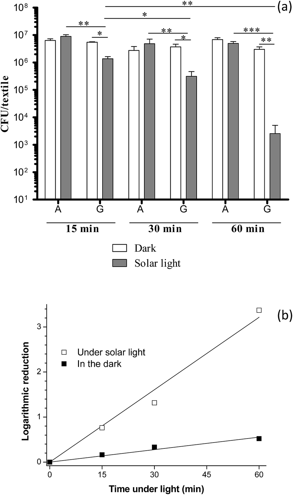 | ||
| Fig. 5 (a and b) Kinetic of the antibacterial effect against E. coli bacteria under solar light of the multilayered PES–KOH/PEI/(PSS/TiO2)10 textile (G). (A) Corresponds to control performed over the bare PES–KOH textile. Kinetic has been determined in the dark as well. | ||
4. Conclusions
Solar light active antibacterial textiles have been prepared by functionalization of polyester textiles with sprayed layer-by-layer assembly of TiO2 photocatalyst using PEI polyelectrolyte as polycation layer in alternation with a dispersion of negatively-charged TiO2 particles, or PSS polyelectrolyte as polyanion layer in alternation with a dispersion of positively-charged TiO2 particles.The high self-decontaminating activity of (PEI/TiO2)n functionalized textiles was assigned to a synergistic effect within the multilayer system resulting from the association of both biocidal photocatalytic activity of TiO2 layers and biocidal activity of PEI polyelectrolyte layers. The application of a single layer of TiO2 at 50 μg cm−2 to get 5log of cell viability reduction turns out to be very promising for targeting a realistic application as self-disinfecting textiles.
PEI/(PSS/TiO2)n functionalized textiles were of high interest for evidencing the sole solar light photocatalytic antibacterial activity of multilayered textiles by masking the biocidal activity of PEI.
Acknowledgements
The authors are grateful to DGA (Direction Générale de l'Armement) and Alsace regional council for financially supporting this work in the frame of the PhD grant of G. Carré. The authors acknowledge the support of the French Agence Nationale de la Recherche (ANR) under reference ANR-09-SECU-10. We thank Th. Romero for performing SEM analysis.Notes and references
- J. Szostak-Kotowa, Int. Biodeterior. Biodegrad., 2004, 53, 165 CrossRef CAS.
- C. Callewaert, E. De Maeseneire, F. M. Kerckhof, A. Verliefde, T. Van de Wiele and N. Boon, Appl. Environ. Microbiol., 2014, 80, 6611 CrossRef CAS PubMed.
- C. Rodriguez, A. Di Cara, F. N. R. Renaud, J. Freney, N. Horvais, R. Borel, E. Puzenat and C. Guillard, Catal. Today, 2014, 230, 41 CrossRef CAS PubMed.
- J. M. Nordstrom, K. A. Reynolds and C. P. Gerba, Am. J. Infect. Control, 2012, 40, 539 CrossRef PubMed.
- S. Fijan and S. Sostar-Turk, Int. J. Environ. Res. Public Health, 2012, 9, 3330 CrossRef PubMed.
- A. Toomey, in Medical and Healthcare Textiles, ed. S. C. Anand, J. F. Kennedy, M. Miraftab and S. Rajendran, Woodhead Publishing, 2010, pp. 357–367 Search PubMed.
- A. Mitchell, M. Spencer and C. Edmiston, J. Hosp. Infect., 2015 DOI:10.1016/j.jhin.2015.02.017.
- G. Borkow and J. Gabbay, Med. Hypotheses, 2008, 70, 990 CrossRef CAS PubMed.
- A. N. Neely, J. Burn Care Rehabil., 2000, 21, 523 CrossRef CAS PubMed.
- A. N. Neely and M. P. Maley, J. Clin. Microbiol., 2000, 38, 724 CAS.
- V. J. Colclasure, T. J. Soderquist, T. Lynch, N. Schubert, D. S. McCormick, E. Urrutia, C. Knickerbocker, D. McCord and J. H. Kavouras, Am. J. Infect. Control, 2015, 43, 154 CrossRef CAS PubMed.
- K. Takai, T. Ohtsuka, Y. Senda, M. Nakao, K. Yamamoto, J. Matsuoka and Y. Hirai, Microbiol. Immunol., 2002, 46, 75 CrossRef CAS PubMed.
- L. Windler, M. Height and B. Nowack, Environ. Int., 2013, 53, 62 CrossRef CAS PubMed.
- F. Piccinno, F. Gottschalk, S. Seeger and B. Nowack, J. Nanopart. Res., 2012, 14, 1109 CrossRef.
- M. S. Sataev, S. T. Koshkarbaeva, A. B. Tleuova, S. Perni, S. B. Aidarova and P. Prokopovic, Colloids Surf., A, 2014, 442, 146 CrossRef CAS PubMed.
- M. A. Radzig, V. A. Nadtochenko, O. A. Koksharova, J. Kiwi, V. A. Lipasova and I. A. Khmel, Colloids Surf., B, 2013, 102, 300 CrossRef CAS PubMed.
- W.-G. Kwak, M. H. Oh and M.-S. Gong, Carbohydr. Polym., 2015, 115, 317 CrossRef CAS PubMed.
- S. Koechler, J. Farasin, J. Cleiss-Arnold and F. Arsène-Ploetze, Res. Microbiol., 2015 DOI:10.1016/j.resmic.2015.03.008.
- R. Dastjerdi and M. Montazer, Colloids Surf., B, 2010, 79, 5 CrossRef CAS PubMed.
- J. Bauer, K. Kowal, S. A. M. Tofail and H. Podbielska, in Biological Interactions with Surface Charge in Biomaterials, Royal Society of Chemistry, 2012, p. 193 Search PubMed.
- K. Nakata and A. Fujishima, J. Photochem. Photobiol., C, 2012, 13(3), 169 CrossRef CAS PubMed.
- P. C. Maness, S. Smolinski, D. M. Blake, Z. Huang, E. J. Wolfrum and W. A. Jacoby, Appl. Environ. Microbiol., 1999, 65, 4094 CAS.
- Z. Huang, P.-C. Maness, D. M. Blake, E. J. Wolfrum, S. L. Smolinski and W. A. Jacoby, J. Photochem. Photobiol., A, 2000, 130, 163 CrossRef CAS.
- J.-M. Herrmann, J. Photochem. Photobiol., A, 2010, 216, 85 CrossRef CAS PubMed.
- M. R. Hoffmann, S. T. Martin, W. Choi and D. W. Bahnemann, Chem. Rev., 1995, 95, 69 CrossRef CAS.
- G. Gogniat, M. Thyssen, M. Denis, C. Pulgarin and S. Dukan, FEMS Microbiol. Lett., 2006, 258, 18 CrossRef CAS PubMed.
- S. Josset, N. Keller, M. C. Lett, M. J. Ledoux and V. Keller, Chem. Soc. Rev., 2008, 37, 744 RSC.
- D. Wu, M. Long, J. Zhou, W. Cai, X. Zhu, C. Chen and Y. Wu, Surf. Coat. Technol., 2009, 203, 3728 CrossRef CAS PubMed.
- J. Kiwi and C. Pulgarin, Catal. Today, 2010, 151, 2 CrossRef CAS PubMed.
- O. Baghriche, S. Rtimi, C. Pulgarin, C. Roussel and J. Kiwi, Appl. Catal., B, 2013, 130–131, 65 CrossRef CAS PubMed.
- M. Montazer and E. Pakdel, J. Photochem. Photobiol., C, 2011, 12, 293 CrossRef CAS PubMed.
- R. Rahal, M. Le Bechec, R. Guyoneaud, T. Pigot, H. Paolacci and S. Lacombe, Catal. Today, 2013, 209, 134–139 CrossRef CAS PubMed.
- S. Matsuzawa, C. Maneerat, Y. Hayata, T. Hirakawa, N. Negishi and T. Sano, Appl. Catal., B, 2008, 83, 39–45 CrossRef CAS PubMed.
- K. Kowal, P. Cronin, E. Dworniczek, J. Zeglinski, P. Tiernan, M. Wawrzynska, H. Podbielska and S. A. M. Tofail, RSC Adv., 2014, 4, 19945 RSC.
- E. Kasuga, Y. Kawakami, T. Matsumoto, E. Hidaka, K. Oana, N. Ogiwara, D. Yamaki, T. Sakurada and T. Honda, Int. J. Nanomed., 2011, 6, 1937 CAS.
- G. Li, H. Liu, H. Zhao, Y. Gao, J. Wang, H. Jiang and R. I. Boughton, J. Colloid Interface Sci., 2011, 358, 307 CrossRef CAS PubMed.
- S. Rtimi, O. Baghriche, R. Sanjines, C. Pulgarin, M. Bensimon and J. Kiwi, J. Photochem. Photobiol., A, 2013, 256, 52 CrossRef CAS PubMed.
- G. Decher, Science, 1997, 277, 1232 CrossRef CAS.
- P. Podsiadlo, Z. Tang, B. S. Shim and N. A. Kotov, Nano Lett., 2007, 7, 1224 CrossRef CAS PubMed.
- G. K. Such, A. P. R. Johnston and F. Caruso, in Comprehensive Nanoscience and Technology, ed. D. L. Andrews, G. D. Scholes and G. P. Wiederrecht, Academic Press, Amsterdam, 2011, pp. 359–377 Search PubMed.
- T. Shibata, N. Sakai, K. Fukuda, Y. Ebina and T. Sasaki, Phys. Chem. Chem. Phys., 2007, 9, 2413 RSC.
- D. N. Priya, J. M. Modak and A. M. Raichur, ACS Appl. Mater. Interfaces, 2009, 1, 2684 CAS.
- D. Dontsova, V. Keller, N. Keller, P. Steffanut, O. Felix and G. Decher, Macromol. Rapid Commun., 2011, 32, 1145 CrossRef CAS PubMed.
- S. T. Dubas, P. Kumlangdudsana and P. Potiyaraj, Colloids Surf., A, 2006, 289, 105 CrossRef CAS PubMed.
- Q. Wang and P. J. Hauser, Carbohydr. Polym., 2010, 81, 491 CrossRef CAS PubMed.
- M. Grandcolas, A. Louvet, N. Keller and V. Keller, Angew. Chem., 2009, 48, 161 CrossRef CAS PubMed.
- M. Grandcolas, L. Sinault, F. Mosset, A. Louvet, N. Keller and V. Keller, Appl. Catal., A, 2011, 391, 455 CrossRef CAS PubMed.
- K. C. Krogman, N. S. Zacharia, D. M. Grillo and P. T. Hammond, Chem. Mater., 2008, 20, 1924 CrossRef CAS.
- S. S. Ugur, M. Sariisik and A. H. Aktas, Nanotechnology, 2010, 21, 325603 CrossRef PubMed.
- B. Ding, J. Kim, E. Kimura and S. Shiratori, Nanotechnology, 2004, 15, 913 CrossRef CAS.
- N. Beyth, D. Kesler Shvero, N. Zaltsman, Y. Houri-Haddad, I. Abramovitz, M. P. Davidi and E. I. Weiss, PLoS One, 2013, 8, e78586 CAS.
- B. Ohtani, O. O. Prieto-Mahaney, D. Li and R. Abe, J. Photochem. Photobiol., A, 2010, 216, 179 CrossRef CAS PubMed.
- M. Takashima, F. Shirai, M. Sageshima, N. Ikeda, Y. Okamoto and Y. Dohi, Am. J. Infect. Control, 2004, 32, 27 CrossRef PubMed.
- A. L. Barry, W. A. Craig, H. Nadler, L. B. Reller, C. C. Sanders and J. M. Swenson, Methods for Determining Bactericidal Activity of Antimicrobial Agents; Approved Guideline, CLSI, 1999 Search PubMed.
- ISO 27447:2009, Fine ceramics (advanced ceramics, advanced technical ceramics) – Test method for antibacterial activity of semiconducting photocatalytic materials, ISO, Geneva, 2009 Search PubMed.
- O. Félix, Z. Zheng, F. Cousin and G. Decher, C. R. Chim., 2009, 12, 225 CrossRef PubMed.
- D. S. Kommireddy, A. A. Patel, T. G. Shutava, D. K. Mills and Y. M. Lvov, J. Nanosci. Nanotechnol., 2005, 5, 1081 CrossRef CAS PubMed.
- L. Rojas-Blanco, M. D. Urzúa, R. Ramírez-Bon and F. J. Espinoza Beltrán, Appl. Surf. Sci., 2012, 258, 2103 CrossRef CAS PubMed.
- M. A. Pfaller, D. J. Sheehan and J. H. Rex, Clin. Microbiol. Rev., 2004, 17, 268 CrossRef CAS.
- N. Beyth, Y. Houri-Haddad, L. Baraness-Hadar, I. Yudovin-Farber, A. J. Domb and E. I. Weiss, Biomaterials, 2008, 29, 4157 CrossRef CAS PubMed.
- I. Yudovin-Farber, J. Golenser, N. Beyth, E. I. Weiss and A. J. Domb, J. Nanomater., 2010 DOI:10.1155/2010/826343.
- J. Lin, S. Qiu, K. Lewis and A. M. Klibanov, Biotechnol. Bioeng., 2003, 83, 168 CrossRef CAS PubMed.
- C. Wiegand, M. Bauer, U. C. Hipler and D. Fischer, Int. J. Pharm., 2013, 456, 165–174 CrossRef CAS PubMed.
- H. Khalil, T. Chen, R. Riffon, R. Wang and Z. Wang, Antimicrob. Agents Chemother., 2008, 52, 1635 CrossRef CAS PubMed.
- E. Kiss, E. T. Heine, K. Hill, Y. C. He, N. Keusgen, C. B. Penzes, D. Schnoller, G. Gyulai, A. Mendrek, H. Keul and M. Moeller, Macromol. Biosci., 2012, 12, 1181 CrossRef CAS PubMed.
- U. Rabuza, S. Sostar-Turk and S. Fijan, Text. Res. J., 2012, 82, 2099 CrossRef PubMed.
- G. Carre, E. Hamon, S. Ennahar, M. Estner, M. C. Lett, P. Horvatovich, J. P. Gies, V. Keller, N. Keller and P. Andre, Appl. Environ. Microbiol., 2014, 80, 2573 CrossRef CAS PubMed.
- A. M. Carmona-Ribeiro and L. D. de Melo Carrasco, Int. J. Mol. Sci., 2013, 14, 9906 CrossRef PubMed.
- I. M. Helander, H. L. Alakomi, K. Latva-Kala and P. Koski, Microbiology, 1997, 143, 3193 CrossRef CAS PubMed.
- S. Brosel-Oliu, N. Abramova, A. Bratov, N. Vigues, J. Mas and F. X. Munoz, Electroanalysis, 2015, 27, 656 CrossRef CAS PubMed.
- K. I. Ishibashi, A. Fujishima, T. Watanabe and K. Hashimoto, J. Phys. Chem. B, 2000, 104, 4934 CrossRef CAS.
- G. Carre, D. Benhamida, J. Peluso, C. D. Muller, M. C. Lett, J. P. Gies, V. Keller, N. Keller and P. Andre, Photochem. Photobiol. Sci., 2013, 12, 610 CAS.
Footnote |
| † Electronic supplementary information (ESI) available. See DOI: 10.1039/c5ra05541e |
| This journal is © The Royal Society of Chemistry 2015 |