Mesoporous NaYF4:Yb,Er@Au–Pt(IV)-FA nanospheres for dual-modal imaging and synergistic photothermal/chemo-anti-cancer therapy

Ruichan Lv, Guixin Yang, Fei He, Yunlu Dai, Shili Gai* and Piaoping Yang*
Key Laboratory of Superlight Materials and Surface Technology, Ministry of Education, College of Material Science and Chemical Engineering, Harbin Engineering University, Harbin 150001, P. R. China. E-mail: yangpiaoping@hrbeu.edu.cn; gaishili@hrbeu.edu.cn

Received 27th March 2015 , Accepted 10th April 2015

First published on 10th April 2015


Abstract

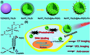
In this report, mesoporous NaYF4:Yb,Er@Au–Pt(IV)-FA up-conversion nanoparticles (UCNPs) have been designed by attaching Au NPs and Pt(IV) pro-drugs on the surface of PEI hydrogel modified mesoporous NaYF4:Yb,Er nanospheres. Finally the molecules modified with folic acid (FA) improve the receptor-mediated endocytosis. Because of the doped rare earth ions in the host matrix, the as-synthesized platform exhibits excellent up-conversion luminescence (UCL) imaging and computed X-ray tomography (CT) imaging properties. Diverse methods including MTT assay, hemolysis experiments, and live/dead cell analysis were employed to evaluate the biocompatibility and ablation efficacy of the as-synthesized platform. It was found that the cytotoxicity of the platform can be tuned by eliminating the axial ligands reductively during intracellular endocytosis. Especially, under 980 nm near-infrared (NIR) irradiation, the platform shows excellent inhibition toward cancer cells due to the synergistic photothermal injury to enzymes and membrane integrity combined with the DNA binding of activated Pt(II) to avoid cell proliferation. The developed nanocomposite may thus be a promising imaging-guided synergistic anti-cancer platform.


1. Introduction

For drug delivery systems (DDSs), when referring to photosensitive therapy, two irrelevant light wavelengths are commonly utilized to achieve diagnosis and effective therapy because of the limited penetration depth of conventional ultraviolet and visible light, thus it is difficult to achieve real-time assessment of the anti-cancer therapeutic effect.1–3 Multi-modal imaging is an effective tool to detect and manage malignant cancer sites which could synergistically combine the penetration, sensitivity and resolution necessary for diagnosis.4,5 CT imaging is one of the most common clinical diagnostic techniques due to the deep tissue penetration and high-resolution 3D structure detail.6,7 Although the high resolution of CT imaging is important, there is a limitation due to its low sensitivity, especially when used for tumor-imaging with small density differences. With single UCL fluorescence, it is sensitive but with poor tissue penetration. Thus, finding an effective, synergistic imaging-guided anti-cancer platform and strategy induced by a single light source is an extremely important subject in this field.8–11

The novel NIR irradiation located at the optical transmission window with lower absorbance of endogenous chromophores (hemoglobin and melanin) by living subjects also has significant advantages such as deep penetration, high detection sensitivity, and decreased background signal.12–17 Meanwhile, NIR irradiation has appealing properties for drug delivery applications which could emit visible light to track the tumor site through a two-/multi-photon energy transfer process, and the obtained high energy of the donors could transfer to the photosensitive agents.18–21 This amazing photo-conversion process allows up-conversion nanoparticles (UCNPs) to serve as effective single NIR light irradiated mediators for their extensive anti-cancer therapeutic applications with remote regulation of photosensitive activation, including bio-imaging and targeted delivery of drug molecules to cancer sites.22–24

cis-Dichlorodiammineplatinum(II) (denoted as cisplatin) is one of the most widely used anti-cancer drugs which is effective and could be clinically used to inhibit head and neck, esophageal, bladder, cervical, and non-small cell lung cancer, etc.25–28 Just like other chemo-therapeutic drug molecules, small molecular cisplatin which lacks controlled release with a very low intracellular reaction efficiency may have serious side effects to normal cells and thus could introduce complications, such as nausea, hearing damage, peripheral nerve impairment, and irreversible nephrotoxicity.29,30 Researchers have tried to find new drug candidates and introduce DDSs to decrease the chemotherapeutic side effects and achieve the targeted anti-tumor therapy.31,32 Pt(IV) compounds have been proposed as an attractive alternative to Pt(II) complexes because of their inertness in normal cells and body fluids. However, it seems the anti-cancer efficiency is not as high as predicted because Pt(IV) pro-drugs can be changed to Pt(II) species merely by reduction in the presence of intracellular biological reducing agents, such as glutathione and ascorbic acid or via acid hydrolysis. Meanwhile, the single chemo-therapeutic mode improves the body’s resistance to drugs, and the medicines lose drug potency with prolonging time.33 Thus, versatile synergistic photothermal/photodynamic anti-cancer platforms were developed.34–42 Gold nanoparticles could receive the broad UCL emissions by fluorescence resonance energy transfer (FRET) from the UCL hosts because there is an intense absorption located in the visible region.41–46 Meanwhile, they demonstrate strong absorption with a relatively higher molar extinction coefficient and photothermal conversion efficiency.47–51 When Au nanostructures were utilized, the generated intensive photothermal effect could destruct the cell membrane integrity and enzymes to interfere with DNA synthesis.52,53 If the photothermal effect is combined with the DNA binding caused by the chemo-effect of Pt(II) species through fabricating a functional structure, it is expected to obtain outstanding therapeutic efficiency because it could yield a synergistic efficacy which is higher than the sum of individual efficacy.

Herein, a dual-modal imaging-guided platform of NaYF4:Yb,Er@Au–Pt(IV)-FA UCNPs was designed for synergistic photothermal/chemo-therapy with non-toxicity to normal cells. Pt(IV) as the targeted anti-cancer pro-drug was synthesized through initial oxidization and subsequent reaction with succinic anhydride. Mesoporous NaYF4:Yb,Er nanospheres were prepared through a hydrothermal process using Y(OH)CO3:Yb,Er as the precursor. A large amount of PEI hydrogel on the surface of 90 nm NaYF4:Yb,Er serves as the drug carrier which can covalently conjugate the negative Au nanospheres and Pt(IV) molecules through physical attraction and chemical functional conjunction. MTT assay, hemolysis experiments, and live/dead cell analysis were employed to evaluate the drug molecule and carrier for the viability of normal cells and cytotoxicity to cancer cells. Moreover, the doped rare earth ions in the composite cause the material to have good UCL imaging and CT imaging properties. The results proved that the NaYF4:Yb,Er@Au–Pt(IV)-FA platform has dual-modal imaging properties and a synergistic photothermal and chemo-therapeutic effect on cancer cells.

2. Experimental section

2.1 Chemicals and materials

All the reagents used in this research were purchased and utilized directly without any further purification, including Y2O3, Yb2O3, and Er2O3 (Sinopharm Chemical Reagent Co., Ltd., China), hydrogen peroxide (H2O2), urea, nitric acid (HNO3), HAuCl4·3H2O, sodium citrate (Na3Cit), tannin acid, dimethyl sulfoxide (DMSO) (from Beijing Chemical Corporation, China), sodium chloride (NaCl), phosphate buffered saline (PBS), glutaraldehyde (from Tianjin Kermel Chemical Co., Ltd., China), cisplatin, succinic anhydride, polyethyleneimine (PEI), 1-(3-dimethylaminopropyl)-3-ethylcarbodiimide hydrochloride (EDC), and N-hydroxysuccinimide (NHS), folic acid (FA), 3-4,5-dimethylthiazol-2-yl-2,5-diphenyl tetrazolium bromide (MTT), 4′,6-diamidino-2-phenylindole (DAPI), calcein AM, propidium iodide (PI), trypan blue (from Sigma-Aldrich).

2.2 Synthesis

Synthesis of gold nanospheres. The Au nanospheres with a mean size of 10 nm were synthesized according to the literature.54 Instead of using the strong reduction of NaBH4, the HAuCl4·3H2O was reduced by citrate in the presence of tannic acid. Briefly, 5 mL of HAuCl4·3H2O (25 mM) was mixed with 30 mL deionized water, and then sodium citrate (0.2 g) and tannic acid (0.03 g) were added swiftly. The colorless transparent solution changed to dark purple red immediately, and then the solution was kept stirring at 60 °C for 4 h. The solution was then kept cold and used within several days.
Synthesis of Pt(IV) pro-drug. The Pt(IV) pro-drug of c,c,t-Pt(NH3)2Cl2(OOCCH2CH2COOH)2 (denoted as Pt(IV))was synthesized through a two-step process according to the literature.55 The intermediate product of c,c,t-Pt(NH3)2Cl2(OH)2 was firstly synthesized. Typically, cisplatin (2.5 g) was suspended into 125 mL of deionized water, 87.5 mL of H2O2 (30%) was added and stirred at 50 °C for 1 h. The product was then isolated and recrystallized. Then, c,c,t-Pt(NH3)2Cl2(OH)2 was obtained after washing with cold deionized water, ethanol and acetone. 0.7 g of synthesized c,c,t-Pt(NH3)2Cl2(OH)2 was dissolved in DMSO (5 mL) with succinic anhydride (2 g) added, and the mixture was stirred at 70 °C for 24 h. Finally, the pale yellow c,c,t-Pt(NH3)2Cl2(OOCCH2CH2COOH)2 was obtained.
Synthesis of Y(OH)CO3:Yb/Er. The precursor of Y(OH)CO3:Yb/Er was synthesized through a co-precipitation method.56 Note that the size could be easily controlled by adjusting the initial pH values. Briefly, 1 mol L−1 Ln(NO3)3 (Ln = Y, Yb, and Er) solutions were firstly prepared by dissolving rare earth oxides into nitric acid with continuous heating. Then, the calculated Ln(NO3)3 (81%Y, 18%Yb, and 1%Er) was added to 50 mL of deionized water, and then 3 g of urea was added with stirring. When the solution was transparent, the mixture in the beaker was kept heating in a water bath at 90 °C for 3 h, and the precipitate was collected after centrifugation and drying at 60 °C for 12 h.
Synthesis of mesoporous NaYF4:Yb/Er-PEI. In order to further obtain the better UCL host of a lanthanide fluoride with favourable dispersity, porous structure, and functional groups, the precursor was dissolved into 20 mL of deionized water, pre-mixed with 5 mL of PEI (50 mg mL−1) under ultrasonic treatment (10 min) and stirred for 2 h. After that, 4 mmol of NaBF4 was added into the mixture, and they were kept stirring for another 30 min. The uniform solution was sealed in a 40 mL kettle and kept at 150 °C for 3 h. NaYF4:Yb,Er with a PEI hydrogel shell was obtained with centrifugation and drying.
Synthesis of NaYF4:Yb/Er@Au–Pt(IV). 1 mL of the as-prepared gold nanosphere solution was firstly added to 0.15 mg of NaYF4:Yb,Er dispersed in 20 mL of deionized water. After stirring for 2 h, the mixture was washed three times with deionized water and centrifuged at 6000 rpm for 5 min. The pale purple precipitation was acquired due to the successful conjunction of gold nanospheres. Then, the precipitation was dispersed into 20 mL of water and mixed with 30 mg of the as-prepared Pt(IV) pro-drug, before NaYF4:Yb/Er@Au–Pt(IV) was obtained after final centrifugation. The supernatant was kept for further ICP-MS in order to calculate the loading amount of Pt(IV).
FA modified NaYF4:Yb/Er@Au–Pt(IV)-FA. Typically, 0.15 g of NaYF4:Yb/Er@Au–Pt(IV) was dissolved into 20 mL of deionized water. 1 mL of FA (10 mg mL−1), 1 mL of NHS (2 mg mL−1), and 1 mL of EDC (6 mg mL−1) were kept stirring for 2 h in darkness, and then the solution was added to the solution of NaYF4:Yb/Er@Au–Pt(IV) with continuous stirring for another 12 h in darkness. The pale brown product was collected by centrifugation and washed with water three times to remove the free FA molecules. After drying at 60 °C for 12 h, the yellow brown powder was the NaYF4:Yb/Er@Au–Pt(IV)-FA UCNPs.

2.3 Characterization

Powder X-ray diffraction (XRD) measurements were carried out on a Rigaku D/max TTR-III diffractometer with graphite monochromatized Cu Kα radiation (λ = 0.15405 nm), and the scanning rate was 15° min−1 in the 2θ range from 20° to 80°. Images were obtained digitally using transmission electron microscopy (TEM, FEI Tecnai G2 S-Twin). Fourier Transform Infrared Spectroscopy (FT-IR) spectra were measured with a Perkin-Elmer 580B IR spectrophotometer using a KBr pellet. N2 adsorption/desorption isotherms were acquired using Micromeritics ASAP Tristar II 3020 apparatus, and the pore size distribution was calculated with the Barrete–Jonere–Halenda (BJH) method. UCL emission spectra were obtained on Edinburgh FLS980 apparatus using a 980 nm laser diode Module (K98D08M-30W) as the irradiation source and recorded in the visible light region. The absorbance of red blood cells was measured on the microplate reader at the wavelength of 405 nm. The loading of Au nanoparticles and Pt(IV) pro-drugs on the materials was detected and calculated using ICP-MS. The amount was calculated as follows:
Mloading = (MaddedCSupernatant × VSupernatant),
where CSupernatant is the concentration of the detected supernatant solution and VSupernatant means the volume of the solution.

2.4 In vitro viability of drug carrier and complex

L929 fibroblast cells (about 7000 cells per well) were put in a 96-well (8 × 12) plate and incubated with 5% CO2 at 37 °C, and one row (12 wells) was left with culture only as the blank control, and then incubated for 24 h to make cells attach to the wells. Then NaYF4:Yb,Er@Au–Pt(IV) was dispersed in the cultures and diluted into various concentrations of 500, 250, 125, 62.5, 31.3, and 15.63 μg mL−1, while the concentrations of the corresponding cisplatin, Pt(IV) pro-drug and complexes were diluted to 75, 37.5, 18.8, 9.4, 4.7, and 2.4 μg mL−1, respectively. Then the solutions with different concentrations were added to the wells and incubated with the L929 cells for another 24 h. After that, 20 μL of the MTT solution (5 mg mL−1) was added to each well and the cells were incubated for another 4 h at 37 °C. MTT could be reduced into formazan by the live cells, and the formazan was dissolved by DMSO. Then, DMSO (150 μL) was added to each well after the solution in the well was discarded. The absorbance at 490 nm was measured using a microplate reader for further calculation. Note that NaYF4:Yb,Er@Au–Pt(IV) has a similar absorbance to the formazan solution, and the absorbance of NaYF4:Yb,Er@Au–Pt(IV) in a culture without cells should be detected by the microplate reader at 490 nm to remove background interference from the measurement of viability.

2.5 Hemolysis assay of drug carrier and complex

Human blood stabilized by EDTA.K2 was kindly provided by Harbin Blood Center, and used in compliance with the regulation of blood products by the State Food and Drug Administration. A hemolysis assay of NaYF4:Yb,Er@Au, cisplatin, and the NaYF4:Yb,Er@Au–Pt(IV) was carried out to evaluate their potential biocompatibility with the blood stream. Typically, red blood cells were obtained by removing the serum from human blood after washing with 0.9% (w/w) saline and centrifuging several times until the supernatant was colourless. Subsequently, the red blood cells were diluted with saline. 0.4 mL of the diluted cells suspension was then mixed with 1.6 mL of PBS (as a negative control), 1.6 mL of deionized water (as a positive control), and 1.6 mL of UCNPs suspensions with varying concentrations of 500, 250, 125, 62.5, 31.3, and 15.63 μg mL−1. The eight samples were shaken for a while, then kept standing for 2 h. Finally, the mixtures were centrifuged at 4000 rpm and the absorbance values of the upper supernatants were measured using a micro-plate reader at a wavelength of 405 nm. The hemolysis percentage was calculated as follows:
Hemolysis (%) = (AsampleAcontrol(−))/(Acontrol(+)Acontrol(−)),
where A is the absorbance.

2.6 In vitro cellular uptake and UCL microscopy (UCLM) observation

HeLa cancer cell lines were utilized to detect the cellular uptake process using a confocal laser scanning microscope (CLSM, Leica SP8). In a 6-well culture plate, the HeLa cells (about 105 per well) were seeded and incubated with one coverslip placed in each well overnight to obtain monolayer cells. After that, NaYF4:Yb,Er@Au–Pt(IV)-FA (1 mg mL−1) were added to the different wells at 37 °C with different incubation times of 1 h and 3 h, respectively. After each incubation time, the cells were flushed with PBS three times, and fixed with 1 mL glutaraldehyde (2.5% w/w in PBS) at 37 °C for 10 min, then rinsed with PBS three times again. 1 mL of DAPI solution (20 μg mL−1 in PBS) was added and kept for another 10 min, and then rinsed three times for further detection using CLSM. For the UCL microscopy observation, the coverslip was executed with the same process except that it was detected using inverted fluorescence microscopy.

2.7 In vitro and in vivo X-ray CT imaging

Female Kunming mice (25–35 g) were purchased from Harbin Veterinary Research Institute, Chinese Academy of Agricultural Sciences (Harbin, China), and all the mouse experiments were performed in compliance with the criterions of The National Regulation of China for Care and Use of Laboratory Animals. The in vitro CT imaging experiments were performed using a Philips 64-slice CT scanner with a voltage of 120 kV. NaYF4:Yb,Er@Au–Pt(IV)-FA was dispersed in saline and diluted to a series of concentrations of 40, 20, 10, 5, 2.5, and 1.25 mg mL−1 and then placed in a line for CT imaging. The mice were first anesthetized with 10% chloral hydrate (0.03 mL g−1 of mouse) by intra-peritoneal injection to perform in vivo CT imaging. Then, 100 μL of NaYF4:Yb,Er@Au–Pt(IV)-FA (40 mg mL−1) was intratumorally injected into the tumor-bearing mice in situ for better scanning.

2.8 In vitro cytotoxicity of drug carrier and complex

The in vitro cytotoxicity of different materials and drugs were performed on HeLa cells using an MTT assay. HeLa cells were incubated with cisplatin, the Pt(IV) pro-drug, the NaYF4:Yb,Er@Au–Pt(IV)-FA nanoparticles, and NaYF4:Yb,Er@Au–Pt(IV)-FA under NIR irradiation. The MTT assay process was carried out similarly to the viability assay of the L929 cells. For the live/dead cell detection process, HeLa cells were seeded in a 6-well culture plate and grown overnight as a monolayer. Then, they were incubated with different treatments: with NaYF4:Yb,Er@Au-FA, culture only under NIR irradiation, Pt(IV) pro-drug, NaYF4:Yb,Er@Au–Pt(IV)-FA, cisplatin, and NaYF4:Yb,Er@Au–Pt(IV)-FA under NIR irradiation. The pump power of the NIR irradiation was 0.72 W cm−2. Note that the materials were added and incubated for 6 h in order to complete the cell uptake, and then the irradiation was carried out just before the final dyeing process. After the treatment, the wells were rinsed with PBS, dyed with both calcein AM and PI, and visualized using CLSM.

3. Results and discussion

3.1 Phase, morphology, and luminescence properties

The formation of the Pt(IV) pro-drug, the synthesis process of NaYF4:Yb,Er@Au–Pt(IV), and the dual-modal imaging-guided photothermal/chemo-anti-tumor therapy application under single NIR irradiation are depicted in Scheme 1. Fig. 1 presents the XRD patterns of the Y(OH)CO3:Yb,Er precursor, as-synthesized NaYF4:Yb,Er after the second hydrothermal process, and NaYF4:Yb,Er@Au–Pt(IV). As shown, there is no obvious peak in the precursor which indicates that the precursor of Y(OH)CO3:Yb,Er is amorphous. After the second-step reaction of the precursor with NaBF4, the XRD pattern of the sample is consistent with the cubic NaYF4:Yb,Er (JCPDS: 06-0342), and the peaks are apparently strong without impurity, indicating that the NaYF4:Yb,Er has good crystallinity. After the Au nanospheres and Pt(IV) pro-drug are conjugated, the XRD pattern of NaYF4:Yb,Er@Au–Pt(IV) shows a new weaker peak assigned to the (111) lattice plane of cubic gold (JCPDS: 04-0784).
image file: c5ra05437k-s1.tif
Scheme 1 Schematic representation of the synthesis of the Pt(IV) pro-drug, the anti-cancer platform, and the imaging-guided synergistic photothermal and chemo-anti-cancer process.

image file: c5ra05437k-f1.tif
Fig. 1 XRD patterns of the precursor, NaYF4:Yb,Er, NaYF4:Yb,Er@Au, and NaYF4:Yb,Er@Au–Pt(IV).

TEM images of the corresponding Y(OH)CO3:Yb,Er, NaYF4:Yb,Er and NaYF4:Yb,Er@Au are presented in Fig. 2. As shown in Fig. 2A and B, the precursor is nearly mono-dispersed with a smooth surface, and the average size of the precursor is 90 nm. After the hydrothermal fluorination, uniform nanospheres with obvious pores and channels inside are obtained (Fig. 2C and D). The generation of this structure is due to the ion-exchange by the amorphous precursor with the gradually erosive NaBF4 under high pressure and at a temperature of 150 °C. Meanwhile, a large amount of –NH– and –NH2– functional groups were generated naturally due to the added PEI which is easily conjugated with other groups. After that, the gold nanospheres with 10 nm size were conjugated because lots of –COOH and –OH groups were lined on the surface of the Au nanospheres during the synthesis. TEM and HRTEM images of the Au spheres are shown in Fig. S1, and there is no difference between the mono-dispersed Au nanospheres and the Au particles after modification due to the good chemical and physical stability of Au noble metal. Meanwhile, besides the particles modified on the surface of NaYF4:Yb,Er (Fig. 2E and F), there are almost no free particles, indicating the strong electronic absorbance between the positive NaYF4:Yb,Er nanospheres and the negative Au nanoparticles. Note that there are not many attached Au spheres considering comprehensive properties of up-conversion luminescence and photothermal property.51


image file: c5ra05437k-f2.tif
Fig. 2 TEM images with different magnification of (A and B) the precursor, (C and D) NaYF4:Yb,Er, and (E and F) NaYF4:Yb,Er@Au.

Fig. 3A further presents a TEM image of NaYF4:Yb,Er@Au with high magnification. As shown, there is an apparent thin PEI hydrogel on the surface of NaYF4:Yb,Er with the mean size of 3–4 nm. Even after Au nanospheres were modified (with a small amount of Au solution), there was still a large amount of PEI groups left for the following conjugation of the Pt(IV) pro-drugs. The pores and channels inside of the nanospheres also played a significant role for the next modification which is attributed to the design of the nanoscale particles from both the chemical and physical perspectives. A high resolution transmission electron microscopy (HRTEM) image is shown in Fig. 3B with the corresponding fast Fourier transform (FFT) images of the corresponding regions (Fig. 3b1 and b2). The 10 nm-sized nanosphere is a Au particle in the b1 region which has an obvious distance of 0.24 nm assigned to the (111) lattice plane of Au spheres. The apparent distance of 0.32 nm is observed in the b2 region which is coincident with the (111) lattice plane of cubic NaYF4:Yb,Er. The results further prove the successful conjunction of the Au nanospheres on the surface of NaYF4:Yb,Er. The energy dispersive spectroscopy (EDS) image in Fig. 3C indicates that there are Na, Y, F, Au, C, and O elements present in the resultant sample. Additionally, the EDS line profile (Fig. 3D) and EDS mapping (Fig. 3E) indicate that the Au nanoparticles and Pt(IV) pro-drugs were well dispersed in the surface and inside of NaYF4:Yb,Er.


image file: c5ra05437k-f3.tif
Fig. 3 (A) TEM image, (B) HRTEM image with FFT images, (C) EDS analysis, (D) EDS line profile, and (E) EDS mapping of NaYF4:Yb,Er@Au NPs.

Fig. 4 demonstrates the nitrogen adsorption/desorption isotherms and the corresponding pore volume versus pore size distributions of NaYF4:Yb,Er@Au, and NaYF4:Yb,Er@Au with the Pt(IV) pro-drug loaded. Both of the two samples exhibit typical IV-type isotherms with the main H1 hysteresis loop in the medium relative pressure range (0.3–0.8), which indicates the mesoporous channels and pores were dispersed in the samples. Meanwhile, the BJH desorption surface of the two samples are 20.7 and 10.4 m2 g−1, and the average pore sizes are 15.6 nm and 10.5 nm, respectively. There is an obvious decrease in the surface area between the NaYF4:Yb,Er@Au with and without the Pt(IV) pro-drug loaded, which indicates that the modified Pt(IV) pro-drug molecules entered the mesopores, attracted by physical attraction and chemical conjunction.


image file: c5ra05437k-f4.tif
Fig. 4 Nitrogen adsorption/desorption curves and the corresponding pore size distribution (inset) of (A) NaYF4:Yb,Er@Au, and (B) NaYF4:Yb,Er@Au–Pt(IV).

FT-IR spectra of NaYF4:Yb,Er, NaYF4:Yb,Er@Au, NaYF4:Yb,Er@Au–Pt(IV), and the final NaYF4:Yb,Er@Au–Pt(IV)-FA to further prove the successful conjugation of each step are shown in Fig. 5. Similar bands at 3438 cm−1 and 1075 cm−1 in the four spectra are attributed to the hydroxyl group stretching and the –YF. The spectrum of NaYF4:Yb,Er has typical peaks at 1500–1650 cm−1 and 2840–2962 cm−1, which are assigned to amide bonds (–NH, –NH2) and –CH2 stretching vibrations from the PEI hydrogel, respectively.57 After the gold nanospheres and Pt(IV) pro-drug were modified, the typical peak at 1530 cm−1 (–NH2) disappeared, while new peaks at 1631 and 1404 cm−1 appeared assigned to the formed –CONH– groups between the amine groups of NaYF4:Yb,Er and the carboxyl groups and carbonyl groups of Au–Cit and c,c,t-Pt(NH3)2Cl2(OOCCH2CH2COOH)2.58 The increased intensity of the two peaks in NaYF4:Yb,Er@Au–Pt(IV) compared with NaYF4:Yb,Er@Au indicates that more –CONH– groups were generated. After the FA molecules were modified, obvious peaks at 770–1696 cm−1 and 2700–3700 cm−1 assigned to the characteristic groups of FA were observed.59 These results further demonstrate that the as-synthesized gold nanospheres, Pt(IV) pro-drug, and the FA molecules successfully conjugated on the NaYF4:Yb,Er nanospheres.


image file: c5ra05437k-f5.tif
Fig. 5 FT-IR spectra of NaYF4:Yb,Er, NaYF4:Yb,Er@Au, NaYF4:Yb,Er@Au–Pt(IV), and (D) NaYF4:Yb,Er@Au–Pt(IV)-FA.

Fig. 6A shows the emission spectra of NaYF4:Yb,Er and NaYF4:Yb,Er@Au samples under 980 nm excitation and the absorbance spectrum of the Au nanosphere solution was also detected with UV-vis spectroscopy. Three strong luminescent emissions at 520, 539, and 654 nm are assigned to the energy transfer process of 2H11/24I15/2, 4S3/24I15/2, and 4F9/24I15/2, respectively.60–65 Meanwhile, there is a weaker peak at 409 nm which corresponds to the 2H9/24I15/2 energy transfer process. Generally, the 409 nm due to the three- or four-photon up-conversion transfer can not be easily obtained because of the low efficiency and strong scattering, and this result indicates that NaYF4:Yb,Er is an excellent host serving as the energy donor.64 In Fig. 6A, an obvious decrease in emission intensity occurs in the green region of NaYF4:Yb,Er@Au because there is an obvious wide peak at 490–570 nm in the Au absorbance spectrum which crosses with the UCL emission of NaYF4:Yb,Er. There is a Förster resonance energy transfer (FRET) between the two materials, and the emission intensity of NaYF4:Yb,Er as the donor decreased after the Au particles as the accepter were added. Thus, a following photothermal effect was generated. The energy transfer process of NaYF4:Yb,Er and the FRET between it and Au using the energy levels are shown in Fig. 6B. In fact, there is also a self-photothermal effect (from room temperature to 41.6 °C) of the Au sphere solution at a wavelength of 980 nm, and the infrared thermal photograph is shown in Fig. S2, which may be caused by the local electronic field enhancement. The UCL spectra of NaYF4:Yb,Er@Au–Pt(IV) and the following FA modified sample are shown in Fig. 6C. As shown, there is no great change among the three samples because there is almost no absorbance in the visible regions of the Pt(IV) pro-drug and FA molecules. Photographs under daylight (left in each image) and the corresponding infrared thermal images (right in each image) of the PBS, NaYF4:Yb,Er–Pt(IV)-FA, and NaYF4:Yb,Er@Au–Pt(IV)-FA solutions are shown in Fig. 6D, and the corresponding increased temperature curves are demonstrated in Fig. S3. As indicated, the solution of the final NaYF4:Yb,Er@Au–Pt(IV)-FA sample after 5 min irradiation reached the highest temperature (48.5 °C) compared with the PBS (36.7 °C) and NaYF4:Yb,Er–Pt(IV)-FA without Au spheres modification (42.1 °C).


image file: c5ra05437k-f6.tif
Fig. 6 (A) UCL emission spectra of NaYF4:Yb,Er with and without attached Au NPs under 980 nm irradiation and the absorbance spectrum of the Au NPs solution. (B) Energy transfer process of NaYF4:Yb,Er and the FRET process with Au. (C) UCL emission spectra of NaYF4:Yb,Er@Au and NaYF4:Yb,Er@Au with Pt(IV) and FA conjugation. (D) The photographs under daylight (left) and infrared thermal images (right) of saline, NaYF4:Yb,Er–Pt(IV)-FA, and NaYF4:Yb,Er@Au–Pt(IV)-FA solutions.

3.2 Biocompatibility, bio-imaging, and anti-cancer efficiency

The proposed ideal Pt(IV) pro-drug and complex should have sufficient stability without decomposition in body fluids or the blood stream, and good biocompatibility should be guaranteed before it reaches the cancer cell. Here, a standard MTT assay was carried out to evaluate the short-term biocompatibility of the drug carrier (NaYF4:Yb,Er@Au) and the Pt(IV) complex compared with the commercially used cisplatin. Before this, the amount of Pt(IV) carried on the NaYF4:Yb,Er@Au was calculated using ICP-MS during the drug-loading process. Fig. 7A–D present the viability of L929 cells incubated with concentrations of drug carrier from 15.63 to 500 μg mL−1, and the corresponding concentrations of drugs range from 2.4 to 75 μg mL−1. The viability of cells incubated with drug carrier NaYF4:Yb,Er@Au of different concentrations is as high as 96.7–112.4%. Even under the highest concentration of 500 μg mL−1, still 100.6% of the cells survived which indicates that NaYF4:Yb,Er@Au has no potential toxicity to normal cells. The viability of L929 cells incubated with the commercial drug cisplatin at different concentrations indicates that there is a stepped decreased viability from 94.1% to 30.4% when the concentrations increase from 9.4 μg mL−1 to 75 μg mL−1, this result indicates that conventional Pt(II) drugs may have serious side effects to normal cells of patients. While for Pt(IV) pro-drugs, the viability of the cells is up to 87.2–126.0% which indicates that small molecular platinum(IV) has no obvious inhibition toward the normal cells. When the drug carrier was loaded with the nontoxic Pt(IV) pro-drugs, NaYF4:Yb,Er@Au–Pt(IV) still kept much higher viability (67.9–101.8%) of the normal cells compared with cisplatin. It seems there is cytotoxicity when the concentration is as high as 500 μg mL−1 (67.9%). However, this concentration is much higher than the amount used practically.
image file: c5ra05437k-f7.tif
Fig. 7 Viability of L929 cells incubated with (A) NaYF4:Yb,Er@Au, (B) cisplatin, (C) Pt(IV) pro-drug, and (D) NaYF4:Yb,Er@Au–Pt(IV). (E) The hemolysis of cisplatin, NaYF4:Yb,Er@Au, and NaYF4:Yb,Er@Au–Pt(IV). The microscope images under the bright field and fluorescence field (405 nm) of L929 cells incubated (F) with culture only, and (G) with NaYF4:Yb,Er@Au dyed by DAPI and trypan blue.

Meanwhile, it is proposed that the clinical drugs and carriers should be steady in the blood stream. Thus, the hemolysis properties of the clinical drug cisplatin, the drug carrier NaYF4:Yb,Er@Au, and the NaYF4:Yb,Er@Au–Pt(IV) complex are shown in Fig. 7E. The highest hemolytic efficiency of each of the three samples is 0.13%, 0.23%, and 0.22% with a material concentration of 15.63–500 μg mL−1 and the corresponding drug concentration of 2.4–75 μg mL−1, indicating that almost no hemolysis occurs to the drugs and materials.

Furthermore, the L929 cells were incubated with 1 mg mL−1 of NaYF4:Yb,Er@Au and NaYF4:Yb,Er@Au–Pt(IV) for 24 h and then dyed with DAPI and trypan blue. Compared with cells incubated with the culture only, there are almost no dead cells observed after incubation with NaYF4:Yb,Er@Au for 24 h (Fig. 7F and G), and there are few observed dead cells after incubation with NaYF4:Yb,Er@Au–Pt(IV) for 24 h (Fig. S4). These results reveal that both the drug carrier and the molecular drugs cause no hemolysis of the human red blood cells, and the as-synthesized Pt(IV) pro-drugs and complexes also have much better bio-compatibility to the normal cells than that of the conventional cisplatin. Thus, although the cisplatin anti-cancer drug molecules are associated with higher reactivity for anti-cancer, the lower biological stability limits its clinical application. The Pt(IV) pro-drug and complex candidates with high biocompatibility and less injury to normal cells, may have more extensive applications in the anti-cancer field.

In order to achieve tumor targeting endocytosis to assure the anti-cancer therapeutic efficiency, NaYF4:Yb/Er@Au–Pt(IV) was modified by FA molecules.56 HeLa cells were seeded in the 6-well plate and incubated with free FA and the FA modified NaYF4:Yb,Er@Au–Pt(IV) UCNPs for 3 h under the same conditions at 37 °C with 5% CO2. As shown in Fig. S5, the red channel is from the NaYF4:Yb,Er@Au–Pt(IV) UCNPs which were excited at 552 nm to track the Pt(IV) complex, and the blue emissions (dyed by DAPI) were used to mark the nuclei. The red emissions may be caused by a non-irradiative or down-conversion process due to the Yb,Er co-dopant (depicted in the energy level diagram in Fig. 6B). The FA modified NaYF4:Yb,Er@Au–Pt(IV) present stronger red luminescence than the cells incubated with particles without FA, suggesting more nanoparticles have been taken up by the HeLa cells. This result reveals there is a positive receptor-mediated endocytosis because of FA conjunction.

Fig. 8 presents the CLSM photographs of HeLa cancer cells incubated with NaYF4:Yb,Er@Au–Pt(IV)-FA for 1 h and 3 h in order to detect the cell uptake process. The overlay of the two channels of red emission from the UCNPs and blue emission from DAPI is shown correspondingly. In the initial 1 h, little red emission is found, indicating only a few of the particles have been taken up by the cells, and stronger red fluorescence emission is found which suggests more particles are localized in the cells gradually. Meanwhile, we can see most of the red emissions focus in the cytoplasm instead of in the nuclei, indicating that the NaYF4:Yb,Er@Au–Pt(IV)-FA UCNPs undergo an endocytosis process instead of passive diffusion.


image file: c5ra05437k-f8.tif
Fig. 8 Confocal laser scanning microscopy images of HeLa cells incubated with NaYF4:Yb,Er@Au–Pt(IV)-FA for 1 h and 3 h. All the scale bars are 50 μm.

Yb doped particles can be used as contrast agents for CT imaging which is one of the most common clinical diagnostic techniques. Here, CT images of the NaYF4:Yb,Er@Au–Pt(IV)-FA UCNPs in vitro and in vivo were taken. As shown in Fig. 9A, the CT signal increases obviously with enhanced concentration, and the values show high positive contrast enhancement as a function of the concentrations with a large slope of 18.64. The in vivo CT imaging of mice is shown in Fig. 9B and C, and the CT value in the tumor site is enhanced from 54.2 HU (Hounsfield units) to 1993.5 HU after injection. The high CT value indicates that the platform could be used as a CT imaging contrast agent for in vitro and in vivo imaging. Although the high resolution of CT imaging is important, there is a limitation because of low sensitivity, especially when it is used in tumor-imaging with small density differences. Fig. 9D–H present the inverted florescence microscope images of HeLa cells incubated with the NaYF4:Yb,Er@Au–Pt(IV)-FA UCNPs (1 mg mL−1). It is obvious NaYF4:Yb,Er@Au–Pt(IV)-FA in the cells emits weaker blue and brighter green and red emissions upon 980 nm NIR irradiation. Also, UCL light is focused on the cytoplasm, which indicates the drug carriers are taken up by endocytosis inside endosomes and lysosomes instead of passive adsorption.66,67 These dual-modal imaging properties reveal that the UCNPs are effective agents for real-time diagnosis.


image file: c5ra05437k-f9.tif
Fig. 9 (A) In vitro CT values as a function of the particle concentrations, (B and C) in vivo CT images before and after intratumoral injection. UCL microscopy images of HeLa cells incubated with NaYF4:Yb,Er@Au–Pt(IV)-FA: (D) bright field, (E) blue region, (F) green region, (G) red region, and (H) overlay of the above channels under NIR irradiation. All the scale bars are 50 μm.

The standard MTT assay and live/dead cell analysis of HeLa cells were employed to evaluate the anti-cancer therapeutic efficiency. The viability of HeLa cells incubated with drug molecules of cisplatin, the Pt(IV) pro-drug, NaYF4:Yb,Er@Au–Pt(IV)-FA, and NaYF4:Yb,Er@Au–Pt(IV)-FA under NIR irradiation are demonstrated in Fig. 10A. As shown, for the small molecular active cisplatin, the viability of cancer cells shows a concentration-dependent result, and only 29.7% of cells survive with the highest concentration of 75 μg mL−1. While for the Pt(IV) pro-drug, the viability is as high as 73.7–111.5%, indicating the nontoxic character of small molecular Pt(IV). The reason for these different results between molecular cisplatin and the Pt(IV) pro-drug is that Pt(IV) drugs can inhibit cell growth only under the condition that they are reduced to Pt(II). A few cancer cells are killed because some of the pro-drug molecules are reactive from the acidic environment due to the characteristic cancer cell environment. While for NaYF4:Yb,Er@Au–Pt(IV)-FA, the viability of cancer cells decrease a lot (51.5–76.4%) compared with the molecular Pt(IV). The difference between the Pt(IV) pro-drug and complex is caused by the different cell uptake processes. As depicted above, the drug carriers enter the cells by endocytosis instead of passive diffusion. That means, the nanoparticles have priority to enter the early endosomes and late endosomes (pH 5.0–6.0), then coalesce with the lysosomes (pH 4.5–5.0) via endocytosis by cancer cells.68 Additionally, FA improves the material’s ability to recognize and enter the cancer cells through receptor-mediated endocytosis. Under intracellular milieu, the Pt(IV) pro-drug species could be reduced in this acidic micro-environment to yield the cytotoxic Pt(II) molecules through elimination of the axial ligands reductively. As known, the obtained Pt(II) drugs reduced from the pro-drugs could kill about a half of the cancer cells through DNA binding which seems not so satisfactory.69,70 While for cells incubated with NaYF4:Yb,Er@Au-FA (without Pt(IV) pro-drugs loaded) under NIR irradiation, there is only the photothermal effect, and the viability is 58.0% with a concentration of 500 μg mL−1. In order to improve the anti-cancer effect of NaYF4:Yb,Er@Au–Pt(IV)-FA and simultaneously decrease the drug-resistance, a photothermal effect is introduced with imaging guidance by the designed platform under NIR irradiation. As depicted in Fig. 6, there is an obvious resulting photothermal effect due to the plastic resonance and FRET within the platform. After NIR laser irradiation, HeLa cells are restrained markedly with a survived cells ratio of 24.3%. Compared with that of NaYF4:Yb,Er@Au–Pt(IV) (no FA) with NIR irradiation, the inhibition efficiency of NaYF4:Yb,Er@Au–Pt(IV)-FA with NIR irradiation is higher which further indicates the targeting effect of FA molecules. The IC50 values of cisplatin and NaYF4:Yb,Er@Au–Pt(IV)-FA + NIR are 31.6 μg mL−1 and 5.48 μg mL−1, respectively. This indicates that there is a higher toxicity caused by the synergistic photothermal/chemo-effect than any single therapy.


image file: c5ra05437k-f10.tif
Fig. 10 (A) The cell viability of HeLa cancer cells incubated separately with small molecular cisplatin and the Pt(IV) pro-drug, with UCNPs separately with NaYF4:Yb,Er@Au-FA and NaYF4:Yb,Er@Au–Pt(IV), and with UCNPs under NIR irradiation. (B) CLSM images of HeLa cells incubated with different groups. All the cells were dyed with calcein AM and PI.

Calcein AM (living cells dyed with green color) and PI (dead cells dyed with red color) were utilized to distinguish the live/dead state of HeLa cancer cells under different treatment conditions detected by CLSM (Fig. 10B). Here, the concentration of all the added nanoparticles was 1 mg mL−1 and the corresponding molecular platinum concentration was 150 μg mL−1. When cisplatin was introduced, almost all of the cancer cells were killed due to the high inhibition effect with high dose of Pt(II) drugs. When there was a small amount of molecular Pt(IV) pro-drug added, a few of the cells died. When only NaYF4:Yb,Er@Au was added, almost no dead cells appeared, and the NIR irradiation had no obvious inhibition toward cancer cells. When only NaYF4:Yb,Er@Au–Pt(IV)-FA was added without NIR irradiation, the inhibition effect was not as high as the MTT assay result in Fig. 10A which could be attributed to many premature dead cells washed away by PBS. This is the reason why the MTT assay is essential. Additionally, when there was no chemo-effect of Pt(IV), some of the cells were incubated with NaYF4:Yb,Er@Au-FA under NIR irradiation for 5 min (pump power of 0.72 W cm−2) and more cells were killed after 10 min irradiation (Fig. S6). With a pump power of 1.44 cm−2, almost no cells survived. However, the higher pump power may induce overheating which is not utilized in the clinical application. Meanwhile, without FA conjugation, the inhibition effect toward the cancer cells incubated with NaYF4:Yb,Er@Au–Pt(IV) (no FA) with NIR irradiation is not so satisfactory. Finally and amazingly, for the cancer cells incubated with the NaYF4:Yb,Er@Au–Pt(IV)-FA nanoparticles under NIR irradiation (0.72 W cm−2), almost no cells survive which is even similar to the cisplatin cytotoxicity with high dose. There may be a synergistic effect because the high temperature could cause the irreversible destruction of the enzymes used in DNA synthesis and membrane integrity, while the Pt(II) could bind to the DNA to avoid cell proliferation, and this inhibition efficiency is obviously higher than that of the individual chemo- or photothermal modes. To summarize, the highly-biocompatible NaYF4:Yb,Er@Au–Pt(IV)-FA UCNPs have a better anti-cancer therapeutic effect resulting from the synergistic photothermal/chemo-function (due to the Au nanoparticles and Pt(IV) molecules), the special endocytosis (due to the drug carriers), and the receptor-mediated effect (due to FA molecules) under NIR irradiation.

4. Conclusions

In summary, a mesoporous 90 nm NaYF4:Yb,Er@Au–Pt(IV) anti-cancer platform was constructed by functionalizing PEI hydrogel, Au nanoparticles, and Pt(IV) molecules sequentially through physical attraction and chemical functional conjugation. The finally conjugated FA is to achieve the receptor-mediated endocytosis into cancer cells. The results of the MTT assay, hemolysis experiments, and live/dead cell analysis indicate that the as-synthesized NaYF4:Yb,Er@Au–Pt(IV)-FA has high biocompatibility, good up-conversion luminescence (UCL) imaging and CT imaging properties, and especially high anti-cancer efficacy. This may be caused by a synergistic photothermal/chemo-effect because the high temperature derived from the attached Au nanoparticles could cause the irreversible destruction of the enzymes used in DNA synthesis and membrane integrity, while the attached Pt(II) could bind the DNA to avoid cell proliferation.

Acknowledgements

Financial support from the National Natural Science Foundation of China (NSFC 21271053, 21401032, and 51472058), Research Fund for the Doctoral Program of Higher Education of China (2011230411002), Natural Science Foundation of Heilongjiang Province (B201403), Harbin Sci.-Tech. Innovation Foundation (2014RFQXJ019) and Fundamental Research Funds for the Central Universities of China (HEUCF1412) are greatly acknowledged.

Notes and references

  1. Y. S. Liu, D. T. Tu, H. M. Zhu and X. Y. Chen, Chem. Soc. Rev., 2013, 42, 6942–6958 Search PubMed.
  2. F. Wang, D. Banerjee, Y. Liu, X. Chen and X. Liu, Analyst, 2010, 135, 1839–1854 RSC.
  3. L. D. Sun, Y. F. Wang and C. H. Yan, Acc. Chem. Res., 2014, 47, 1001–1009 CrossRef CAS PubMed.
  4. D. T. Tu, L. Q. Liu, Q. Ju, Y. S. Liu, H. M. Zhu, R. F. Li and X. Y. Chen, Angew. Chem., Int. Ed, 2011, 50, 6306–6310 CrossRef CAS PubMed.
  5. Z. Zhao, H. Fan, G. Zhou, H. Bai, H. Liang, R. Wang, X. Zhang and W. Tan, J. Am. Chem. Soc., 2014, 136, 11220–11223 CrossRef CAS PubMed.
  6. H. Xing, W. Bu, S. Zhang, X. Zheng, M. Li, F. Chen, Q. He, L. Zhou, W. Peng, Y. Hua and J. Shi, Biomaterials, 2012, 33, 1079–1089 CrossRef CAS PubMed.
  7. Y. Wu, Y. Sun, X. Zhu, Q. Liu, T. Cao, J. Peng, Y. Yang, W. Feng and F. Li, Biomaterials, 2014, 35, 4699–4705 CrossRef CAS PubMed.
  8. Y. Tao, E. Ju, Z. Liu, K. Dong, J. Ren and X. Qu, Biomaterials, 2014, 35, 6646–6656 CrossRef CAS PubMed.
  9. J. Kim, Y. Piao and T. Hyeon, Chem. Soc. Rev., 2009, 38, 372–390 RSC.
  10. Y. Sun, X. Zhu, J. Peng and F. Li, ACS Nano, 2013, 7, 11290–11300 CrossRef CAS PubMed.
  11. C. Li, D. Yang, P. A. Ma, Y. Chen, Y. Wu, Z. Hou, Y. Dai, J. Zhao, C. Sui and J. Lin, Small, 2013, 9, 4150–4159 CrossRef CAS PubMed.
  12. L. D. Carlos, R. A. S. Ferreira, V. d. Z. Bermudez, B. Julian-Lopez and P. Escribano, Chem. Soc. Rev., 2011, 40, 536–549 RSC.
  13. A. D. Ostrowski, E. M. Chan, D. J. Gargas, E. M. Katz, G. Han, P. J. Schuck, D. J. Milliron and B. E. Cohen, ACS Nano, 2012, 6, 2686–2692 CrossRef CAS PubMed.
  14. S. Zeng, Z. Yi, W. Lu, C. Qian, H. Wang, L. Rao, T. Zeng, H. Liu, H. Liu, B. Fei and J. Hao, Adv. Funct. Mater., 2014, 24, 4051–4059 CrossRef CAS.
  15. A. Hlavacek, A. Sedlmeier, P. Skladal and H. H. Gorris, ACS Appl. Mater. Interface, 2014, 6, 6930–6935 CrossRef CAS PubMed.
  16. F. C. J. M. van Veggel, Chem. Mater., 2014, 26, 111–122 CrossRef CAS.
  17. Z. Chen, Q. Wang, H. Wang, L. Zhang, G. Song, L. Song, J. Hu, H. Wang, J. Liu and M. Zhu, Adv. Mater., 2013, 25, 2095–2100 CrossRef CAS PubMed.
  18. J. Liu, W. Bu, L. Pan and J. Shi, Angew. Chem., Int. Ed., 2013, 52, 4375–4379 CrossRef CAS PubMed.
  19. S. Zeng, M.-K. Tsang, C.-F. Chan, K.-L. Wong, B. Fei and J. Hao, Nanoscale, 2012, 4, 5118–5124 RSC.
  20. H. H. Gorris and O. S. Wolfbeis, Angew. Chem., Int. Ed., 2013, 52, 3584–3600 CrossRef CAS PubMed.
  21. X. Huang, S. Han, W. Huang and X. Liu, Chem. Soc. Rev., 2013, 42, 173–201 RSC.
  22. L. Xia, X. Kong, X. Liu, L. Tu, Y. Zhang, Y. Chang, K. Liu, D. Shen, H. Zhao and H. Zhang, Biomaterials, 2014, 35, 4146–4156 CrossRef CAS PubMed.
  23. F. Liu, Q. Zhao, H. You and Z. Wang, Nanoscale, 2013, 5, 1047–1053 RSC.
  24. J. V. Garcia, J. Yang, D. Shen, C. Yao, X. Li, R. Wang, G. D. Stucky, D. Zhao, P. C. Ford and F. Zhang, Small, 2012, 8, 3800–3805 CrossRef CAS PubMed.
  25. R. P. Feazell, N. Nakayama-Ratchford, H. Dai and S. J. Lippard, J. Am. Chem. Soc., 2007, 129, 8438–8439 CrossRef CAS PubMed.
  26. S. Dhar, W. L. Daniel, D. A. Giljohann, C. A. Mirkin and S. J. Lippard, J. Am. Chem. Soc., 2009, 131, 14652–14653 CrossRef CAS PubMed.
  27. Y. Jung and S. J. Lippard, Chem. Rev., 2007, 107, 1387–1407 CrossRef CAS PubMed.
  28. P. M. Takahara, A. C. Rosenzweig, C. A. Frederick and S. J. Lippard, Nature, 1995, 377, 649–652 CrossRef CAS PubMed.
  29. P. J. Bednarski, F. S. Mackay and P. J. Sadler, Anti-Cancer Agents Med. Chem., 2007, 7, 75–93 CrossRef CAS PubMed.
  30. J. Turkson, S. M. Zhang, L. B. Mora, A. Burns, S. Sebti and R. Jove, J. Biol. Chem., 2005, 280, 32979–32988 CrossRef CAS PubMed.
  31. K. R. Barnes, A. Kutikov and S. J. Lippard, Chem. Biol., 2004, 11, 557–564 CrossRef CAS PubMed.
  32. L. R. Kelland, C. F. Barnard, K. J. Mellish, M. Jones, P. M. Goddard, M. Valenti, A. Bryant, B. A. Murrer and K. R. Harrap, Cancer Res., 1994, 54, 5618–5622 CAS.
  33. H. Xiao, G. T. Noble, J. F. Stefanick, R. Qi, T. Kiziltepe, X. Jing and B. Bilgicer, J. Controlled Release, 2014, 173, 11–17 CrossRef CAS PubMed.
  34. W. J. Rieter, K. M. Pott, K. M. L. Taylor and W. Lin, J. Am. Chem. Soc., 2008, 130, 11584–11585 CrossRef CAS PubMed.
  35. Y. Min, J. Li, F. Liu, E. K. L. Yeow and B. Xing, Angew. Chem., Int. Ed., 2014, 53, 1012–1016 CrossRef CAS PubMed.
  36. W. Fang, S. Tang, P. Liu, X. Fang, J. Gong and N. Zheng, Small, 2012, 8, 3816–3822 CrossRef CAS PubMed.
  37. X. Wang, C. Wang, L. Cheng, S.-T. Lee and Z. Liu, J. Am. Chem. Soc., 2012, 134, 7414–7422 CrossRef CAS PubMed.
  38. A. Kumar, S. Kumar, W.-K. Rhim, G.-H. Kim and J.-M. Nam, J. Am. Chem. Soc., 2014, 136, 16317–16325 CrossRef CAS PubMed.
  39. W. Chen, R. Yu, L. Li, A. Wang, Q. Peng and Y. Li, Angew. Chem., Int. Ed., 2010, 49, 2917–2921 CrossRef CAS PubMed.
  40. R. Lv, P. Yang, F. He, S. Gai, C. Li, Y. Dai, G. Yang and J. Lin, ACS Nano, 2015, 9, 1630–1647 CrossRef CAS PubMed.
  41. X. Wang, K. Liu, G. Yang, L. Cheng, L. He, Y. Liu, Y. Li, L. Guo and Z. Liu, Nanoscale, 2014, 6, 9198–9205 RSC.
  42. L. Cheng, C. Wang and Z. Liu, Nanoscale, 2013, 5, 23–37 RSC.
  43. A. Priyam, N. M. Idris and Y. Zhang, J. Mater. Chem., 2012, 22, 960–965 RSC.
  44. S. Xu, S. Xu, Y. Zhu, W. Xu, P. Zhou, C. Zhou, B. Dong and H. Song, Nanoscale, 2014, 6, 12573–12579 RSC.
  45. G. Wang, Q. Peng and Y. Li, Acc. Chem. Res., 2011, 44, 322–332 CrossRef CAS PubMed.
  46. J. Liu, J. Bu, W. Bu, S. Zhang, L. Pan, W. Fan, F. Chen, L. Zhou, W. Peng, K. Zhao, J. Du and J. Shi, Angew. Chem., Int. Ed., 2014, 53, 4551–4555 CrossRef CAS PubMed.
  47. L. Wang, H. Dong, Y. Li, C. Xue, L.-D. Sun, C.-H. Yan and Q. Li, J. Am. Chem. Soc., 2014, 136, 4480–4483 CrossRef CAS PubMed.
  48. J.-W. Oh, D.-K. Lim, G.-H. Kim, Y. D. Suh and J.-M. Nam, J. Am. Chem. Soc., 2014, 136, 14052–14059 CrossRef CAS PubMed.
  49. X. Chen, Y. Chen, M. Yan and M. Qiu, ACS Nano, 2012, 6, 2550–2557 CrossRef CAS PubMed.
  50. M. A. Mahmoud and M. A. El-Sayed, ChemCatChem, 2014, 6, 3540–3546 CAS.
  51. M. Chen, S. Tang, Z. Guo, X. Wang, S. Mo, X. Huang, G. Liu and N. Zheng, Adv. Mater., 2014, 26, 8210–8216 CrossRef CAS PubMed.
  52. E. C. Dreaden, M. A. Mackey, X. Huang, B. Kang and M. A. El-Sayed, Chem. Soc. Rev., 2011, 40, 3391–3404 RSC.
  53. H. Chen, X. Zhang, S. Dai, Y. Ma, S. Cui, S. Achilefu and Y. Gu, Theranostics, 2013, 3, 633–649 CrossRef PubMed.
  54. R. Lv, P. Yang, Y. Dai, S. Gai, F. He and J. Lin, ACS Appl. Mater. Interfaces, 2014, 6, 15550–15563 CAS.
  55. J. Yang, W. Liu, M. Sui, J. Tang and Y. Shen, Biomaterials, 2011, 32, 9136–9143 CrossRef CAS PubMed.
  56. R. Lv, P. Yang, F. He, S. Gai, G. Yang and J. Lin, Chem. Mater., 2015, 27, 483–496 CrossRef CAS.
  57. H.-T. Wong, M.-K. Tsang, C.-F. Chan, K.-L. Wong, B. Fei and J. Hao, Nanoscale, 2013, 5, 3465–3473 RSC.
  58. Y. Dai, X. Kang, D. Yang, X. Li, X. Zhang, C. Li, Z. Hou, Z. Cheng, P. a. Ma and J. Lin, Adv. Drug Delivery Rev., 2013, 2, 562–567 CAS.
  59. K. Y. Lee, E. Seow, Y. Zhang and Y. C. Lim, Biomaterials, 2013, 34, 4860–4871 CrossRef CAS PubMed.
  60. J.-C. Boyer, L. A. Cuccia and J. A. Capobianco, Nano Lett., 2007, 7, 847–852 CrossRef CAS PubMed.
  61. J. Zhao, Z. Lu, Y. Yin, C. McRae, J. A. Piper, J. M. Dawes, D. Jin and E. M. Goldys, Nanoscale, 2013, 5, 944–952 RSC.
  62. X. H. He and B. Yan, J. Mater. Chem. C, 2014, 2, 2368–2374 RSC.
  63. J. L. Zhuang, J. Wang, X. F. Yang, I. D. Williams, W. Zhang, Q. Y. Zhang, Z. M. Feng, Z. M. Yang, C. L. Liang, M. M. Wu and Q. Su, Chem. Mater., 2009, 21, 160–168 CrossRef CAS.
  64. F. Liu, X. He, L. Liu, H. You, H. Zhang and Z. Wang, Biomaterials, 2013, 34, 5218–5225 CrossRef CAS PubMed.
  65. M. Haase and H. Schaefer, Angew. Chem., Int. Ed., 2011, 50, 5808–5829 CrossRef CAS PubMed.
  66. M. K. G. Jayakumar, A. Bansal, K. Huang, R. Yao, B. N. Li and Y. Zhang, ACS Nano, 2014, 8, 4848–4858 CrossRef CAS PubMed.
  67. Q. He and J. Shi, Adv. Mater., 2014, 26, 391–411 CrossRef CAS PubMed.
  68. D. Schmaljohann, Adv. Drug Delivery Rev., 2006, 58, 1655–1670 CrossRef CAS PubMed.
  69. S. Dhar, Z. Liu, J. Thomale, H. Dai and S. J. Lippard, J. Am. Chem. Soc., 2008, 130, 11467–11476 CrossRef CAS PubMed.
  70. A. C. Plooy, M. van Dijk and P. H. Lohman, Cancer Res., 1984, 44, 2043–2051 CAS.

Footnote

Electronic supplementary information (ESI) available: a TEM image and HRTEM image of the Au NPs. A digital photograph and the infrared thermal image of a Au NP solution after NIR irradiation for 5 min. Temperature curves of the different solutions as a function of NIR irradiation time. Microscope images under the bright field and fluorescence field (405 nm) of L929 cells incubated with NaYF4:Yb,Er@Au–Pt(IV) dyed by DAPI and trypan blue. Microscopy images of HeLa cells incubated with NaYF4:Yb,Er@Au–Pt(IV) with and without FA modification for 1 h. CLSM images of HeLa cells incubated with NaYF4:Yb,Er@Au-FA under NIR irradiation for 10 min with different pump powers dyed by calcein AM and PI. See DOI: 10.1039/c5ra05437k

This journal is © The Royal Society of Chemistry 2015