Amorphous carbon framework stabilized SnO2 porous nanowires as high performance Li-ion battery anode materials

Banghong Zhoua, Shuanglei Yanga, Laidi Wua, Wei Wub, Weifeng Weia, Libao Chena, Hongbo Zhanga, Jun Pan*a and Xiang Xiong*a
aState Key Laboratory for Powder Metallurgy, Central South University, Changsha 410083, P. R. China. E-mail: jun.pan@csu.edu.cn; xiongx@csu.edu.cn
bLaboratory of Functional Nanomaterials and Printing Electronics, School of Printing and Packaging, Wuhan University, Wuhan 430072, P. R. China

Received 26th March 2015 , Accepted 26th May 2015

First published on 27th May 2015


Abstract

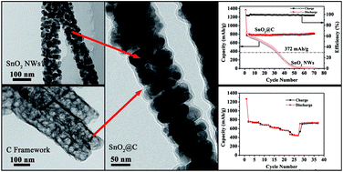
Amorphous carbon framework stabilized SnO2 porous nanowires (SnO2@C nanocomposites) were successfully synthesized through a hydrothermal-calcining method. The in situ formed amorphous carbon framework not only enhances the electron conductivity but also accommodates the volume expansion during the electrochemical cycling process. Benefiting from the structure and amorphous carbon framework, SnO2@C nanocomposites show ultra-excellent cycling performance, rate capability and high coulombic efficiency. At a rate of 782 mA g−1, their reversible capacity is as high as 751 mA h g−1 with no capacity fading after 160 cycles. These experimental findings may provide some insights to further improve the cyclability and rate capability of anode materials, paving the way for the next-generation high performance Li-ion batteries.


1 Introduction

Lithium ion batteries (LIBs), characteristic of relatively high operating voltage, high energy and power density and long cycle-life, have been widely used in various electronic devices and electric tools.1 Graphite-based materials have been utilized widely as the most important anode materials in commercial LIBs due to their low cost and good capacity retention (theoretical capacity 372 mA h g−1). However, with the increasing demands, especially for power-grid applications and electric or hybrid vehicles, new anode materials with higher specific capacity and/or higher power density are urgently needed to replace conventional graphite-based materials.2–8

As an alternative competition, SnO2 becomes a promising candidate material due to its higher theoretical capacity of 782 mA h g−1.3 However, much lager volume change (over 200%) occurred upon lithium insertion–extraction causes the pulverization phenomena,9,10 leading to a fast capacity fading during long-term electrochemical cycles, especially under high current density charge–discharge process. Nanostructuration and porous structures showing evident advantages to effectively mitigate the pulverization phenomena, have been considered as promising strategy to prepare high performance SnO2-based anode materials.11–14 For instance, Fan et al. prepared porous SnO2 nanowire bundles via a wet chemical method and achieved a corresponding specific capacity as higher as 566 mA h g−1 after 20 cycles under a current density of 180 mA h g−1.15 Moreover, Yin et al. reported mesoporous SnO2 nanospheres with a specific capacity of 557 mA h g−1 after 40 cycles.16 Despite the cycling performance was improved by nanostructuration and porous structures to some extent, the capacity retention and rate capability of SnO2 is still a challenge due to the destruction of the nanostructures and poor conductivity of SnO2. Hence, porous nanocomposites with a highly conductive and flexible phase would be the most promising candidate materials to achieve superior cycle performance and remarkable rate capability simultaneously. Recently, amorphous carbon is consider to be suitable as the second component and it has been found that the amorphous carbon can accommodate the volume expansion and effectively improved the conductivity of the composites.17,18

Hence, a novel nanocomposite contains SnO2 porous nanowires and amorphous carbon is considered to be the most feasible strategy to solve the above problems. The SnO2 porous nanowires are filled by in situ formed amorphous carbon framework and encapsulated by amorphous carbon layer, which not only stabilizes the structure of SnO2 porous nanowires, but also improves the electron conductivity between the SnO2@C nanocomposites and collector.

In this study, we report an effective method to synthesize amorphous carbon framework stabilized SnO2 porous nanowires (SnO2@C nanocomposites) for lithium ion battery anode materials, and investigate the electrochemical performance systematically. Moreover, we attempted to present a clear portrait of the key factor in determining the enhanced electrochemical performance.

2 Experimental section

Characterization

The transmission electron microscopy (TEM, JEOL JEM-2100F) and the scanning electron microscopy (SEM, FEI Nova-400) were used to characterise the morphology of all samples. Powder X-ray diffraction (XRD) patterns of the samples were collected using a D/ruax2550PC (Japan, Cu Kα radiation (λ = 0.1542 nm)). The SnO2 content in SnO2@C nanocomposites was estimated by thermogravimetric analysis (TGA, SNTQ600). The specific surface area of the SnO2 porous nanowires and SnO2@C nanocomposites were measured using a BET analyzer (ASAP 2020M) at 77 K. Electrochemical performance were tested on an electrochemical analyzer (LAND 2001A) using coin-type cells (CR2025) with Li metal as a counter and reference electrode at room temperature. The commercial electrolyte was used in the test, which contains 1.0 mol L−1 solution of LiPF6 in EC–EMC–DMC (1[thin space (1/6-em)]:[thin space (1/6-em)]1[thin space (1/6-em)]:[thin space (1/6-em)]1, volume ratio).

Synthesis of SnO2 porous nanowires

SnO2 porous nanowires (SnO2 NWs) were fabricated according to a previously reported method with slightly modified.19 Briefly, 5 g oxalic acid dehydrate was dispersed completely in 50 mL polyethylene glycol mixture (PEG400[thin space (1/6-em)]:[thin space (1/6-em)]PEG200 = 9[thin space (1/6-em)]:[thin space (1/6-em)]1, volume ratio) under magnetic stirring. After several minutes stirring, a certain amount of SnCl2·2H2O dissolved in ethanol and ethylene glycol (1[thin space (1/6-em)]:[thin space (1/6-em)]1, volume ratio) was added to the above solution, and continuously stirred for another 20 min. Then 2 mL deionized water was added into the solution, a white suspension was yielded after stirring for half an hour. After stirring for another 15 min, the as-prepared precipitation (SnC2O4 precursor) was separated by centrifugation, washed several times alternately with deionized water and absolute ethanol, and then dried in electric oven at 60 °C. Then the as-obtained SnC2O4 powder was calcined at 800 °C for 2.5 h, the SnO2 porous nanowires were synthesized.

Synthesis of SnO2@C nanocomposites

Carbon framework stabilized SnO2 nanocomposites were synthesized via a simple hydrothermal-calcining process. Typically, 0.3 g as-prepared SnO2 NWs was dispersed in ethanol/water solution which contains 30 mL ethanol and 25 mL water. Then 6 g D-glucose was added into the solution under vigorous stirring. After stirring for 15 min, the solution was transfer into a 100 mL Teflon-lined stainless steel autoclave, followed by heated to 180 °C and maintained for 12 h. After cooling down, the precipitate was separated by filter and washed with deionized water. SnO2 NWs with carbon-rich polysaccharide framework were obtained by drying the precipitate in electric oven at 90 °C over night. After being calcined at 600 °C in the argon flow for 3 h, the SnO2@C nanocomposites were successfully synthesized.

3 Results and discussion

The synthesis procedures are illustrated in Scheme 1. SnO2 porous nanowires were firstly prepared by calcining the stannous oxalate (SnC2O4) precursor in the air. The heating rate is about 5 °C min−1 and influences the pores structure of the SnO2 nanowires (Fig. S1). After a simple hydrothermal process, glucose-derived carbon-rich polysaccharide (GCP) framework were formed. Then, GCP framework was in situ converted into amorphous carbon framework after calcining in argon flow. After being grinding, SnO2@C nanocomposites were successfully obtained.
image file: c5ra05372b-s1.tif
Scheme 1 Schematic of the synthesis procedures of SnO2@C nanocomposites.

Fig. 1 shows the XRD patterns of the samples. It is easy to confirm that SnO2 nanowires contain two phases. The main reflections for all the samples at 26.60°, 33.90°, 37.97° and 51.81°, corresponding to (110), (101), (200) and (211) planes, are well match the major phase, which were indexed as rutile-structured SnO2 (PDF#70-4177). The diffraction peaks at 29.92° and 34.34°, corresponding to the (111) and (021) of SnO2 (PDF#29-1484), are in good agreement with synthesis condition. The two peaks around 32° are identified to metallic tin (Sn). The reason for these peaks is mainly ascribed to the carbon-thermal reduction of SnO2 during the calcining process.20 There are no peaks of carbon were observed, it means that the carbon framework is amorphous.


image file: c5ra05372b-f1.tif
Fig. 1 XRD patterns of SnO2 porous nanowires, SnO2@GCP and SnO2@C samples.

As shown in Fig. 2, X-ray photoelectron spectroscopy (XPS) was used to further confirm the state of carbon framework and SnO2 NWs. Fig. 2a shows the full survey spectrum (0–1200 eV). It is obvious that only the typical peaks of C (C 1s), O (O 1s) and Sn (Sn 4p, Sn 3d, Sn 3p) are observed. The Sn 3d peaks of the sample are shown in Fig. 2b. These two peaks are centered at 487.0 eV and 495.4 eV, respectively, which are attributed to Sn 3d5/2 and Sn 3d3/2 in SnO2.21 The gap between the Sn 3d5/2 and Sn 3d3/2 level is 8.5 eV and the area ratio of this two peaks is about 3/2, which means the chemical states of Sn is mostly in the form of SnO2 and there is little amount of SnO2 was reduced by the carbon-thermal reaction. The O 1s peaks is shown in Fig. 2c, which can be deconvoluted into four peaks corresponding to 531.0 eV (SnO2), 532.3 eV (H2O),21 531.5 eV (C–O) and 533.5 eV (C[double bond, length as m-dash]O),17 respectively. Fig. 2d show the XPS spectra of C 1s, which can be fitted into three peaks centered at 284.7, 286.2 and 288.6 eV. The peak centered at 284.7 eV is dominated by amorphous carbon and the other two peaks located at 286.2 and 288.6 eV are corresponding to carbon bonds in C–O and C[double bond, length as m-dash]O, respectively.17,21,22


image file: c5ra05372b-f2.tif
Fig. 2 XPS spectrum of SnO2@C nanocomposites.

The morphology and structure evolution of SnC2O4 precursor, SnO2 porous nanowires, SnO2@C nanocomposites and carbon framework were examined with transmission electron microscopy (TEM). SnC2O4 precursor (Fig. 3a) was converted into SnO2 porous nanowires (Fig. 3b) with a diameter of about 90 nm after calcining at 800 °C in air. Obviously, there exist many interconnected pores within SnO2 nanowires. The selected area electron diffraction (SAED) pattern (inset in Fig. 3b) and the lattice fringes (inset in Fig. 3c and d) reveal the polycrystalline nature of the SnO2 porous nanowires. The morphology of the SnO2@C nanocomposites (Fig. 3c and d) confirms that the porous structure of SnO2 nanowires was conserved during the fabrication process. The thickness of the outer carbon layer were estimated to be ∼3 nm (Fig. 3c, contrast sample) and ∼8 nm (Fig. 3d), respectively. The interplanar distances were estimated to be 0.335 and 0.264 nm, corresponding to (110) and (101) planes of the rutile SnO2 (PDF#70-4177). Fig. 3e shows the SEM image of SnO2@C nanocomposites, it is obvious that SnO2 nanowires were completely embedded in amorphous carbon framework. After removing SnO2 from SnO2@C nanocomposites by using thermal evaporation method, amorphous carbon framework (Fig. 3f) was obtained. It is evident that the interconnected pores of SnO2 porous nanowires were filled by the amorphous carbon framework. And the framework was connected with the outer carbon layer. This result indicates that carbon framework stabilized SnO2 nanocomposites were successfully synthesized.


image file: c5ra05372b-f3.tif
Fig. 3 Transmission electron microscopy (TEM) images of (a) SnC2O4 precursor, (b) porous SnO2 nanowires, and (c), (d) SnO2@C nanocomposites with different thickness of outer carbon layer. (e) SEM image of SnO2@C and (e) in situ formed carbon framework after SnO2 removed from SnO2@C nanocomposites. The inset in (b) was the selected area electron diffraction of porous SnO2 nanowires. The insets (c) and (d) were the high-resolution TEM images of the SnO2@C nanocomposites.

The mass percentages of amorphous carbon framework were estimated by the thermogravimetry analysis (TGA). As shown in Fig. 4, the weight loss processes are mainly observed between 400 °C and 630 °C primarily because of the removal of amorphous carbon. According to the TGA curve, the contents of amorphous carbon framework are about 41.8% (SnO2@C) and 19.6% (contrast sample) respectively.


image file: c5ra05372b-f4.tif
Fig. 4 TGA curves of SnO2@C nanocomposites.

To further study the fine structure of SnO2 porous nanowires and SnO2@C nanocomposites, Brunner–Emmett–Teller (BET) method was used to analyze the specific surface and pore size. Fig. 5a shows the nitrogen adsorption–desorption isotherms, it is noted that the SnO2@C nanocomposites deliver a large surface area of 197.5 m2 g−1, which is an order of magnitude higher than that of SnO2 nanowires (13.5 m2 g−1) and indicates that the major surface area is coming from amorphous carbon framework. Fig. 5b shows the pore size distribution. Clearly, a broad pore size distribution ranging from 20 to 40 nm exists in the SnO2 porous nanowires. After the amorphous carbon framework formed, the broad peak around 20–40 nm disappeared in the SnO2@C nanocomposites, while another peak appeared around 3.5 nm instead (insets in Fig. 5b) and revealed the existence of mesopores. This result further confirms that the SnO2 porous nanowires were filled with amorphous carbon framework and void space during the hydrothermal-calcining process.


image file: c5ra05372b-f5.tif
Fig. 5 (a) Nitrogen adsorption–desorption isotherms; (b) pore size distribution of the SnO2 and SnO2@C nanocomposites, insets are the corresponding magnified curves for SnO2 and SnO2@C, respectively.

The cyclic voltammograms (CV) test was carried out at a scan rate of 0.5 mV s−1 with in a potential range of 0.01–3 V (versus Li/Li+) at room temperature. Fig. 6 displays the first three cycles of cyclic voltammograms curve. Generally, the reactions of SnO2 with Li ions are generalized as follows:21

 
SnO2 + 4Li+ + 4e → Li2O + Sn (1)
 
Sn + xLi + xe ↔ LixSn (0 ≤ x ≤ 4.4) (2)


image file: c5ra05372b-f6.tif
Fig. 6 The cyclic voltammograms curves of the SnO2@C nanocomposites.

The reduction peak occurred around 0.61 V is observed only in the first cycle, which can be assigned to the formation of SEI films and the reduction of SnO2 to metallic tin (reaction (1)). And the peaks observed between 0.6 V–0.01 V in the cathodic sweep are related to the formation of LixSn alloys (reaction (2)). The corresponding anodic peak around 0.57 V appears in all the three cycles without voltage shift is the dealloying process of LixSn alloys (reaction (2)), indicating that the dealloying process of LixSn is completely reversible. Another peak at 1.25 V also appears in all three cycles, which corresponds to the oxidation of Sn to SnO, SnO2 and suggests the redox reaction of Sn is partially reversible.21,23 All the results demonstrated that the electrochemistry mechanism of the SnO2@C nanocomposites is well consistent with that of SnO2 anode materials.24 The peak around 1.9 V on the anodic sweep is attributed to the lithium ion diffusion out/into the interspace between carbon framework and SnO2 nanoparticles.

Fig. 7a presents the charge–discharge curves of the SnO2@C nanocomposites for the 1st, 2nd and 70th cycles. The initial discharge and charge capacities are 1371 and 775 mA h g−1 for SnO2@C nanocomposites (the specific capacity is calculated based on the total mass of SnO2@C nanocomposites). It is clear showed that all the charge–discharge plateaus are well consistent with the CV peaks in Fig. 6 and the first coulombic efficiency is estimated about 56.7%. The large initial irreversible capacity of 576 mA h g−1 is mainly owing to the decomposition of SnOx (0 ≤ x ≤ 2) and the formation of solid electrolyte interphase (SEI) films during the first discharge process.21 Generally, the larger the surface is, the more irreversible capacity will be. Hence, the large specific surface area is the main reason for the high initial irreversible capacity of SnO2@C nanocomposites.


image file: c5ra05372b-f7.tif
Fig. 7 (a) The charge–discharge curves of SnO2@C nanocomposites of the 1st, 2nd and 70th cycles between 0.01 V and 3 V at 0.5 C; (b) the cycling performance of SnO2 porous nanowires and SnO2@C nanocomposites (at 0.5 C); (c) the rate performances of SnO2@C nanocomposites at variable rates (0.2 C to 5 C); (d) the cycling performance of the SnO2@C nanocomposites and contrast sample at 1 C.

Fig. 7b shows the comparison of the cycling performance of the SnO2 porous nanowires and SnO2@C nanocomposites at a current density of 0.5 C. Obviously, SnO2@C nanocomposites present much better cycling stability and higher capacity. For the SnO2 porous nanowires electrode, the initial charge\discharge capacities are higher but fade fast. After 30 cycles, the capacities dropped vastly down to about 10 mA h g−1. As to SnO2@C nanocomposites, the capacities increase slightly over cycling. After 70 cycles, it delivered a reversible capacity of 811 mA h g−1. The increase of charge capacity is not fresh in tin oxides nanocomposites and other transition metal oxide nanomaterials/nanocomposites anode materials.21,25–30 As mentioned in the inset of Fig. 5d, the sharp peak of ∼3.5 nm suggests that interior mesopores were formed between carbon framework/layer and SnO2 nanoparticles due to the contraction of the GCP during the carbonized process. Then the solid electrolyte interfaces (SEI) layer formed at the interior mesopores, as a result of decomposition of electrolyte in the first cycle, assured the connection between SnO2 NPs and the carbon framework/layer and offered excess capacity through interfacial Li storage phenomena.28,31–33 Here, it's worth noting that the capacity of SnO2@C nanocomposites is slightly higher than the theoretical capacity of SnO2 (782 mA h g−1, calculated on Li4.4Sn). This phenomena was also reported by He's group and Guan's group.34 The main reason for the phenomena could be ascribed to side reactions occurred in the charge–discharge process, such as the oxide reaction of metallic Sn. As shown in Fig. 6, the peaks at 1.25 V appears in all three cycles are closely related to the oxidation of Sn to SnO, SnO2, suggesting that these side reactions are partially reversible.21 Because of the stabilization of amorphous carbon framework, the coulombic efficiency of SnO2@C electrode maintains above 97.8% except the first cycle (initial coulombic efficiency is 56.6%). The carbon framework accommodates the volume change and inhibits the formation of new surface during the cycling process. Hence, the side reactions between electrode and electrode are reduced, resulting the stabilization of coulombic efficiency.

Fig. 7c shows the rate performance of the SnO2@C electrode (from 0.2 C to 5 C) which is crucial for high performance Li-ion batteries. Due to the high electron conductivity and ion transport velocity, SnO2@C nanocomposites have inherent advantages in high-rate applications. The SnO2@C electrode delivered a capacity of 753 mA g−1 at 0.2 C, which is very close to the theory capacity of SnO2. After cycling at 0.2 C for 5 cycles, the current density increased stepwise to 5 C. With the increase of current density, the reversible capacity of SnO2@C electrode are 685 mA h g−1 at 0.5 C, 625 mA h g−1 at 1 C, 560 mA h g−1 at 2 C, respectively. Even at ultra-high current density of 5 C (3900 mA g−1), the reversible capacity is reached to about 470 mA h g−1. When the current density was returned to 0.2 C, a high reversible capacity of ∼730 mA h g−1 was recovered, implying that the reversibility of SnO2@C electrode is quite well and the structure of SnO2@C nanocomposites is durable for high rate charge/discharge process. These results indicate that the amorphous carbon framework enhanced SnO2 nanocomposites are of good electron conductivity and fast ion diffusion paths due to their unique structure.

Fig. 7d shows the cycling performance of the SnO2@C and the contrast samples at 1 C. SnO2@C electrode delivered an initial capacity of 706 mA h g−1. After cycling for 160 cycles, the reversible capacity increased gradually to a maximum of 751 mA h g−1. As a comparison, the initial capacity of contrast sample is as high as 912 mA h g−1. After 40 cycles, the capacity of contrast sample fades fast to about 30 mA h g−1. However, the cycling performance of contrast sample is much better than that of pure SnO2 porous nanowires. All the results demonstrate that the amorphous carbon framework plays a key role in the performance of SnO2 based electrode. Without carbon framework, the cycling performance of SnO2 porous nanowires is very poor. However, even there exist carbon framework in electrode materials, the thickness of the outer carbon layer is crucial.20 On the one hand, the outer carbon layer could accommodate the volume change of Li insertion/desertion in SnO2 nanocrystals, and the flexible carbon framework prevent the aggregation of the nanosized SnO2 active materials during charger–discharge cycling.20,35 On the other hand, the outer carbon layer and carbon framework formed a three-dimensional (3D) network with high electronic conductivity, and thus facilitated the charge transfer from active materials down to the collector.36,37 Meanwhile, one-dimensional (1D) porous nanowire feature could also offer short Li+ diffusion distance and large interfacial contact area between electrode and electrolyte for the fast transport of Li+. The resistance of SnO2@C electrodes was lower than that of SnO2 nanowires, and the SnO2@C electrode (with ∼8 nm out carbon layer) own the lowest resistance (Fig. S2). These results show that the presence of carbon framework in the composite reduces the resistance of charge transport. Benefiting from all features of this special structure, the SnO2@C electrode shows the excellent cycling performance and rate capability.

4 Conclusion

In summary, amorphous carbon framework stabilized SnO2 nanowires were successfully synthesized by a facile approach. The SnO2@C nanocomposites showed very excellent cycling performance and rate capability. The bifunctional amorphous carbon framework not only enhances the electron conductivity but also accommodates the large volume expansion during the charge–discharge process. It is anticipated that the experimental findings may provide some insights to further improve the cyclability and rate capability of anode materials, paving the path for the next-generation high performance Li-ion batteries.

Acknowledgements

We gratefully acknowledge financial support by the National Science Foundation of China (51302325), Science Fund for Distinguished Young Scholars of Hunan Province (2015JJ1016), Program for New Century Excellent Talents in University (NECT-12-0553), the fund of the State Key Laboratory of Advanced Technologies for Comprehensive Utilization of Platinum Metals (SKL-SPM-201507), the Scientific Research Foundation for the Returned Oversea China Scholars, the Hunan Youth Innovation Platform and Program for Shenghua Overseas Talent (90600-903030005; 90600-996010162) from Central South University (CSU).

References

  1. B. Dunn, H. Kamath and J. M. Tarascon, Science, 2011, 334, 928 CrossRef CAS PubMed .
  2. M. Armand and J. M. Tarascon, Nature, 2008, 451, 652 CrossRef CAS PubMed .
  3. Y. Idota, Science, 1997, 276, 1395 CrossRef CAS .
  4. C. K. Chan, H. Peng, G. Liu, K. McIlwrath, X. F. Zhang, R. A. Huggins and Y. Cui, Nat. Nanotechnol., 2008, 3, 31 CrossRef CAS PubMed .
  5. L. Zhang, H. B. Wu, R. Xu and X. W. Lou, CrystEngComm, 2013, 15, 9332 RSC .
  6. L. Yu, H. B. Wu and X. W. Lou, Adv. Mater., 2013, 25, 2296 CrossRef CAS PubMed .
  7. N. Mahmood, C. Zhang, F. Liu, J. Zhu and Y. Hou, ACS Nano, 2013, 7, 10307 CrossRef CAS PubMed .
  8. J. Xiao, D. W. Choi, L. Cosimbescu, P. Koech, J. Liu and J. P. Lemmon, Chem. Mater., 2010, 22, 4522 CrossRef CAS .
  9. C. M. Wang, W. Xu, J. Liu, J. G. Zhang, L. V. Saraf, B. W. Arey, D. Choi, Z. G. Yang, J. Xiao, S. Thevuthasan and D. R. Baer, Nano Lett., 2011, 11, 1874 CrossRef CAS PubMed .
  10. S. Ding, J. S. Chen, G. Qi, X. Duan, Z. Wang, E. P. Giannelis, L. A. Archer and X. W. Lou, J. Am. Chem. Soc., 2011, 133, 21 CrossRef CAS PubMed .
  11. X. Song, Nanotechnology, 2013, 24, 205401 CrossRef PubMed .
  12. A. Vu, Y. Q. Qian and A. Stein, Adv. Energy Mater., 2012, 2, 1056 CrossRef CAS PubMed .
  13. H. X. Chen, Q. B. Zhang, J. X. Wang, Q. Wang, X. Zhou, X. H. Li, Y. Yang and K. L. Zhang, Nano Energy, 2014, 10, 245 CrossRef CAS PubMed .
  14. Q. Zhang, J. Wang, J. Dong, F. Ding, X. Li, B. Zhang, S. Yang and K. Zhang, Nano Energy, 2015, 13, 77 CrossRef CAS PubMed .
  15. Y. T. Han, X. Wu, Y. L. Ma, L. H. Gong, F. Y. Qu and H. J. Fan, CrystEngComm, 2011, 13, 3506 RSC .
  16. X. K. Wang, Z. Q. Li, Q. Li, C. B. Wang, A. L. Chen, Z. W. Zhang, R. H. Fan and L. W. Yin, CrystEngComm, 2013, 15, 3696 RSC .
  17. X. Wang, H. Fan, P. Ren and M. Li, RSC Adv., 2014, 4, 10284 RSC .
  18. L. Zhang, G. Q. Zhang, H. B. Wu, L. Yu and X. W. Lou, Adv. Mater., 2013, 25, 2589 CrossRef CAS PubMed .
  19. Y.-X. Yin, L.-Y. Jiang, L.-J. Wan, C.-J. Li and Y.-G. Guo, Nanoscale, 2011, 3, 1802 RSC .
  20. X. Y. Yu, S. Y. Yang, B. H. Zhang, D. Shao, X. M. Dong, Y. P. Fang, Z. S. Li and H. Q. Wang, J. Mater. Chem., 2011, 21, 12295 RSC .
  21. M. He, L. Yuan, X. Hu, W. Zhang, J. Shu and Y. Huang, Nanoscale, 2013, 5, 3298 RSC .
  22. F. H. Li, J. F. Song, H. F. Yang, S. Y. Gan, Q. X. Zhang, D. X. Han, A. Ivaska and L. Niu, Nanotechnology, 2009, 20, 6 Search PubMed .
  23. F. Han, W.-C. Li, M.-R. Li and A.-H. Lu, J. Mater. Chem., 2012, 22, 9645 RSC .
  24. M. S. Park, G. X. Wang, Y. M. Kang, D. Wexler, S. X. Dou and H. K. Liu, Angew. Chem., Int. Ed., 2007, 46, 750 CrossRef CAS PubMed .
  25. Y. Yu, C. H. Chen and Y. Shi, Adv. Mater., 2007, 19, 993 CrossRef CAS PubMed .
  26. H.-X. Zhang, C. Feng, Y.-C. Zhai, K.-L. Jiang, Q.-Q. Li and S.-S. Fan, Adv. Mater., 2009, 21, 2299 CrossRef CAS PubMed .
  27. J. Wang, Q. Zhang, X. Li, B. Zhang, L. Mai and K. Zhang, Nano Energy, 2015, 12, 437 CrossRef CAS PubMed .
  28. Z. Y. Wang, D. Y. Luan, S. Madhavi, Y. Hu and X. W. Lou, Energy Environ. Sci., 2012, 5, 5252 CAS .
  29. Q. Zhang, H. Chen, J. Wang, D. Xu, X. Li, Y. Yang and K. Zhang, ChemSusChem, 2014, 7, 2325 CrossRef CAS PubMed .
  30. Y. Shi, B. Guo, S. A. Corr, Q. Shi, Y. S. Hu, K. R. Heier, L. Chen, R. Seshadri and G. D. Stucky, Nano Lett., 2009, 9, 4215 CrossRef CAS PubMed .
  31. P. Balaya, H. Li, L. Kienle and J. Maier, Adv. Funct. Mater., 2003, 13, 621 CrossRef CAS PubMed .
  32. J. Y. Shin, D. Samuelis and J. Maier, Adv. Funct. Mater., 2011, 21, 3464 CrossRef CAS PubMed .
  33. Y. F. Zhukovskii, P. Balaya, E. A. Kotomin and J. Maier, Phys. Rev. Lett., 2006, 96, 058302 CrossRef .
  34. J. Li, Y. Zhao, N. Wang and L. Guan, Chem. Commun., 2011, 47, 5238 RSC .
  35. P. Wu, N. Du, H. Zhang, J. X. Yu and D. R. Yang, J. Phys. Chem. C, 2011, 115, 3612 CAS .
  36. S. W. Kim, D. H. Seo, H. Gwon, J. Kim and K. Kang, Adv. Mater., 2010, 22, 5260 CrossRef CAS PubMed .
  37. P. Wu, N. Du, H. Zhang, J. Yu, Y. Qi and D. Yang, Nanoscale, 2011, 3, 746 RSC .

Footnote

Electronic supplementary information (ESI) available. See DOI: 10.1039/c5ra05372b

This journal is © The Royal Society of Chemistry 2015
Click here to see how this site uses Cookies. View our privacy policy here.