DOI:
10.1039/C5RA05350A
(Paper)
RSC Adv., 2015,
5, 43824-43830
Coumarin-modified gold nanoprobes for the sensitive detection of caspase-3
Received
26th March 2015
, Accepted 23rd April 2015
First published on 23rd April 2015
Abstract
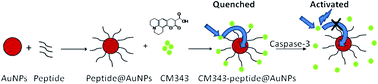
Caspase-3 has been identified as a key mediator and a well-established cellular marker of apoptosis. To increase the sensitivity, coumarin-functionalized gold nanoparticles (AuNPs) connected to polypeptide chains containing specific sequences (DEVD) were designed and synthesized for the sensing of caspase-3, because there was a large overlap between the emission of coumarin-343 and the absorption of the AuNPs. The fluorescence of coumarin 343 was quenched due to the energy transfer process by the gold nanoparticles. The fluorescence could be restored after the particular polypeptide sequence (DEVD) was cut off by caspase-3. Based on this mechanism, the caspase enzyme activity in vitro could be detected by a fluorescence assay with a high sensitivity. The effect of the different lengths of polypeptide chains on the luminescence quenching efficiency and sensing ability was also studied, which is of great importance in designing FRET-based sensing platforms. This kind of sensitive luminescent functional coumarin 343-modified gold nanoprobe is suitable for caspase-3 sensing in biological applications.
Introduction
Recently, gold nanoparticles (AuNPs) have become one of the most widely studied materials due to the advantages such including small size, ease of manufacture, the resonant nature having ion conductivity and biocompatibility, localized surface plasma resonance, special optical activity and surface modification characteristics.1,2 The applications of Au nanoparticles in bioanalysis have drawn great attention due to their high extinction and strong size- and distance-dependent optical properties.3–10 The appealing feature of high extinction coefficients in the visible region enables them to function as efficient quenchers for most fluorophores in the design of biological sensors such as proteases, DNA and so on.11–16
Apoptosis is an important physiological mechanism to maintain homeostasis of multicellular organisms by elimination of infected or damaged cells and regulation of cell numbers.17,18 Caspases, which were discovered recently, are a group of cysteine proteases existing in cytoplasmic sol. The proteases can catalytically crack (ADP ribose) polymerase and lead to cell apoptosis.19 There are 13 known species, including caspase-3 and caspase-7, which have similar substrate and inhibitor specificities.20,21 They can cut off the aspartic acid residue of a peptide bond (DEVD) in a specific way and degrade the PARP (DNA repair enzyme) and DFF-45 (DNA fragmentation factor), which results in the inhibition of DNA repair and the starts the degradation of DNA. Caspase-3 is activated in apoptotic cells both by extrinsic and intrinsic pathways, and has been identified as a key mediator and a well-established cellular marker of apoptosis.22 Accordingly, the development of a highly sensitive and specific detection system for caspase activities will provide new insights into the roles of proteases in biological events. Based on the special activity of caspase-3, various caspase assays were developed by utilizing fluorescence resonance energy transfer (FRET) between a donor and acceptor pair, which involved conventional organic dyes,23–25 fluorescent proteins,26–30 or quantum dots (QDs)31 that are paired with quencher moieties. A simple strategy for the detection of proteolytic activity was designed by the use of peptide substrates containing a FRET pair, fluorescein and tetramethylrhodamine.25 Genetically encoded fluorescent indicators that include green-fluorescent protein (GFP) derivatives have also become valuable tools for studying temporal caspase activity in single living cells.13,14 A near-infrared fluorescence dye was attached to the AuNP surface through the bridge of the peptide substrate (DEVD) to fabricate an apoptosis imaging probe.32 A specific FRET-based protease sensor for caspase-3 was also developed based on quantum dots (QDs) as the energy donors and the fluorescent protein mCherry as the energy acceptor.31 Recently, some hybrid nanomaterial-based sensors and probes were successfully developed for the detection of caspase-3 in our work, in which [Ru(bpy)3]2+-encapsulated silica nanoparticles (SiNPs) were used as fluorescence energy donors and gold nanoparticles (AuNPs) were used as energy acceptors.33 However, further development of a highly sensitive luminescent probe for caspase-3 sensing in analytical- and bio-systems is still a challenge.
To further increase the sensitivity, a FRET system with highly fluorescent coumarin 343 (CM343) as the energy donor and AuNPs as the energy acceptors has been designed and applied for caspase-3 detection. Because the fluorescence of the coumarin 343 has a large overlap with the absorption band of the AuNPs, the fluorescence quenching property of the gold nanoparticles was utilized to efficiently quench the fluorescence of coumarin 343. Another advantage of coumarin 343 as an emission donor is the much higher quantum yield compared to luminescent transition metal-based complexes, although the lifetime is shorter.34 Two kinds of CM343–peptide@AuNP probes were designed and synthesized with two different lengths of peptide chains linking CM343 and the AuNPs, to tune the distance between the donor and the acceptor. The peptide chains contained a recognition sequence, DEVD, specific for active caspase-3 cleavage.35 A difference in distance between the two building blocks resulted in a difference in FRET efficiency on account of the different lengths of the peptide chains. The synthesis and principle of the sensor is shown in Scheme 1. The fluorescence was quenched in physiological conditions due to the quenching effect of the AuNPs. When caspase-3 triggers the cleavage of the linkers and the system releases coumarin 343 from the Au nanoparticles, the quenched fluorescence was recovered and the fluorescence efficiency increased. Based on this FRET mechanism, caspase-3 could be detected in a simple and sensitive way, which might be significant in bioanalysis and biodetection.
 |
| Scheme 1 Schematic diagram of the synthesis and principle of the functionalized AuNPs. | |
Experiments
Materials and chemicals
Coumarin 343 was synthesized in our group according to a previous report.36 The peptides (95%, target sequence 1: Gly-Asp-Glu-Val-Asp-Cys; target sequence 2: Gly-Gly-Ala-Asp-Glu-Val-Asp-Gly-Cys) were purchased from Sangon Biotech (Shanghai) Co., Ltd. Caspase-3 (3000 pmol min−1 μg−1) was purchased from R&D Systems. Chloroauric acid (HAuCl4) and sodium citrate were purchased from Sigma-Aldrich. Other chemicals were of analytical reagent grade and used as received.
Experimental apparatus
The fluorescent and UV-vis spectra were obtained on a Hitachi F-4600 spectrofluorometer and a Perkin Elmer LAMBDA 750 UV-vis spectrometer, respectively. Raman spectra were obtained on a Renishaw InVia Raman spectrometer. Transmission electron microscopy (TEM) images were obtained using a Tecnai G2F20 High-Resolution Transmission Electron Microscope (Manufacturer: FEI, Ltd., America). Dynamic light scattering (DLS) experiments were carried out on a Malvern Zetasizer NanoZS90.
Synthesis of AuNPs
Aqueous dispersions of citrate-stabilized AuNPs were prepared by the citrate reduction of chloroauric acid as described in detail elsewhere.37 Briefly, a 50 mL aqueous solution of HAuCl4 (0.25 mM) was heated to boiling point under vigorous stirring, and then 0.5 mL of trisodium citrate solution (1%) was quickly added. The reaction was allowed to continue for another 20 min. The prepared citrate-stabilized AuNPs generally appeared red in color. Then, the solution was cooled to room temperature and stored at 4 °C in the refrigerator for further use. The AuNPs were characterized by TEM and DLS experiments.
Functionalization of the AuNPs
The peptides (target sequence 1: Gly-Asp-Glu-Val-Asp-Cys; target sequence 2: Gly-Gly-Ala-Asp-Glu-Val-Asp-Gly-Cys) were conjugated to the AuNPs via the Au–S bond, by a slight modification of a method previously described in the literature.38 Briefly, 375 μL of 15.7 mM peptide solution was added to 20 mL of 5.9 nM AuNPs, followed by incubation for 4 h. Excess peptides were then removed by centrifugation at 12
000 rpm for 30 min. The resultant peptide-modified AuNPs (peptide@AuNPs) were washed with DI water and redispersed in phosphate buffer solution (PBS, 10 mM, pH 7.2). The peptide-modified gold colloids appeared wine red after being centrifuged, suggesting sufficient steric stabilization.
Carbodiimide chemistry was employed to functionalize the AuNPs with CM343 by modification of a method perviously described in the literature.39 Briefly, 180 μL of 13 mM CM343 was suspended in 10 mL of 0.05 M 4-(2-hydroxyethyl)-1-piperazineethanesulfonic acid (HEPES, pH 7.2) buffer containing 1.20 mM N-hydroxysuccinimide (NHS) and 1.20 mM 1-ethyl-3-(3-dimethyl-aminopropyl)carbodiimide (EDC) to activate the carboxylic groups. After 1 h, 20 mL of 5.9 nM peptide@AuNPs were added to the mixture, and it was stirred for 12 h at room temperature. Excess CM343 was then removed by centrifugation at 12
000 rpm for 30 min. The AuNPs modified by CM343 (CM343–peptide@AuNPs) were washed with 10 mM PBS (pH 7.2) and then resuspended in 10 mM PBS (pH 7.2). At last, the AuNPs modified by CM343 (CM343–peptide@AuNPs) appeared deep red and were stored in the refrigerator at 4 °C for further use. Then the functionalized AuNPs were characterized by Raman, UV-vis absorption and fluorescent spectra.
Sensing application
For caspase-3 sensing, the functionalized CM343–peptide@AuNPs (5.9 nmol, 2 mL) were incubated with caspase-3 (final concentrations: 0.004–1.8 ng mL−1) in assay buffer (total volume 2.5 mL, pH 7.4, 50 mM HEPES, 100 mM NaCl, 1 mM EDTA, 10% v/v glycerol and 0.1% v/v CHAPS) for 0–150 min at 37 °C. Then the emission intensity was recorded on the F-4600 spectrofluorometer. The optimum response time was about 30 min in our experiments. The control experiment was also carried out for the CM343–peptide@AuNP system in the absence of caspase-3. To evaluate the selectivity of the functionalized AuNPs toward caspase-3, a series of proteases including trypsin, chymotrypsin and esterase, and ions including K+, Na+, Cl, were examined for their possible interference in caspase-3 detection.
Caspase-3 determination with the nanohybrid sensor was also conducted in cell extracts.40 Hela299 cells were obtained from the Type Culture Collection of the Chinese Academy of Sciences (CAS, Shanghai, China), and were cultured in DMEM (supplemented with 15% fetal bovine serum) at 37 °C in a humidified 5% CO2 atmosphere. 1.0 × 10⁵ cells were collected in the exponential phase of growth, and were then dispensed in an EP tube. After washing twice with ice-cold PBS, cells were resuspended in 200 μL of ice-cold CHAPS buffer. The suspension was subjected to sonication in an ice bath (KQ3200DB sonicator amplitude set at 70%) for 30 min and centrifuged at 12
000 rpm for 20 min. The supernatant was carefully transferred to a new EP tube, and was used immediately for caspase-3 detection after 6-fold dilution with assay buffer or stored at −70 °C. 250 μL diluted cell extracts incubated with the functionalized CM343–peptide@AuNPs (5.9 nmol, 2 mL) in buffer solution (total volume 2.5 mL) for 30 min at 37 °C were used to determine the caspase-3 in cell extracts. In the standard addition experiment, 250 μL diluted cell extracts and 10 μL standard caspase-3 solution (0.2 μg mL−1) incubated with the functionalized CM343–peptide@AuNPs (5.9 nmol, 2 mL) in buffer solution (total volume 2.5 mL) for 30 min at 37 °C were used to determine the total concentration of caspase-3.
Results and discussion
Synthesis and functionalization of the AuNPs
The AuNPs were obtained by a citrate reduction of chloroauric acid.37 Fig. 1A displays a typical TEM image of the AuNPs, where the AuNPs are regular, monodisperse, and spherical in shape. The dynamic light scattering (DLS) experiment shows a diameter of ∼13 nm (Fig. 1B) and the Zeta potential is −39.33 mV. Peptides were functionalized onto the AuNPs via the Au–S chains (peptide@AuNPs), and the peptide-modified AuNPs generally appeared wine red suggesting sufficient steric protection by the peptides. The further functionalization of the peptide@AuNPs with CM343 was carried out using carbodiimide chemistry, where cross-linking reactions between free carboxylic groups of coumarin 343 and amine groups on the peptides were initiated by adding a solution of EDC and NHS.41 Fig. 2 shows the Raman spectra of CM343 and AuNPs functionalized by two kinds of peptides. The CM343–peptide@AuNP probes show similar features in the Raman spectra relative to CM343, similar to a previous report.42 The phenyl ring breathing motions are at 633–785 cm−1, the CH2 rock motions and C–C stretching vibrations are at 1200 cm−1, C
C, C–N and C
C stretching vibrations are at 1370–1574 cm−1, and the 1654 and 1728 cm−1 are C
O stretching modes. The results demonstrated that coumarin 343 had been successfully modified on the peptides of the gold nanoparticle surface by an amide bond.
 |
| Fig. 1 TEM images of the AuNPs (A) and the particle size distribution from the DLS experiments (B). | |
 |
| Fig. 2 Raman spectra of CM343 (A), CM343–peptide1@AuNPs (B), and CM343–peptide2@AuNPs (C). | |
The optical properties of the AuNPs were examined by UV-vis absorption spectra. As shown in Fig. 3, a characteristic surface plasmon resonance (SPR) peak is located at 520 nm. Compared to the AuNPs, the CM343–peptide1@AuNPs and CM343–peptide2@AuNPs showed a slight red shift of ca. 5 nm in the UV-vis absorption bands, which demonstrated that the conjugation of CM343 on the AuNP surfaces by polypeptide chains had a tiny effect on the aggregation of the AuNPs. The functionalized gold colloids generally appeared deep red after being centrifuged, which suggested sufficient steric stabilization after it was protected by the peptides.
 |
| Fig. 3 UV-vis absorption spectra of bare AuNPs (a), CM343 (b), CM343–peptide1@AuNPs (c), and CM343–peptide2@AuNPs (d). | |
The emission spectra of the CM343–peptide1@AuNPs, CM343–peptide2@AuNPs and CM343 in aqueous solution are also shown in Fig. 4. The CM343 gave rise to a strong emission at ca. 478 nm, and the CM343–peptide1@AuNPs and CM343–peptide2@AuNPs showed weak emissions at about 490 nm with a small red shift of ca. 12 nm in the emission wavelength. The excess adsorption of CM343 on the AuNP surface was removed by washing and centrifuging during the preparation and the partial CM343 was conjugated on the AuNP surface. The fluorescence of the conjugated CM343 was efficiently quenched by the AuNPs due to the large overlap between the fluorescence of CM343 and the absorption band of the functionalized AuNPs based on the FRET process from CM343 to the AuNPs. The above results further suggested that CM343 had successfully been modified on the peptide@AuNPs.
 |
| Fig. 4 Fluorescence spectra of CM343 (a), CM343–peptide1@AuNPs (b), and CM343–peptide2@AuNPs (c). | |
Sensing application of the CM343–peptide@AuNPs
Except for the overlap of the emission spectra of a donor and the absorption band of an acceptor, the FRET process also depends on the distance between the donor and the acceptor. In our assembled CM343–peptide@AuNPs nanohybrid, the distance between the AuNPs and CM343 can be tuned through engineering the bridging molecules. This property can be utilized for developing new sensing strategies through employing FRET technology, which is a distance-dependent energy transfer phenomenon. A change in the distance between the AuNPs and CM343 will result in a change in FRET efficiency and cause a ratiometric change in emission. Because the proteases can recognize and cleave a specific substrate peptide (DEVD) to produce two separated fragments and thus change the distance between the two ends of the peptide, it is possible to explore the biological sensing ability for caspase-3 and compare the the sensing ability for caspase-3 by the two different peptide links of the functionalized gold nanoparticles, in which both contain a specific sequence of DEVD. As shown in Fig. 5, when caspase-3 was introduced into these probe systems, a dramatic fluorescence recovery of the CM343–peptide@AuNP system was observed within 30 min, although a small fluorescence enhancement was detected for the CM343–peptide@AuNP systems without caspase-3 due to the gold nanoparticles’ electrostatic incorporation. Upon addition of caspase-3, the emission increased in intensity and reached saturation at about 1.6 ng mL−1 and 4.0 ng mL−1 of caspase-3 for the CM343–peptide1@AuNPs and CM343–peptide2@AuNPs, respectively. And the emission intensity was enhanced about 1-fold and 0.4-fold for the CM343–peptide1@AuNPs and CM343–peptide2@AuNPs, respectively, 30 mins after the additon of caspase-3. The results showed that the shorter linker between the fluorophore and the AuNP quencher would result in a higher FRET efficiency and the recovery of emission would be more obvious after the linkage was cleaved by caspase-3.
 |
| Fig. 5 (A) Fluorescence intensity of the CM343–peptide1@AuNPs in the presence of various concentrations of caspase-3 (0, 0.004, 0.02, 0.04, 0.08, 0.2, 0.4, 0.6, 0.8, 1.2, 1.6, 1.8 ng mL−1) as a function of incubation time. (B) Fluorescence intensity of the CM343–peptide2@AuNPs in the presence of various concentrations of caspase-3 (0, 0.04, 0.2, 0.4, 0.8, 1.6, 3.2, 4.0, 4.8, 6.0 ng mL−1) as a function of incubation time. | |
After the addition of caspase-3 to the CM343–peptide1@AuNP probes in a buffer solution, the fluorescence intensity increased significantly in 30 min. As shown in Fig. 6A, the fluorescence recovery of the CM343–peptide1@AuNP system was observed after only 0.004 ng mL−1 (∼1.2 pM) of caspase-3 was added, and the fluorescence intensity increased gradually with the increase of the caspase-3 concentration from 0.004 to 1.8 ng mL−1. The fluorescence recovery linearly correlated to the concentration of caspase-3 over the range of 0.08–1.2 ng mL−1 (inset of Fig. 7A). For the CM343–peptide2@AuNP probes, the fluorescence recovery of the CM343–peptide2@AuNP system was obtained after the addition of 0.04 ng mL−1 (∼12 pM) of caspase-3, and the fluorescence intensity increased gradually with the increase of the caspase-3 concentration from 0.04 to 6 ng mL−1 (Fig. 6B). The fluorescence recovery linearly correlated to the concentration of caspase-3 over the range of 0.20–3.2 ng mL−1 (Fig. 7B). The results further demonstrated that the shorter linkage between the donor and the acceptor would result in a higher FRET efficiency, which would cause more significant changes in the emission spectra after the addition of caspase-3 and show a higher sensitivity toward caspase-3.
 |
| Fig. 6 (A) Fluorescence spectra of CM343–peptide1@AuNPs in the presence of various concentrations of caspase-3 (0, 0.004, 0.02, 0.04, 0.0.08, 0.2, 0.4, 0.6, 0.8, 1.2, 1.6, 1.8 ng mL−1) after incubation for 30 min. (B) Fluorescence spectra of CM343–peptide2@AuNPs in the presence of various concentrations of caspase-3 (0, 0.04, 0.2, 0.4, 0.8, 1.6, 3.2, 4.0, 4.8, 6.0 ng mL−1) after incubation for 30 min. | |
 |
| Fig. 7 (A) Fluorescence intensity of CM343–peptide1@AuNPs as a function of caspase-3 concentration. (B) Fluorescence intensity of CM343–peptide2@AuNPs as a function of caspase-3 concentration. | |
The results show that the CM343–peptide1@AuNP system has a higher sensitivity toward caspase-3 by monitoring the fluorescence change of the peptide-bridged nanohybrid system. The limit of detection (LOD) of the CM343–peptide1@AuNP system toward caspase-3 was about 4 pg mL−1, which is more sensitive than in previous reports,32,33,43,44 probably due to a large overlap between the emission of coumarin 343 and the absorption of the AuNPs, which resulted in a high efficient energy transfer process between the donor and the acceptor in our system. The results showed that it will be potentially suitable for DNA sensing in bioanalytical applications.
Selectivity
To evaluate the selectivity of the functionalized AuNPs toward caspase-3, a series of proteases and ions were examined for their possible interference in caspase-3 detection. In this study, typical proteases including trypsin, chymotrypsin and esterase, and ions including K+, Na+, Cl−, have been evaluated for their interference to caspase-3 detection. As shown in Fig. 8, no obvious fluorescence recovery was observed for any inspected substances except for caspase-3. All the results indicated that the peptide linker containing a DEVD sequence shows a good specificity for caspase-3 cleavage, and suggested that the CM343–peptide@AuNP system demonstrated a high selectivity toward caspase-3 detection.
 |
| Fig. 8 Specificity test for caspase-3, where F0 represents the fluorescence intensity of the CM343–peptide1@AuNPs (A) and CM343–peptide2@AuNPs (B) and F is the fluorescence intensity of the functionalized AuNPs plus inspected species. | |
Determination of caspase-3 in samples
Caspase-3 determination with the functionalized AuNP sensor was also conducted in cell extracts. The CM343–peptide1@AuNP probe was used in this determination. Using the calibration curve obtained in the aqueous solution, the diluted caspase-3 concentration in cell extracts was determined as 0.26 ng mL−1. A standard addition experiment was also carried out, and the recovery was found to be 106.6%, with the RSD (Relative Standard Deviation) at around 1% (Table 1). The results demonstrated the sensing capabilities of the functionalized AuNP FRET system in a complex biological environment and the potential of this kind of nanosensor in bioanalysis and biodetection, which might be significant in disease diagnosis in the future.
Table 1 Analytical results of the determination of Caspase-3 in the cell extract sample
Sample |
Standard caspase-3 spiked (ng mL−1) |
Caspase-3 measured (ng mL−1) |
Recovery (%) |
RSD (%) (n = 3) |
Cell extracts |
0 |
0.26 ± 0.45 |
|
6.8 |
0.8 |
1.13 ± 0.18 |
106.6 |
1.1 |
Conclusion
In summary, CM343–peptide@AuNP probes have been fabricated through peptide-bridged assembly in a controllable way, and the functionalized AuNP system has been successfully employed for caspase-3 detection by tuning the FRET efficiency between CM343 and the AuNPs. A peptide containing recognition sequence, DEVD, specific for active caspase-3 cleavage was utilized to compose a functionalized AuNP biosensor based on a FRET process. Caspase-3 cleaves the peptide-bridge and releases CM343 from the AuNPs, which forms the basis of caspase-3 recognition. The CM343 functionalized AuNP biosensor by the proper linkage is highly sensitive and specific towards caspase-3 determination, which might be significant in bioanalysis and biodetection and could have potential applications in disease diagnosis in the future.
Acknowledgements
This work was supported by the National Scientific Foundation of China (no. 21171038 and no. J1103303), the Program for Changjiang Scholars and Innovative Research Team in University (no. IRT1116) and the Basic Research Program of Shenzhen Science and Technology Innovation Plan (JC201105201055A).
References
- S. R. Johnson, S. D. Evans and R. Brydson, Langmuir, 1998, 14, 6639 CrossRef CAS.
- J. Chen, Q. Huang and Q. G. Du, RSC Adv., 2014, 4, 57574 RSC.
- J. Liu and Y. Lu, Nat. Protoc., 2006, 1, 246 CrossRef CAS PubMed.
- S. Zeng, X. Yu, W.-C. Law, Y. Zhang, R. Hu, X.-Q. Dinh, H.-P. Ho and K.-T. Yong, Sens. Actuators, B, 2013, 176, 1128 CrossRef CAS PubMed.
- Q. H. Zeng, Y. L. Zhang, X. M. Liu, L. P. Tu, X. G. Kong and H. Zhang, Chem. Commun., 2012, 48, 1781 RSC.
- M. Xue, X. Wang, L. L. Duan, W. Gao, L. F. Ji and B. Tang, Biosens. Bioelectron., 2012, 36, 168 CrossRef CAS PubMed.
- C. A. Mirkin, R. L. Letsinger, R. C. Mucic and J. J. Storhoff, Nature, 1996, 382, 607 CrossRef CAS PubMed.
- K. Sato, K. Hosokawa and M. Maeda, J. Am. Chem. Soc., 2003, 125, 8102 CrossRef CAS PubMed.
- I. I. Lim, E. Crew, P. N. Njoki, D. Mott, C. J. Zhong, Y. Pan and S. Zhou, Langmuir, 2007, 23, 826 CrossRef CAS PubMed.
- M. Li, Q. Y. Wang, X. D. Shi, L. A. Hornak and N. Q. Wu, Anal. Chem., 2011, 83, 7061 CrossRef CAS PubMed.
- Y. Jiang, H. Zhao, Y. Lin, N. Zhu, Y. Ma and L. Mao, Angew. Chem., Int. Ed., 2010, 49, 4800 CrossRef CAS PubMed.
- M. J. Li, X. Liu, M. J. Nie, Z. Z. Wu, C. Yi, G. Chen and V. W. W. Yanm, Organometallics, 2012, 31, 4459 CrossRef CAS.
- M. De, P. S. Ghosh and V. M. Rotello, Adv. Mater., 2008, 20, 4225 CrossRef CAS PubMed.
- M.-J. Li, M.-J. Nie, Z.-Z. Wu, X. Liu and G.-N. Chen, Biosens. Bioelectron., 2011, 29, 109 CrossRef CAS PubMed.
- Y. Zhou, S. X. Wang, K. Zhang and X. Y. Jiang, Angew. Chem., Int. Ed., 2008, 47, 7454 CrossRef CAS PubMed.
- F. Y. Yeh, I. H. Tseng, S. H. Chuang and C. S. Lin, RSC Adv., 2014, 4, 22266 RSC.
- A. Ishaque and M. Al-Rubeai, in Cell Engineering, ed. M. Al-Rubeai and M. Fussenegger, Kluwer Academic Publishers, Dordrecht, 2005, pp. 281–306 Search PubMed.
- D. L. Vaux and S. J. Korsmeyer, Cell, 1999, 96, 245 CrossRef CAS.
- R. C. Taylor, S. P. Cullen and S. J. Martin, Nat. Rev. Mol. Cell Biol., 2008, 9, 231 CrossRef CAS PubMed.
- N. A. Thornberry, J. Biol. Chem., 1997, 272, 17907 CrossRef CAS PubMed.
- S. Lien, R. Pastor, R. Sutherlin and H. Lowman, Protein J., 2004, 23, 413 CrossRef CAS.
- K. Kim, M. Lee, H. Park, J. H. Kim, S. Kim, H. Chung, K. Choi, I. S. Kim, B. L. Seong and I. C. Kwon, J. Am. Chem. Soc., 2006, 128, 3490 CrossRef CAS PubMed.
- Y. Kushida, K. Hanaoka, T. Komatsu, T. Terai, T. Ueno, K. Yoshida, M. Uchiyama and T. Nagano, Bioorg. Med. Chem. Lett., 2012, 22, 3908 CrossRef CAS PubMed.
- V. Gurtu, S. R. Kain and G. H. Zhang, Anal. Biochem., 1997, 251, 98 CrossRef CAS PubMed.
- E. Reits, A. Griekspoor, J. Neijssen, T. Groothuis, K. Jalink, P. van Veelen, H. Janssen, J. Calafat, J. W. Drijfhout and J. Neefjes, Immunity, 2003, 18, 97 CrossRef CAS.
- T. Nagai and A. Miyawaki, Biochem. Biophys. Res. Commun., 2004, 319, 72 CrossRef CAS PubMed.
- K. Takemoto, T. Nagai, A. Miyawaki and M. Miura, J. Cell Biol., 2003, 160, 235 CrossRef CAS PubMed.
- X. Xu, A. L. V. Gerard, B. C. B. Huang, D. C. Anderson, D. G. Payan and Y. Luo, Nucleic Acids Res., 1998, 26, 2034 CrossRef CAS PubMed.
- N. P. Mahajan, D. C. Harrison-Shostak, J. Michaux and B. Herman, Chem. Biol., 1999, 6, 401 CrossRef CAS.
- S. Sakamoto, M. Terauchi, A. Hugo, T. Kim, Y. Araki and T. Wada, Chem. Commun., 2013, 49, 10323 RSC.
- K. Boeneman, B. C. Mei, A. M. Dennis, G. Bao, J. R. Deschamps, H. Mattoussi and I. L. Medintz, J. Am. Chem. Soc., 2009, 131, 3828 CrossRef CAS PubMed.
- I. C. Sun, S. Lee and H. Koo, et al., Bioconjugate Chem., 2010, 21(11), 1939 CrossRef CAS PubMed.
- Y. Shi, C. Yi, Z. Zhang, H. Zhang, M. J. Li, M. Yang and J. Qing, ACS Appl. Mater. Interfaces, 2013, 5, 6494 CAS.
- K. Huang, C. Jiang and A. A. Marti, J. Phys. Chem. A, 2014, 118, 10353 CrossRef CAS PubMed.
- G. M. Cohen, Biochem. J., 1997, 326, 1 CAS.
- M.-J. Li, K. M.-C. Wong, C. Yi and V. W.-W. Yam, Chem. - Eur. J., 2012, 18, 8724 CrossRef CAS PubMed.
- S. K. Ghosh, A. Pal and S. Kundu, et al., Chem. Phys. Lett., 2004, 395, 366 CrossRef CAS PubMed.
- W. Gao, L. F. Ji, L. Li, G. W. Cui, K. H. Xu, P. Li and B. Tang, Biomaterials, 2012, 33, 3710 CrossRef CAS PubMed.
- H. J. Chung, H. K. Kim and J. J. Yoon, et al., Pharm. Res., 2006, 23(8), 1835 CrossRef CAS PubMed.
- Y. P. Li, Microchim. Acta, 2012, 177, 443 CrossRef CAS.
- T. Greg, Bioconjugate Techniques, Academic, New York, 2nd edn, 1996, vol. 219, p. 598 Search PubMed.
- R. R. Frontiera, J. Dasgupta and R. A. Mathies, J. Am. Chem. Soc., 2009, 131, 15630 CrossRef CAS PubMed.
- H. Shi, R. T. K. Kwok and J. Liu, et al., J. Am. Chem. Soc., 2012, 134, 17972 CrossRef CAS PubMed.
- K. E. Bullok, D. Maxwell and A. H. Kesarwala, et al., Biochemistry, 2007, 46, 4055 CrossRef CAS PubMed.
|
This journal is © The Royal Society of Chemistry 2015 |
Click here to see how this site uses Cookies. View our privacy policy here.