Luísa C. R. Carvalhoa,
Daniela Ribeirob,
Raquel S. G. R. Seixasc,
Artur M. S. Silvac,
Mariana Navea,
Ana C. Martinsa,
Stefan Erhardtd,
Eduarda Fernandes*b,
Eurico J. Cabrita*e and
M. Manuel B. Marques*a
aLAQV@REQUIMTE, Departamento de Química, Faculdade de Ciências e Tecnologia, Universidade Nova de Lisboa, Campus de Caparica, 2829-516 Caparica, Portugal. E-mail: mmbmarques@fct.unl.pt
bUCIBIO@REQUIMTE, Departamento de Ciências Químicas, Laboratório de Química Aplicada, Faculdade de Farmácia, Universidade do Porto, Rua de Jorge Viterbo Ferreira no. 228, 4050-313 Porto, Portugal
cQOPNA & Departamento de Química, Universidade de Aveiro, 3810-193 Aveiro, Portugal
dSchool of Life, Sport and Social Sciences, Edinburgh Napier University, Sighthill Campus, Edinburgh EH11 4BN, UK
eUCIBIO@REQUIMTE, Departamento de Química, Faculdade de Ciências e Tecnologia, Universidade Nova de Lisboa, Campus de Caparica, 2829-516 Caparica, Portugal
First published on 27th May 2015
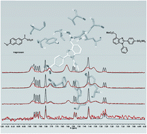
Non-steroidal anti-inflammatory drugs exert their pharmacological activity through inhibition of cyclooxygenase 1 and 2 (COX-1 and COX-2). Recent research suggests that a balanced inhibition of both COX-1 and COX-2 is the key to reduce the side-effects exhibited by COX inhibitors. We developed new benzimidazole-based compounds that showed a balanced COX inhibition, supported by molecular docking screening. The human whole blood assays demonstrated that the ester derivatives were potent inhibitors. Competitive saturation transfer difference (STD)-NMR experiments, in the presence of COX-2, using naproxen and diclofenac demonstrated that ester derivatives do not compete with diclofenac for the same binding site, but compete with the allosteric inhibitor naproxen. Combination of NMR spectroscopy with molecular docking has permitted us to detect a new naproxen-like inhibitor, which could be used for future drug development.
The unravelling of COX structure, and the main differences between COX-1 and COX-2 lead to the development of potent and highly selective COX-2 inhibitors.5 When compound DuP 697 was reported as the first selective COX-2 inhibitor in 1990, it was rapidly perceived that its shape, composed of a 1,2-diaryl heterocycle template, was an important feature to accommodate the drug inside the COX-2 active site.6 This observation formed the basis of the early work in the field of selective COX-2 inhibitors (usually known as coxibs). In the late 90s, several coxibs were released to the market, such as celecoxib, rofecoxib, valdecoxib and etoricoxib (Fig. 1).7 Coxibs selectively inhibit COX-2 and hamper the beneficial vascular effects of prostacyclin (PGI2) without blocking thromboxane A2 (TXA2) formation produced by COX-1. The loss of the antiplatelet and vasodilatory effects of PGI2, causes a relative excess of TXA2, promoting vasoconstriction, platelet aggregation and thrombosis.8 Consequently, some coxibs demonstrated acute cardiovascular side effects, and some of these drugs, like rofecoxib and valdecoxib, were removed from the market.9
Several studies indicate that prolonged NSAIDs use is also associated with a small increase in cardiovascular risk and that myocardial infarctions are comparable between coxibs and the traditional NSAIDs ibuprofen and diclofenac.10 Surprisingly, naproxen was not associated with an increase in cardiovascular events. The divergence between naproxen and other COX inhibitors (selective and nonselective) is not clear but may be related to its longer half-life compared to ibuprofen or diclofenac.11 Prolonged inhibition of COX-1 would prevent the formation of the prothrombotic TXA2, which might counterbalance the effect of inhibition of COX-2 that is involved in PGI2 biosynthesis. Therefore and in view of these findings, some groups support that the key to regulate thrombotic events can rely on a balanced inhibition of both COX-1 and COX-2 isoenzymes to ensure a prostacyclin/thromboxane balance in the body.8a
Following our previous studies on COXs inhibition,12 we aimed to develop a new class of inhibitors that present a balanced COX-1/COX-2 inhibition in order to control the severe side-effects resulting from selective COX inhibition.
We decided to investigate the behavior of structures possessing both a coxib's shape and an acetic acid chain, which is usually associated with non-selective inhibitors. With these hybrid structures we aimed to achieve a balanced COX-1/COX-2 inhibition and elucidate COX-ligand molecular interaction. So far, structural modifications of NSAIDs have been performed to improve their safety profile, some of these modifications include derivatization of the carboxylate function of NSAIDs.13
Intensive studies have been developed in order to disclose COX inhibition mechanism and regulation.14 Despite the efforts on the design of new COX inhibitors and X-ray crystallographic studies developed so far, the structural requirements for COX inhibition and selectivity are still not fully understood.
Mechanistically, COXs inhibitors have been classified according to two major categories: time-dependent and time-independent/rapidly reversible inhibitors.14a
Recently, it was proposed that COX-2 acts as a conformational heterodimer that possesses a catalytic (Ecat) and an allosteric monomer (Eallo).15 Eallo can allosterically influence the catalytic efficiency of the partner Ecat subunit, i.e. Ecat is regulated by Eallo in a manner-related way the ligand is bound to Eallo. It was also proposed the existence of time-independent inhibitors, such as ibuprofen, that bind both Eallo and Ecat; time-dependent inhibitors that bind the COX site of Ecat (e.g. celecoxib, diclofenac); and time-dependent substrates (e.g. naproxen, flurbiprofen) that function by binding Eallo.
The design of new anti-inflammatory drugs requires a deep understanding of the COX regulation and Ecat/Eallo interplay and the influence of structural features on COX inhibition. The primary source of information towards the rational drug design of novel COXs inhibitors rely on X-ray crystallography, molecular modeling and kinetic studies, which are the most used methods to study the mode of action of COXs inhibitors. Recently, we reported an STD-NMR study of known NSAIDs, such as ibuprofen, diclofenac and ketorolac, to characterize their binding to both COX-1 and COX-2. The encouraging results proved that STD-NMR can be a powerful technique to embrace this challenging quest.
To establish the structural determinants for effective interaction with COXs, SAR and molecular docking studies were conducted. The data collected revealed that the target molecule should contain an arylsulfonamide on a diarylheterocyclic scaffold and the presence of a carboxylic moiety. In order to design a library of potent compounds, the best results were obtained for the benzimidazole core possessing two aromatic units at N1 and C2 positions. A heterocyclic core, larger than the known coxibs, was chosen on the expectation that this could be relevant for a balanced COX inhibition. The presence of halogen atoms and CF3 group were considered at N1-aryl moiety. Additionally, a methylsulfone and a sulfonamide bound to the aromatic ring at C2 were also evaluated. The existence of an acetic acid chain was investigated as an important anchor for key interactions with COX active site residues. The best results were obtained for a small benzimidazole library that demonstrated an orientation in COX active site similar to SC-558 (Fig. 1). Notwithstanding promising results,16 the benzimidazole scaffold has been scarcely explored as COX inhibitor.
Herein we present the synthesis, biological evaluation and STD-NMR experiments of novel benzimidazole derivatives and the data rationalization recurring also to the molecular docking models. We found novel potent inhibitors and evidence of their binding mode.
The introduction of the ester or acid moiety in the position 4 of the benzimidazole ring – which could be a challenging task, was attained using a commercially available heterocyclic ring. Thus, despite the not so obvious application, 7-Br isatin 1 was chosen as starting material. It was anticipated that its reduction to 7-Br oxindole (not shown) followed by a basic hydrolysis would give the key synthetic intermediate 2, which contains all the functional groups necessary for the assembly of the benzimidazole core properly substituted at C4. Consequently, the first synthetic step involved the reduction of the 7-Br isatin using the usual Wolff–Kishner conditions. However, it was envisioned that instead of the 7-Br oxindole, one could obtain directly compound 2. Since the method involves basic conditions, it was verified that using an excess of KOH (17 equiv.), the ring opening product 2 was easily obtained in excellent yield.
The amine moiety of compound 2 was readily modified by oxidation to the nitro group using oxone as the oxidizing agent. Further esterification with MeOH in aqueous HCl gave product 3 in 67% yield. Compounds 4a–e were attained in good yields using a Pd-catalyzed C–N cross-coupling with several anilines. In this step several catalytic systems were tested, including the Buchwald conditions.18 However, the Pd2db3/BINAP system combined with Cs2CO3 in toluene presented the best results. Subsequently, the reduction of nitro derivatives 4a–e was performed using Pd/C 10% under H2 atmosphere in toluene. It is worth mentioning, that the deliverance of a free amine group can promote the intramolecular nucleophilic attack of amine to the methyl ester group, yielding the corresponding oxindole derivative. Indeed, despite a preliminary optimization of reaction conditions, the aminated products were attained, in some cases, with a small amount of the corresponding oxindole structures. These compounds were directly carried onto the next step without purification since both of them presented the same retention time, impeding their isolation. Benzoylation with sulfamoylbenzoyl chloride without the use of any base, promoted the cyclization to achieve the corresponding benzimidazole structures 5a–e in good yields. In this step the corresponding oxindoles were also observed as minor products.
The acid derivatives were obtained by hydrolysis with LiOH·H2O, a fast and smooth process that yielded the target 1,2-diarylated benzimidazoles 6a–e, quantitatively. All the final ester and acid based derivatives were either crystallized or purified by chromatography. The structures of all intermediates and target compounds were confirmed and fully characterized by NMR spectroscopy and mass spectrometry. The compounds chosen for biological evaluation were all subjected to HPLC analysis (UV detection at 270 nm) in order to establish their purity (≥95%, if not otherwise denoted). Detailed experimental conditions and spectral data for all synthesized compounds are provided in ESI.†
The biological evaluation required a thromboxane synthase inhibitor (TXBSI), (E)-7-phenyl-7-(pyridin-3-yl)hept-6-enoic acid, that was prepared via a modification of a reported procedure (see ESI†).19
In order to understand whether the prepared compounds possess a balanced inhibition between both COX isoforms, the inhibitory activity of the synthesized compounds was tested against COX-1 and COX-2 using the human whole blood (HWB) assay.20 The inhibitory activity studies were performed for the ester and acid compounds (5a–e and 6a–e, respectively) at different concentrations, starting at 50 μM and gradually decreasing the tested concentrations until the compounds showed inexpressive inhibitory activity. Inhibition of COXs is expressed as the percent inhibition of control COX-1 or COX-2 activity. The known COX inhibitors indomethacin and celecoxib were used as positive controls.
Tables 1 and 2 show the percentage of inhibition of prostaglandin E2 (PGE2) production via COX-1 and COX-2, for all the studied compounds. As observed, the majority of the tested compounds were found to inhibit both COX-1 and COX-2. However, it was neatly observed that the ester derivatives 5a–e showed higher inhibition values when compared to acid related structures 6a–e, inhibiting both COX-1 and COX-2 in a concentration-dependent manner.
| Compound | 50 μM | 12.5 μM | 5 μM | 2.5 μM | 1 μM | 0.625 μM |
|---|---|---|---|---|---|---|
| 5a | 98 ± 1 | 93 ± 2 | 80 ± 3 | 60 ± 4 | — | 15.2 ± 0.4 |
| 5b | 77 ± 6 | 61 ± 3 | NA | — | — | — |
| 5c | 97 ± 1 | 81 ± 6 | 68 ± 6 | 42 ± 11 | — | 32 ± 5 |
| 5d | 98 ± 1 | 94 ± 2 | 86 ± 5 | 64 ± 8 | — | 41 ± 14 |
| 5e | 74 ± 5 | 24 ± 2 | NA | — | — | — |
| 6a | 52 ± 9 | — | NA | — | — | — |
| 6b | 25 ± 2 | — | NA | — | — | — |
| 6c | — | — | NA | — | — | — |
| 6d | 23 ± 8 | — | NA | — | — | — |
| 6e | — | — | NA | — | — | — |
| Indomethacin | — | — | — | — | 88 ± 8 | 55 ± 12 |
| Compound | 50 μM | 12.5 μM | 5 μM | 1.25 μM | 0.5 μM | 0.125 μM |
|---|---|---|---|---|---|---|
| 5a | 88 ± 3 | — | 81 ± 5 | 59 ± 4 | 30 ± 2 | — |
| 5b | 79 ± 1 | — | 65 ± 7 | 31 ± 3 | NA | — |
| 5c | 88 ± 2 | — | 81 ± 2 | 82 ± 3 | 42 ± 8 | — |
| 5d | 84 ± 3 | — | 81 ± 2 | 83 ± 3 | 60 ± 8 | 21 ± 2 |
| 5e | 83 ± 5 | 40 ± 4 | NA | — | — | — |
| 6a | 30 ± 3 | NA | — | — | — | — |
| 6b | NA | NA | — | — | — | — |
| 6c | NA | NA | — | — | — | — |
| 6d | 18 ± 2 | NA | — | — | — | — |
| 6e | NA | NA | — | — | — | — |
| Celecoxib | — | — | 72 ± 10 | — | — | — |
For COX-1 inhibition (Table 1), a clear distinction was observed for the ester compounds 5a, 5c and 5d when compared to 5b (R = CF3) and 5e (R = OAc). While the first three compounds presented inhibitory activity above 97 ± 1%, at 50 μM, compounds 5b and 5e presented 77 ± 6% and 74 ± 5%, respectively. For lower concentrations and below 5 μM, 5b and 5e displayed inhibition values less than 10%, which were considered not relevant. These facts, allows one to consider 5a, 5c and 5d the most active compounds towards COX-1. Concerning the acids, only 6a, 6b and 6d presented inhibitory activity at 50 μM. These compounds showed high values of SEM making difficult to achieve a concentration-dependent effect.
Similar to COX-1, the same trend was found for COX-2 inhibition for acids and esters (Table 2). Within the acids group, 6a and 6d were the only ones exhibiting inhibitory activities at 50 μM. The other acid compounds displayed inhibition values lower than 10%, which conducted to high values of SEM thus not being considered. Compounds 5a, 5c and 5d were the most potent, showing a similar pattern and inhibiting COX-2 by 88 ± 3%, 88 ± 2% and 84 ± 3 (50 μM), respectively. At the same concentration, compound 5b and 5e exhibited 78.94 ± 0.13% and 83 ± 5%, respectively. However, below 5 μM, 5e displayed inhibition values less than 10%, while for 5b the same was verified below 0.5 μM. Therefore the less potent compound was 5e possessing an OAc group at the meta position. Conversely, compound 5d, which has a chlorine atom, showed the higher potency, inhibiting 81 ± 2% at 5 μM. Remarkably this compound has similar inhibitory potency as celecoxib which showed 72 ± 10% at 5 μM.
The biological activity found for the benzimidazole compounds, suggests that the presence of an ester/carboxylic acid side chain at C4 has a strong influence on COXs inhibition. Clearly the ester functionality has an important role on the inhibition. A different behavior between acid/ester derivatives was previously observed for pyrrole based structures. As reported, pyrroles containing an acetic ester chain at C3 possess an increased activity toward COX-2 than the corresponding acids.21
Our results show that substituents at N1 aryl ring also influence the compounds activity. As previously reported, an halogen at the meta position of the aromatic ring can contribute to an enhanced activity.22 Indeed, the presence of a fluorine or chlorine atom improved the activity. Compound 5a – which does not have any substituent in aromatic ring – had similar values to 5c and 5d, only at higher concentrations. The presence of a m-CF3 or a m-OAc (5b and 5e), seems to have an unfavourable effect, that can be attributed to the groups bulkiness.
STD-NMR is a useful technique to detect binding of small molecules to a biological target.23 This technique was used to investigate the binding mode of the biologically active benzimidazoles to COX-2. Due to the poor solubility of esters in the buffer solution used in the NMR studies, the experiments were performed only for the ester 5a as well as for all the acids 6a–e that were soluble on the studied conditions. The NMR studies were undertaken with ovine COX-2 and human COX-2 with similar results.
All compounds analysed showed STD-NMR responses demonstrating interaction with the protein (see spectra in ESI†) and reversible binding (Fig. 2). Nevertheless, when compared with the strong STD-NMR signals of the acid derivatives 6a–e, the ester compound 5a yielded a poorer STD-NMR spectrum, with a low signal-to-noise ratio, which can be due to its high affinity towards COX-2.
For both 5a and 6a–e, it is possible to integrate the resonances of the resolved protons signals at the aromatic region and determine the relative STD intensities. Due to the strong signals in the aliphatic region from the buffer and the additives that overlaps with the ligand resonance peaks, it was not possible to verify the interaction between the acetic acid anchor and protein, e.g. CH2 STD percentages.14b The STD percentages observed for the aromatic protons of the heterocyclic core are above 86%. It is also seen that the aromatic ring containing the sulfonamide group has lower STD interaction when compared with the other aromatic protons. This epitope mapping indicates that both acids and esters compounds possess a similar interaction when binding to protein. This observation is in agreement with the docking studies that predict the same orientation at the active site for both esters and acids (Fig. 3A and B). The ester and acid groups interact with Arg120 in the same way as non-selective inhibitors such as ibuprofen. The STD results also support this binding mode, since as shown in Fig. 3 protons H6/7 are in close proximity to Leu531 and protons 2′′/3′′ are close to Tyr355, Trp387 and Phe518 thus receiving higher STD than the sulphonamide ring which is located in the selective pocket. The docking studies also predict a slightly stronger binding towards COX-2 for all acid and ester benzimidazoles (see ESI†). However, the true significance can only be established by in vivo and in vitro experiments as the novel inhibitors also fit inside the COX-1 binding pocket.
![]() | ||
| Fig. 3 Docking of compounds in the active site of COX-2: (A) 5a; (B) 6a; and (C) selective inhibitor SC-558 in the active site of COX-2 from X-ray data (pdb 1CX2). | ||
These findings are however in contradiction with the activity found on the HWB assay, since under the tested conditions the acids 6 were not active. It is important to stress that NMR experiments are carried with isolated enzymes while in HWB assays, that recreates physiological conditions, the blood components can affect drug–protein interaction. In fact discrepancies between HWB results and isolated enzymes have already been reported.22 The standard STD-NMR experiment only reports binding events and does not provide information about the specificity of the binding or the binding location. Thus, in order to establish a possible interaction site for these compounds, competitive STD-NMR experiments were undertaken. Due to the higher inhibitory activity for esters 5 detected in the biological assays, our interest was to verify whether this compound binds Ecat and/or Eallo. Diclofenac and naproxen were used as spy molecules to evaluate whether the designed ester compounds have a binding preference for Eallo or Ecat. The choice of these drugs is related to their binding mode to the protein, since these time-dependent inhibitors preferentially bind to a single monomer.15f Diclofenac binds Ecat and competes with AA for the catalytic site which conducts to the complete COX inhibition, while naproxen is an allosteric regulator that binds to Eallo, causing an incomplete inhibition. According to reported IC50 values, diclofenac (IC50 of 38 nM) is a more potent inhibitor of COX-2 than naproxen (IC50 = 28 μM).24
The initial competitive experiments were performed with naproxen (protein
:
ligand, 1
:
50) and 5a at different naproxen
:
5a ratios and differing the order of addition. When adding 5a to a solution already containing naproxen and for a 1
:
0.5 ratio of naproxen
:
5a, it was observed that 5a had a higher STD response than naproxen (Fig. 4A). On the other hand, reversed addition, i.e. adding naproxen to a solution of 5a until the same 1
:
0.5 ratio, resulted in a clear reduction of 5a STD response. Two possible outcomes can explain the results obtained; either naproxen expels this compound from its binding site (Eallo) or naproxen modulates Ecat and thus decreases 5a affinity to this binding site.
In order to clarify this behaviour a competitive STD-NMR experiment was carried with diclofenac (Fig. 4B). When 1 equivalent of diclofenac was added to 5a both compounds showed STD response with similar intensities, suggesting that 5a binds to Eallo, without influence on the binding mode of diclofenac to Ecat.
To rationalize the results obtained for the new benzimidazole library, the docking data was further analysed (Fig. 3). According to the epitope mapping obtained from the STD-NMR, one can conclude that both acids 6 and esters derivatives 5 should have a very similar interaction with COX. Additionally, the lower energy docking structures present an almost identical orientation for both benzimidazoles 5 and 6 (Fig. 3A and B), and very similar to that of the known selective inhibitor SC-558 (Fig. 3C). The presence of an arylsulfonamide group has been pointed as determinant of selectivity of diarylheterocyclic inhibitors. However, it is known that selectivity profile can be affected by other structural differences on COXs outside the selective pocket.4a This might explain the lack of selectivity found for benzimidazole compounds.
The data obtained demonstrates that the presence of the carboxymethyl ester chain at C4 plays a crucial role on inhibition. Inspection of the binding mode of the benzimidazoles 5, reveals that this might be due to an important hydrophobic interaction between the aliphatic chain and the residues Val116, Tyr355 and Leu359. Analogous conclusions have been drawn by others, which proved that the type of ester, e.g. insertion of isopropyl or butyl moiety, have high influence in the inhibitory activity.21 Unfortunately, it was not possible to verify the STD response for the acetic acid chain, which could give valuable information about its interaction with COX-2.
On the other hand, it was expected that the carboxylate group on compounds 6 could particularly interact with Arg120 and Tyr355 residues, as verified for naproxen or indomethacin and thus be a key for activity.25 The molecular docking predicted such orientation and by STD the epitope mapping observed matched the epitope found for compounds 5, suggesting the same orientation within the active site.
:
50; 5 min, 30
:
70; 20 min 10
:
90; 30 min 0
:
100; 35 min, 0
:
100; 38 min 50
:
50. Measured at 270 nm.Melting points were determined using melting point apparatus Reichert Thermovar equipped with a Kofler plate and are uncorrected. IR spectra were recorded using a Perkin-Elmer Spectrum 1000 FTIR. NMR spectra were recorded with a Brucker ARX 400 and Brucker Avance 400 spectrometers using CDCl3, DMSO-d6, acetone-d6 and D2O as solvents using their corresponding CHCl3, DMSO, acetone and water signals as reference, respectively. Mass spectra were obtained on a Micromass AutoSpecQ and a Micromass GTC (MALDI-TOF-MS, Matrix: α-cyano-4-hydroxycinnamic acid).
:
EtOAc (3
:
2). mp: 206 °C (decomp.); IR (KBr, cm−1): 3366, 3301, 1695, 1458, 1251, 1229, 743; 1H NMR (400 MHz, acetone-d6) δ: 7.34 (d, J = 8.0 Hz, ArH, 1H), 7.10 (d, J = 7.4 Hz, ArH, 1H), 6.58 (t, J = 7.7 Hz, ArH, 1H), 4.90 (bs, NH2, 1H), 3.63 (s, CH2, 2H); 13C NMR (100 MHz, acetone-d6) δ: 172.6 (CO), 132.2 (CAr), 131.3 (CAr), 131.0 (CAr), 124.8 (CAr), 119.2 (CAr), 38.8 (CH2).
:
acetone 1
:
1 (8 mL). The mixture was stirred for 4 h in an ice/water bath. The mixture was quenched by a saturated NaHSO4 solution until a suspension was formed. Then, was extracted with EtOAc (3 × 25 mL), washed with water and brine, dried over Na2SO4 and evaporated. The yellow residue was used in the next step without further purification (210 mg, 93%). Rf = 0.2, 2× (hexane
:
EtOAc, 3
:
2). IR (KBr, cm−1): 3075, 2929, 1719, 1534, 1277, 1229, 942; 1H NMR (400 MHz, acetone-d6) δ: 7.78 (d, J = 7.9 Hz, ArH, 1H), 7.61 (d, J = 7.5 Hz, ArH, 1H), 7.53 (t, J = 7.8 Hz, ArH, 1H), 3.77 (s, CH2, 2H); 13C NMR (100 MHz, acetone-d6) δ: 172.3 (CO), 152.7 (CqNO2), 133.9 (CAr), 133.3 (CAr), 133.1 (CAr), 131.3 (CqAr), 113.5 (CqAr), 38.4 (CH2). To the compound (30 mg, 0.078 mmol) solution in DCM (1 mL) in an ice bath, was added dropwise diazomethane (0.25 mL, 0.2 mmol). The reaction mixture was stirred at room temperature for 1 h. The mixture was evaporated and the crude was purified, to give 3 (18 mg, 60%) as a light yellow solid. Rf = 0.7, hexane
:
EtOAc (3
:
2). mp: 53–56 °C; IR (KBr, cm−1): 3075, 2954, 2888, 1737, 1529, 1438, 1368, 1338, 1218, 1162, 1000, 764; 1H NMR (400 MHz, acetone-d6) δ: 7.81 (d, J = 7.0 Hz, ArH, 1H), 7.63–7.54 (m, ArH, 2H), 3.80 (s, CH2, 2H), 3.67 (s, CO2CH3, 3H); 13C NMR (100 MHz, acetone-d6) δ: 170.6 (CO), 152.7 (CqNO2), 134.3 (CAr), 133.2 (2× CAr), 130.3 (CqAr), 113.8 (CqAr), 53.0 (CH3), 37.8 (CH2); HRMS: calcd for C9H8BrNO4 [M + Na]+: 295.9529, found 295.9527.
:
hexane, 1
:
4) to give 4a as red oil (86 mg, 83%). Rf = 0.5, EtOAc
:
hexane (1
:
4); IR (NaCl, cm−1): 3381, 2952, 1738, 1592, 1504, 1280, 1175, 1065, 752; 1H NMR (400 MHz, acetone-d6) δ: 8.15 (bs, NH, 1H), 7.41–7.27 (m, ArH, 6H), 7.12 (t, J = 7.1 Hz, ArH, 1H), 6.91 (d, J = 7.1 Hz, ArH, 1H), 3.93 (s, CH2, 2H), 3.67 (s, CO2CH3, 3H); 13C NMR (100 MHz, acetone-d6) δ: 171.0 (CO), 141.8 (CqAr), 141.0 (CqAr), 133.5 (CAr), 131.7 (CqAr), 130.4 (2× CAr), 124.6 (CAr), 124.2 (CAr), 122.7 (2× CAr), 118.3 (CAr), 52.2 (CO2CH3), 39.6 (CH2); HRMS: calcd for C15H14N2O4 [M + Na]+ 309.0846, found 309.0842.
:
hexane, 1
:
6) to give 4b as a red solid (80 mg, 95%). Rf = 0.5, 2× (Et2O
:
hexane, 1
:
4); mp: 69–73 °C; IR (NaCl, cm−1): 3379, 2957, 1739, 1593, 1503, 1330, 1166, 1123, 782; 1H NMR (400 MHz, acetone-d6) δ: 8.15 (s, NH, 1H), 7.55–7.33 (m, ArH, 6H), 7.07 (d, J = 6.9 Hz, ArH, 1H), 3.92 (s, CH2, 2H), 3.67 (s, CO2CH3, 3H); 13C NMR (100 MHz, acetone-d6) δ: 170.9 (CO), 143.9 (CqAr), 138.9 (CqAr), 133.4 (CAr), 131.3 (CAr), 126.1 (CAr), 124.2 (CAr), 120.3 (CAr), 119.7 (CAr), 117.4 (CAr), 52.3 (CO2CH3), 39.1 (CH2); HRMS: calcd for C16H13F3N2O4 [M + Na]+: 377.0720, found 377.0725.
:
hexane (1
:
6) to (1
:
5) to give 4c as red solid (79 mg, 89%). Rf = 0.5, 2× (Et2O
:
hexane, 1
:
4); mp: 88–91 °C; IR (NaCl, cm−1): 3379, 2949, 1742, 1600, 1505, 1357, 1168, 784; 1H NMR (400 MHz, acetone-d6) δ: 8.03 (1H, bs, NH), 7.48–7.32 (3H, ArH, m), 7.03–6.99 (3H, ArH, m), 6.78 (1H, ArH, m), 3.91 (2H, CH2, s), 3.66 (3H, CO2CH3, s); 13C NMR (100 MHz, acetone-d6) δ: 170.9 (CO), 164.5 (d, J = 243.1 Hz, CF), 144.8 (CqAr), 142.0 (CqAr), 139.2 (CqAr), 133.4 (CqAr, J = 113.0 Hz), 133.4 (CAr), 131.8 (d, J = 9.7 Hz, CAr), 131.3 (CqAr), 125.8 (CAr), 120.3 (CAr), 116.8 (CAr), 110.0 (d, J = 21.4 Hz, CAr), 107.9 (d, J = 24.7 Hz, CAr), 52.3 (CO2CH3), 39.1 (CH2); HRMS: calcd for C15H13FN2O4 [M + Na]+: 327.0752, found 327.0748.
:
hexane
:
MeOH, 1
:
5
:
0.1) to give 4d as a red oil (75 mg, 80%). Rf = 0.5, 2× (EtOAc
:
hexane, 1
:
4); IR (NaCl, cm−1): 3383, 2949, 1739, 1587, 1494, 1352, 1284, 1176, 774; 1H NMR (400 MHz, acetone-d6) δ: 8.03 (s, NH, 1H), 7.48–7.02 (m, ArH, 7H), 3.91 (s, CH2, 2H), 3.66 (s, CO2CH3, 3H); 13C NMR (100 MHz, acetone-d6) δ: 171.0 (CO), 144.3 (CqAr), 141.8 (CqAr), 139.2 (CqAr), 135.4 (CqAr), 133.4 (CAr), 131.7 (CAr), 131.4 (CqAr), 125.8 (CAr), 123.4 (CAr), 121.0 (CAr), 120.2 (CAr), 119.5 (CAr), 52.3 (CO2CH3), 39.2 (CH2); HRMS: calcd for C15H13ClN2O4 [M + Na]+: 343.0456, found 343.0453.
:
hexane, 1
:
3 to 1
:
2) to give 4e as a red oil (105 mg, 66%). Rf = 0.5, EtOAc
:
hexane (2
:
3); IR (NaCl, cm−1): 2954, 1745, 1538, 1368, 1239, 1173, 849; 1H NMR (400 MHz, acetone-d6) δ: 8.08 (bs, 1H), 7.45–7.33 (m, ArH, 3H), 7.12 (dd, J = 7.9, 1.7 Hz, ArH, 1H), 7.02–6.99 (m, ArH, 2H), 6.82 (dd, J = 8.1, 1.5 Hz, ArH, 1H), 3.91 (s, CH2, 2H), 3.66 (s, CO2CH3, 3H), 2.24 (s, OCH3, 3H); 13C NMR (100 MHz, acetone-d6) δ: 171.0 (CO), 169.7 (COCH3), 153.0 (CqAr), 143.4 (CqAr), 140.0 (CqAr), 133.5 (CAr), 131.6 (CqAr), 131.0 (CAr), 125.2 (CAr), 119.4 (CAr), 119.0 (CAr), 117.4 (CAr), 115.3 (CAr), 52.3 (CO2CH3), 39.4 (CH2), 21.1 (COCH3).
:
hexane (2
:
3)] to give 5a as a light pink solid (51 mg, 89%). Rf = 0.2, 2× (EtOAc
:
hexane 2
:
3); mp: 199–200 °C; IR (KBr, cm−1): 3383, 3270, 1724, 1500, 1329, 1170, 767; 1H NMR (400 MHz, acetone-d6) δ: 7.87 (d, J = 8.0 Hz, H2′′, 2H), 7.76 (d, J = 8.0 Hz, H3′′, 2H), 7.64–7.61 (m, H3′, H4′, 3H), 7.52–7.49 (m, H2′, 2H), 7.29–7.27 (m, H4, H5, 2H), 7.16 (m, H6, 1H), 6.69 (bs, NH2, 2H), 4.18 (s, CH2, 2H), 3.70 (s, CO2CH3, 3H); 13C NMR (100 MHz, acetone-d6) δ: 172.3 (COCH3), 151.3 (C1′′), 145.3 (C1), 143.2 (C2), 138.4 (C1′), 137.8 (C7), 134.4 (C4′′), 131.1 (C3′), 130.7 (C3′′), 130.0 (C4′), 128.6 (C2′), 127.3 (C3), 126.9 (C2′′), 124.6 (C5), 124.5 (C4), 110.3 (C6), 52.1 (CO2CH3), 36.2 (CH2); HRMS m/z calcd for C22H20N3O4S [M + H]+: 422.1169, found 422.1162; HPLC purity: 98%, RT = 12.8 min.
:
hexane 1
:
2) and recrystallized from ethanol to give 5b as a white solid (65 mg, 62%). Rf = 0.2, EtOAc
:
hexane (1
:
1); mp: 259–261 °C; IR (KBr, cm−1): 3310, 1716, 1345, 1317, 1172, 1130, 845; 1H NMR (400 MHz, acetone-d6) δ: 7.98 (s, H6′, 1H), 7.95 (dd, J = 7.9 Hz, H4′, 1H), 7.89–7.87 (m, H3′, H2′′, 3H), 7.82 (dd, J = 8.0 Hz, H2′, 1H), 7.77 (d, J = 8.5 Hz, H3′′, 2H), 7.31–7.30 (m, H4, H6, 2H), 7.20–7.18 (m, H5, 1H), 6.72 (bs, NH2, 2H), 4.17 (s, CH2, 2H), 3.70 (s, CO2CH3, 3H); 13C NMR (100 MHz, acetone-d6) δ: 172.2 (CO2CH3), 151.3 (C1′′), 145.8 (C1), 143.2 (C2), 138.5 (C1′), 138.0 (C7), 134.2 (C4′), 132.7 (C2′), 132.6 (q, J = 32.9 Hz, C5′), 132.2 (C3′), 130.8 (C3′′), 127.4 (C3), 126.9 (C2′′), 126.6 (d, J = 3.8 Hz, C4′), 125.5 (d, J = 3.9 Hz, C6′), 124.8 (C5) 124.7 (C4), 124.6 (q, J = 270.7 Hz, CF3), 110.1 (C6), 52.1 (CO2CH3), 36.1 (CH2); HRMS: m/z calcd for C23H19F3N3O4S [M + Na]+: 490.1043, found 490.1051; HPLC purity: 97%, RT = 15.3 min.
:
hexane (2
:
3)] to give 5c as a white solid (36 mg, 59%); Rf = 0.4, hexane
:
EtOAc (2
:
3); mp: 179–181 °C; IR (KBr, cm−1): 3368, 1732, 1596, 1492, 1338, 1166; 1H NMR (400 MHz, acetone-d6) δ: 7.89 (d, J = 8.6 Hz, H2′′, 2H), 7.78 (d, J = 8.6 Hz, H3′′, 2H), 7.67 (dd, J = 14.5, 8.1 Hz, H3′, 1H), 7.43–7.29 (m, H2′, H4′, H6′, 3H), 7.30–7.29 (m, H4, H5, 2H), 7.22–7.20 (m, H6, 1H), 6.72 (bs, NH2, 2H), 4.17 (s, CH2, 2H), 3.70 (s, CO2CH3, 3H); 13C NMR (100 MHz, acetone-d6) δ: 172.2 (CO2CH3), 164.0 (d, J = 247.3 Hz, C5′), 151.2 (C1′′), 145.8 (C1), 143.1 (C2), 139.2 (d, J = 10.1 Hz, C1′), 138.1 (C7), 134.3 (C4′′), 132.7 (d, J = 9.3 Hz C3′), 130.7 (C3′′), 127.4 (C3), 126.9 (C2′′), 124.8 (d, J = 3.3 Hz, C2′), 124.8 (C5), 124.7 (C4), 116.91 (d, J = 21.1 Hz, C6′), 116.01 (d, J = 23.7 Hz, C4′), 110.2 (C6), 52.1 (CO2CH3), 36.1 (CH2); HRMS: m/z calcd for C22H18N3O4FS [M + Na]+: 439.1002, found: 439.1004. HPLC purity: 96%, RT = 12.9 min.
:
hexane (2
:
3)] to give 5d as a white solid (85 mg, 84%). Rf = 0.2, hexane
:
EtOAc (3
:
2); mp: 197–199 °C; IR (KBr, cm−1): 3368, 2949, 1734, 1590, 1339, 1165, 757; 1H NMR (400 MHz, acetone-d6) δ: 7.90 (d, J = 8.7 Hz, H2′′, 2H), 7.80 (d, J = 8.7 Hz, H3′′, 2H), 7.66–7.64 (m, H2′, H3′, H6′, 3H), 7.49–7.47 (m, H4′, 1H), 7.30–7.28 (m, H4, H5, 2H), 7.20–7.18 (m, H6, 1H), 6.73 (bs, NH2, 2H), 4.17 (s, CH2, 2H), 3.69 (s, CO2CH3, 3H); 13C NMR (100 MHz, acetone-d6) δ: 172.2 (CO2CH3), 151.2 (C1′′), 145.8 (C1), 143.2 (C2), 139.1 (C1′), 138.1 (C7), 135.8 (C5′), 134.3 (C4′′), 132.4 (C3′), 130.7 (C3′′), 130.1 (C6′), 128.6 (C2′), 127.4 (C4′, C3), 126.9 (C2′′), 124.8 (C5), 124.7 (C4), 110.2 (C6), 52.0 (CO2CH3), 36.1 (CH2); HRMS: m/z calcd for C22H18N3O4ClS [M + Na]+: 455.0707, found: 455.0717. HPLC purity: 96%, RT = 14.5 min.
:
hexane (2
:
3)] to give 5e as a white solid (95 mg, 73%). Rf = 0.25, hexane
:
EtOAc (3
:
2); mp: 167–169 °C; IR (KBr, cm−1): 3317, 2956, 1768, 1718, 1596, 1342, 1165, 758; 1H NMR (400 MHz, acetone-d6) δ: 7.88 (d, J = 8.4 Hz, H2′′, 2H), 7.80 (d, J = 8.5 Hz, H3′′, 1H), 7.64 (t, J = 8.1 Hz, H3′, 1H), 7.41–7.33 (m, H2′, H4′, H6′, 3H), 7.30–7.27 (m, H4, H5, 2H), 7.20–7.18 (m, H6, 1H), 6.72 (bs, NH2, 1H), 4.17 (s, CH2, 2H), 3.69 (s, CO2CH3, 3H), 2.28 (s, COCH3, 3H); 13C NMR (151 MHz, acetone-d6) δ: 172.2 (CO2CH3), 169.5 (COCH3), 152.8 (C5′), 151.1 (C1′′), 145.6 (C1), 143.1 (C2), 138.4 (C1′), 138.2 (C7), 134.3 (C4′′), 131.7 (C3′), 130.6 (C3′′), 127.3 (C3), 126.8 (C2′′), 125.8 (C5), 124.7 (C4), 124.6 (C4′), 123.4 (C2′), 122.4 (C6′), 110.2 (C6), 52.0 (CO2CH3), 36.1 (CH2), 20.9 (COCH3); HRMS: m/z calcd for C24H21N3O6S [M + Na]+: 479.1151, found: 479.1154. HPLC purity: 94%, RT = 11.1 min.
:
THF
:
water mixture (1
:
1
:
1, 0.5 M), was added LiOH·H2O (5 equiv.). The mixture was stirred at room temperature until total conversion verified by TLC (about 1 h). The mixture was diluted with EtOAc and washed with water and brine. The combined organic layer was dried over Na2SO4 and concentrated in vacuum.
:
hexane (3
:
2); mp: 275–278 °C; IR (KBr, cm−1): 3418, 3331, 1668, 1338, 1160; 1H NMR (400 MHz, acetone-d6) δ: 7.87 (d, J = 8.3 Hz, H2′′, 2H), 7.75 (d, J = 8.3 Hz, H3′′, 2H), 7.63–7.48 (m, H1′, H2′, H3′, 5H), 7.28–7.12 (m, H4, H5, H6, 3H), 7.03 (bs, NH2, 2H), 4.11 (s, CH2, 2H); 1H NMR (600 MHz, D2O) δ: 7.99 (d, J = 8.3 Hz, H2′′, 2H), 7.86 (d, J = 8.4 Hz, H3′′, 2H), 7.70–7.79 (m, H3′, H4′, H5′, 3H), 7.53–7.35 (m, H1′, H6′, H4, H5, H6, 5H), 4.06 (s, CH2, 2H); 13C NMR (100 MHz, acetone-d6) δ: 173.1 (CO2H), 151.1 (C1′′), 145.9 (C1), 143.1 (C2), 138.2 (C1′), 137.7 (C7), 134.2 (C4′′), 130.9 (C3′), 130.4 (C3′′), 129.8 (C4′), 128.4 (C2′), 127.8 (C3), 126.7 (C2′′), 124.4 (C4, C5), 109.9 (C6), 36.5 (CH2); HRMS: m/z calcd for C21H17N3O4S [M + Na]+: 407.0940, found: 407.0922. HPLC purity: 97%, RT = 9.60 min.
:
hexane (3
:
2); mp: 265–268 °C; IR (KBr, cm−1): 3416, 3343, 3078, 1671, 1458, 1338, 1162, 1128, 756; 1H NMR (400 MHz, acetone-d6) δ: 7.99 (s, H6′, 1H), 7.95–7.85 (m, H2′′, H3′, H4′, 4H), 7.82–7.75 (m, H3′′, H2′, 3H), 7.33–7.28 (m, H4, H5, 2H), 7.17 (dd, J = 7.3, 1.8 Hz, H6, 1H), 7.11 (bs, NH2, 2H), 4.11 (s, CH2, 2H); 1H NMR (600 MHz, D2O) δ: 8.08–7.95 (m, H2′′, H6′, 3H), 7.92–7.81 (m, H3′′, H3′, H4′, H2′, 5H), 7.50–7.35 (m, H4, H5, H6, 3H), 4.06 (s, CH2, 2H); 13C NMR (100 MHz, acetone-d6) δ: 173.1 (CO2H), 151.2 (C1′′), 146.1 (C1), 143.1 (C2), 138.4 (C1′), 137.9 (C7), 133.9 (C4′), 132.7 (C2′), 132.5 (q, J = 32.8 Hz, C5′), 132.2 (C3′), 130.6 (C3′′), 128.0 (C3), 126.8 (C2′′), 126.5 (d, J = 3.8 Hz, C4′), 125.4 (d, J = 3.8 Hz, C6′), 124.8 (C5), 124.7 (C4), 109.8 (C6), 36.4 (CH2); HRMS: m/z calcd for C22H17N3F3O4S [M + Na]+: 476.0888, found: 476.0886. HPLC Purity: 98%, RT = 13.25 min.
:
hexane (3
:
2); mp: 256–258 °C; IR (KBr, cm−1): 3420, 3337, 3084, 1676, 1594, 1341, 1162, 754; 1H NMR (400 MHz, acetone-d6) δ: 7.90 (d, J = 8.5 Hz, H2′′, 2H), 7.79 (d, J = 8.5 Hz, H3′′, 2H), 7.68 (dd, J = 14.5, 8.1 Hz, H3′, 1H), 7.45–7.29 (m, H2′, H4′, H6′, H4, H5, 5H), 7.22 (dd, J = 7.3, 1.8 Hz, H6, 1H), 6.70 (bs, NH2, 2H), 4.16 (s, CH2, 2H); 1H NMR (600 MHz, D2O) δ: 8.01 (d, J = 8.4 Hz, H2′′, 2H), 7.87 (d, J = 8.4 Hz, H3′′, 2H), 7.71 (dd, J = 15.0, 8.4 Hz, H3′, 1H), 7.52–7.37 (m, H2′, H4′, H6′, H4, H5, H6, 6H), 4.06 (s, CH2, 2H); 13C NMR (100 MHz, acetone-d6) δ: 172.4 (CO2H), 164.0 (d, J = 247.3 Hz, C5′), 151.1 (C1′′), 145.8 (C1), 143.0 (C2), 139.2 (d, J = 10.2 Hz C1′), 138.0 (C7), 134.2 (C4′′), 132.6 (d, J = 9.4 Hz, C3′), 130.7 (C3′′), 127.5 (C3), 126.9 (C2′′), 124.8 (d, J = 3.2 Hz, C2′), 116.90 (d, J = 21.1 Hz, C6′), 116.0 (d, J = 23.7 Hz, C4′), 110.2 (C6), 36.5 (CH2); HRMS: m/z calcd for C21H16N3O4FS [M + Na]+: 425.0846, found: 425.0843; HPLC purity: 96%, RT = 9.95 min.
:
hexane (3
:
2); mp: 269–270 °C; IR (KBr, cm−1): 3338, 3068, 1671, 1591, 1338, 1160; 1H NMR (400 MHz, acetone-d6) δ: 7.90 (d, J = 8.4 Hz, H2′′, 2H), 7.80 (d, J = 8.4 Hz, H3′′, 2H), 7.68–7.63 (m, H2′, H3′, H6′, 3H), 7.51–7.48 (m, H4′, 1H), 7.34–7.28 (m, H4, H5, 2H), 7.20 (dd, J = 7.3, 1.5 Hz, H6, 1H), 6.71 (bs, NH2, 2H), 4.16 (s, CH2, 2H); 1H NMR (600 MHz, D2O) δ: 8.01 (d, J = 5.9 Hz, H2′′, 2H), 7.87 (d, J = 8.3 Hz, H3′′, 2H), 7.73–7.67 (m, H2′, H3′, 2H), 7.62 (s, H6′, 1H), 7.51–7.49 (m, H4′, H5, 2H), 7.44 (d, J = 7.2 Hz, H4, 1H), 7.37 (d, J = 8.2 Hz, H6, 1H), 4.06 (s, CH2, 2H); 13C NMR (100 MHz, acetone-d6) δ: 172.5 (CO2H), 151.1 (C1′′), 145.8 (C1), 143.0 (C2), 139.0 (C1′), 138.0 (C7), 135.8 (C5′), 134.2 (C4′′), 132.4 (C3′), 130.7 (C3′′), 130.1 (C6′), 128.6 (C2′), 127.6 (C4′), 127.4 (C3), 126.9 (C2′′), 124.8 (C5), 124.8 (C4), 110.1 (C6), 36.5 (CH2); HRMS: m/z calcd for C21H16N3O4ClS [M + H]+: 441.0550, found: 441.0564. HPLC purity: 92%, RT = 12.11 min.
:
hexane (3
:
2); mp: 245–247 °C; IR (KBr, cm−1): 3368, 1596, 1338, 1161, 757; 1H NMR (400 MHz, acetone-d6) δ: 7.89 (d, J = 8.5 Hz, H2′′, 2H), 7.82 (d, J = 8.5 Hz, H3′′, 2H), 7.44 (t, J = 8.3 Hz, H3′, 1H), 7.30–7.29 (m, H4, H5, 2H), 7.20 (dd, J = 6.8, 2.3 Hz, H6, 1H), 7.06 (dd, J = 7.9 Hz, H2′, 1H), 6.96–6.94 (m, H4′, H6′, 2H), 6.71 (bs, NH2, 1H), 4.16 (s, CH2, 2H); 1H NMR (600 MHz, D2O) δ: 7.98 (d, J = 8.2 Hz, H2′′, 2H), 7.88 (d, J = 8.3 Hz, H3′′, 2H), 7.45–7.36 (m, H3′, H4, H5, H6, 4H), 6.93 (d, J = 9.4 Hz, H2′, 1H), 6.78 (s, H6′, 1H), 6.73 (d, J = 8.2 Hz, H4′, 1H), 4.04 (s, CH2, 2H); 13C NMR (100 MHz, acetone-d6) δ: 172.4 (CO2H), 159.7 (C5′), 151.0 (C1′′), 145.6 (C1), 142.9 (C2), 138.6 (C1′), 138.2 (C7), 134.4 (C4′′), 131.8 (C3′), 130.5 (C3′′), 127.3 (C3), 126.8 (C2′′), 124.6 (C5), 124.5 (C4), 119.3 (C4′), 117.1 (C2′), 115.3 (C6′), 110.4 (C6), 36.7 (CH2); HRMS: m/z calcd for C21H17N3O5S [M + H+]+: 423.0889, found: 423.0887. HPLC purity: 95%, RT = 7.5 min.For the COX-2 assay, the samples of fresh heparinized blood were aliquoted to 800 μL, mixed with 10 μL of TXBSI (final concentration = 1 μM) and 50 μL acetylsalicylic acid (final concentration = 10 μg mL−1). The use of TXBSI reduces the amount of LPS needed and its incubation period, and the acetylsalicylic acid exclude any contribution of COX-1. After adding 100 μL of the tested compound (0.125–50 μM) in DMSO/cremophor/ethanol 1% (1
:
10), samples were equilibrated in a humidified incubator (37 °C, 5% CO2). After 15 min, 50 μL of LPS (final concentration = 10 μg mL−1) were added into all caps (except those for basal data; 50 μL DPBS-Gentamicin were added instead) to induce COX-2 expression and samples were incubated for another 5 h. The reactions were stopped by adding an equal volume (1000 μL) of ice cold DPBS-Gentamicin buffer into the aliquots and further cooling on ice for 10 min. The samples were centrifuged (1000×g, 4 °C, 15 min) and the plasma was removed and stored at −20 °C until further examination.
For the COX-1 assay, the samples of fresh heparinized blood were aliquoted to 500 μL, mixed and incubated with 5 μL of TXBSI (final concentration = 1 μM) and 5 μL of the tested compound (0.625–50 μM) in DMSO, in a water bath at 37 °C for 15 min. TXBSI, reduces the amount of A23187 needed, which results in a higher prostaglandin production. During this incubation period, the TXBSI and the tested compounds interact with all the cells and the other components of the blood. Subsequently, 2.5 μL of A23187 (final concentration = 12.2 μg mL−1) was added into all microtubes (except those for basal data where 2.5 μL of DPBS were added instead) and the mixture was incubated for 1 min, to trigger COX-1 activity. The reaction was stopped by cooling the samples at 0 °C for 5 min. The samples were centrifuged (1000×g, 4 °C, 20 min) and plasma was removed and stored at −20 °C until further examination.
:
1 and the solutions were prepared as above.
All STD-NMR experiments were acquired at 37 °C in a Bruker Avance III spectrometer operating at 600 MHz, with a 5 mm triple resonance cryogenic probe head. The STD-NMR spectra were acquired with 1024 transients in a matrix with 32k data points in t2 in a spectral window of 12
019.23 Hz centered at 2814.60 Hz. Excitation sculpting with gradients was employed to suppress the water proton signals. A spin lock filter (T1ρ) with a 2 kHz field and a length of 20 ms was applied to suppress protein background. Selective saturation of protein resonances (on resonance spectrum) was performed by irradiating at −300 Hz using a series of 40 Eburp2.1000 shaped 90° pulses (50 ms, 1 ms delay between pulses) for a total saturation time of 2.0 s. For the reference spectrum (off resonance) the samples were irradiated at 20
000 Hz. Proper control experiments were performed with the reference samples in order to optimize the frequency for protein saturation (−0.5 ppm) and off-resonance irradiation, to ensure that the ligand signals were not affected. The STD effect was calculated using (I0 − ISTD)/I0, in which (I0 − ISTD) is the peak intensity in the STD spectrum and I0 is the peak intensity in the off-resonance spectrum. The STD intensity of the largest STD effect was set to 100% as a reference, and the relative intensities were determined.
To address this problem we have designed and developed a new library of hybrid COX inhibitors based on the benzimidazole core with motifs from both selective and non-selective known-inhibitors. The HBW assay revealed that the ester derivatives 5 were indeed strong inhibitors of both COXs isoforms, and slightly selective for COX-2 at low concentrations. The inhibitory activity found prompted us to explore its binding interaction to COX-2 by employing STD-NMR experiments. The structural requirements that rule activity and binding to COX were investigated. The STD-NMR experiments revealed an interaction with COX-2. Furthermore, the epitope mapping obtained clearly highlighted that arylsulfonamide received less saturation and thus the hydrophobic interaction may rule the binding. Additionally competitive STD-NMR was carried for 5a with known drugs, suggesting that this compound has a naproxen-like behaviour binding to the allosteric monomer. Molecular docking supported the compounds design and data rationalization.
We have experimentally demonstrated that the presence of an ester moiety in cooperation with a coxib shaped molecule containing an aryl sulphonamide provides a balanced COX-1/COX-2 inhibition. Moreover we demonstrated that a properly functionalized benzimidazole core can result in potent COX inhibitors.
These results are important for further investigations on the drug design of novel inhibitors and on the disclosure of allosteric COX regulation.
Footnote |
| † Electronic supplementary information (ESI) available. See DOI: 10.1039/c5ra04984a |
| This journal is © The Royal Society of Chemistry 2015 |