Synthesis of 4-aminobenzoic acid esters of polyethylene glycol and their use for pegylation of therapeutic proteins

D. V. Lonshakov*a, S. V. Sheremet'eva, E. M. Belosludtsevaa, S. A. Korovkinb, A. V. Semchenkoa and A. V. Katlinskija
aLimited Liability Company “FORT” (LLC “FORT”), Ryazan, 390000 Russian Federation. E-mail: londmitrij@yandex.ru
bI.I. Mechnikov Research Institute of Vaccines and Sera, Russian Academy of Medical Sciences, Moscow, 105064 Russian Federation

Received 20th March 2015 , Accepted 6th May 2015

First published on 6th May 2015


Abstract

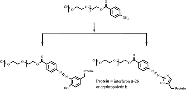
As the majority of reagents commonly used for protein pegylation have certain disadvantages concerning their reactivity, stability, convenience and selectivity of pegylation reaction, etc., the search for new pegylation reagents is of current interest. In the present paper, 4-aminobenzoic acid esters of polyethylene glycol are considered as promising pegylation reagents for chemical modification of molecules of biologically active proteins to prepare their conjugates characterized by improved therapeutic properties. These reagents are highly reactive and stable and make it possible to perform histidine- and tyrosine-targeted pegylation of protein chains. The convenient technique for the synthesis of 4-aminobenzoic acid esters of polyethylene glycol was optimized to prepare these pegylation reagents with actual quantitative loading of the polymer with the functional group (the residue of 4-aminobenzoic acid). The efficiency of the synthesized compounds was shown in the pegylation of such proteins as interferon α-2b and erythropoietin β and it was found that the activity of the prepared interferon α-2b conjugate is comparable with those of its commercial analogs such as Pegintron® and Pegasys®.


Introduction

Pegylation of proteins, that is, covalent binding of their molecules with polyethylene glycol (PEG) is one of the ways to improve the effectiveness of drugs of a protein nature. The presence of this polymer in the structure of such chemically modified drugs makes it possible to reduce their immunogenicity and toxicity, decrease the clearance, and prolong their circulation in the blood, thus, reducing the frequency of drug administration.1–5

The choice of suitable pegylation reagent is one of the important tasks in the preparation of pegylated pharmacologically active compounds. To modify the protein drugs, a wide range of pegylation reagents is currently known. The reagents bear various functional groups introduced into PEG molecule to attach it covalently to the protein molecules.3–7 The conjugation of PEGs with proteins is generally performed via the N-terminal amino groups or lysine ones; the reactions with cysteine, arginine, and tyrosine residues are also used.8–13 However, the majority of these pegylation reagents are characterized by certain disadvantages making them less attractive.4,8,9 Thus, the disadvantages may consist in low reactivity, inconvenient multistage reactions used both for the synthesis of pegylation reagents and for their binding to the protein molecules, low stability of these reagents and final pegylated products as well as increased toxicity. The other important problem in the protein pegylation via amino groups is the formation of a large number of positional isomers with PEG molecules attached to different sites of the protein molecule, with these isomers being characterized by various biological activities.14

Thereby, the search of new pegylation reagents, that is, new active functional groups in PEG molecules used for their conjugation with proteins is the actual problem.

The recently proposed 4-aminobenzoic acid esters of polyethylene glycol 4 seem to be one of very promising pegylation reagents meeting the above requirements.15–18 In contrast to commonly employed reagents affecting the amino groups of polypeptides, the histidine- and tyrosine-targeted pegylation with compounds 4 makes it possible to change not only the place of attachment of the polymer to protein but also the number and the ratio of positional isomers. Although the described routes for the synthesis of 4-aminobenzoic acid esters of polyethylene glycol consist of a small number of simple chemical transformations, they are not always effective and optimal enough. Thus, the synthesis involving acylation of ω-methoxypolyethylene glycol (mPEG) with 4-nitrobenzoyl chloride as a key step leads to only 50% loading of the polymer with functional group (the residue of 4-nitrobenzoic acid).16,17 The similar acylation of mPEG with 4-nitrobenzoyl chloride followed by reduction with palladium on carbon18 and acylation of tosylate derivative of mPEG with 4-aminobenzoic acid19 leads to the total yields of 40–80% and may be somewhat inconvenient. The acylation reaction of Boc-protected 4-aminobenzoic acid in the presence of 1-ethyl-3-(3-dimethylaminopropyl)carbodiimide hydrochloride includes additional synthetic steps for introduction and removal of protecting group and results in 36–86% yields.20 In addition, in mentioned papers, the loadings of polymers with the functional group are not specified.

The use of highly reactive 4-nitrobenzoyl chloride 2 in acylation of mPEGs 1 which allow avoiding the need for expensive water soluble carbodiimides, as well as subsequent rapid and almost quantitative reduction of nitro derivative 3 with titanium(III) chloride instead of expensive palladium on carbon, makes the first of described ways more promising (Scheme 1). Therefore, in the present paper, this synthetic pathway was studied in more detail and the optimal conditions for acylation of mPEGs 1 were found, making it possible to prepare pegylation reagents 4 characterized by the loading with functional group (the residue of 4-aminobenzoic acid) close to 100%.


image file: c5ra04950d-s1.tif
Scheme 1 Synthesis of 4-aminobenzoic acid esters of polyethylene glycol 4 based on mPEGs 1.

Results and discussion

Optimal conditions for the synthesis of 4-aminobenzoic acid esters of polyethylene glycol

Initially, in the first step of the synthesis, corresponding mPEG 1 (0.10–0.15 g mL−1 concentration) was treated with acyl chloride 2 (6–10 eq.) in methylene chloride in the presence of pyridine (10–20 eq.) as a base at room temperature overnight. In the second step, reduction of nitro group was performed in acetate buffer with an excess of the solution of titanium(III) chloride in hydrochloric acid (Scheme 1). However, these reactions supposed to be quantitative resulted in the target compounds 4 characterized by the loading of polymer with the residue of 4-aminobenzoic acid of as little as 40–60% (both chromatographic and spectrophotometric determination of the loading) (entries 1–3 in Table 1).
Table 1 Conditions for acylation of mPEGs 1 with 4-nitrobenzoyl chloride 2 and loadings of resulting polymers 3 and 4 with the residues of corresponding benzoic acids
Entry Concentration of mPEG 1 in solution, g mL−1 Excess of acyl chloride 2 and pyridine, eq. Acylation reaction time, h Loading of polymer 3, % Loading of polymer 4, %
HPLC determination Spectrophotometric determination HPLC determination Spectrophotometric determination
a Products of degradation of polymers are observed in size-exclusion chromatograms.b Reaction was performed in a nitrogen atmosphere.
Compounds 3a, 4a (n = 20)
1 0.10 6.8; 20 24 56.2 ± 1.34 49.5 ± 2.69
[thin space (1/6-em)]
Compounds 3b, 4b (n = 30)
2 0.15 10; 10.1 24 45.5 ± 2.21 40.1 ± 3.04 43.8 ± 1.08 40.4 ± 1.95
3 0.16 10; 10.1 24 50.5 ± 1.46 51.3 ± 2.03
4 0.28 25; 50 108 104 ± 2.91a 106 ± 1.85a 100.6 ± 2.43a
5b 0.28 25; 250 108 103 ± 1.53 103 ± 1.79 95.1 ± 2.85


As after reduction of compounds 3, the loading of amino derivatives 4 was equal to that of the initial nitro derivatives 3 (entry 2 in Table 1), it was concluded that it is acylation of hydroxyl group of compounds 1 which is the limiting step in the synthesis. This circumstance may be due to the influence of steric factors such as a certain arrangement of PEG molecules in the solution and the formation of aggregates preventing hydroxyl groups of polymers from the reaction. Indeed, at the same reaction time (24 h) with the use of a decreased concentration of the initial compound 1a and a lower excess of the acylating agent 2, the loading of product 4a (with a lower molecular weight and, therefore, less ability to aggregate) was not only reduced but even slightly increased in comparison to that of compound 4b (entries 1 and 2 in Table 1). Data in ref. 21 also confirm the discussed effect, namely, the similar acylation of mPEG of 5 kDa molecular weight leads to the quantitative loading of the polymer after 12 h of the reaction.

Substitution of 4-nitrobenzoyl chloride 2 with corresponding more active bromide did not seem to be promising, as releasing hydrogen bromide as a stronger acid (in comparison to hydrogen chloride) could cause an undesired cleavage of PEG molecules through the acid-labile ether bonds.22 Thus, in order to optimize the synthesis and prepare polymers 3 and 4 with the loading close to 100%, the kinetics of the acylation reaction of mPEG 1b was examined (the kinetic curve is shown in Fig. 1). The acylation was expected to be a second-order reaction and a sharp decrease in the reaction rate observed after 4 h is likely to be explained by both a decrease in the concentration of mPEG 1 and acyl chloride 2 (due to its partial hydrolysis to form 4-nitrobenzoic acid) and above-mentioned steric hindrances. Based on the calculated value of the rate constant (5.86 × 10⁻⁵ L mol−1 s−1), the optimal conditions for the synthesis of nitro compound 3 with the loading close to 100% were found. It was proposed to perform the reaction for 108 h with two times increased concentration of the initial compound 1b and 25-fold excess of the acylating agent 2.


image file: c5ra04950d-f1.tif
Fig. 1 Kinetic curve of the dependence of the concentration of product 3b formed in the reaction mixture on the time of acylation of mPEG 1b under conditions given in entry 2 of Table 1 (insert: linearized kinetic curve, where a and b, mol L−1, are the initial concentrations of alcohol 1 and acyl chloride 2, respectively, and x, mol L−1, is the concentration of each of these reagents reacted at time t).

Indeed, the use of these conditions makes it possible to synthesize nitro compound 3b characterized by the loading with the residue of 4-nitrobenzoic acid of approximately 100% (determined via HPLC and spectrophotometry). The loading of the final polymer 4b obtained after reduction of 3b was also about 100% (entry 4 in Table 1). However, according to size-exclusion chromatography, the impurities of lower molecular weights were contained in the prepared samples of 3b and 4b (Fig. 2a). The presence of impurities may be owing to both the peroxide degradation of the polymer under the influence of atmospheric oxygen23,24 and the destruction of ether bonds of PEG molecules in the presence of hydrogen chloride.22 To avoid this undesirable degradation, it was proposed to use a 10-fold excess of pyridine relative to the acyl chloride 2 and carry out the synthesis in a nitrogen atmosphere (entry 5 in Table 1). These conditions were shown to result in the quantitative loading of polymers 4 without any impurities of the degradation products (Fig. 2b).


image file: c5ra04950d-f2.tif
Fig. 2 Size-exclusion chromatograms of compound 4b obtained after using (a) 2-fold excess of pyridine (relative to acylating agent 2) in an air atmosphere (entry 4 in Table 1) and (b) 10-fold excess of pyridine in a nitrogen atmosphere (entry 5 in Table 1); the detection wavelength is at the absorption maximum of polymer 4b (289 nm).

The stability of compounds 4 was estimated at various temperatures. It was found that pegylation reagents 4 are stable and may be stored in an air atmosphere at room temperature in the dark for more than one year, with no degradation and reduction of the loading of polymers being observed during this period.

It also should be mentioned that the use of non-toxic residue of 4-aminobenzoic acid25–27 as a spacer in the molecules of compounds 4 is not expected to lead to toxicity of resulting pegylated proteins.

Protein pegylation with 4-aminobenzoic acid esters of polyethylene glycol

The efficiency of synthesized pegylation reagents 4a,b was shown via the pegylation of proteins of various nature, such as non-glycosylated recombinant human INF α-2b (pI of approximately 6.0 (ref. 28 and 29)) and glycosylated EPO β (pI of approximately 4.5 (ref. 30)), with derivative 4b.

One of important features of 4-aminobenzoic acid esters of polyethylene glycol as pegylation reagents consists in their specific binding with protein molecules through diazotation of amino groups of the firsts and their subsequent azo coupling with the residues of tyrosine and histidine to form azo compounds (similarly to Pauly reaction31,32) (Scheme 2). This approach leads to various advantages of considered pegylation reagents. First, in comparison to the use of “classic” reagents affecting amino groups of polypeptides, it is possible to change the amount and ratio of positional isomers and, thus, the therapeutic activity of conjugates. Moreover, the reactivity of tyrosine and histidine residues depends on pH of the reaction mixture. This circumstance suggests the possibility to change the ratio of positional isomers by varying pH during the pegylation reaction and, as various isomers are characterized by different specific activities,14 adjust (increase or decrease) the activity of conjugates. Thus, under synthetic conditions used in the present work (pH maintained at 8–9), the ratio between pegylated tyrosin and histidine residues of EPO β molecule is estimated based on spectrophotometry data (comparison of Fig. 3a and b) to be 3[thin space (1/6-em)]:[thin space (1/6-em)]2. The structures of positional isomers as well as pharmacokinetics of the prepared conjugates will be studied and published separately.


image file: c5ra04950d-s2.tif
Scheme 2 Pegylation of therapeutic proteins (INF α-2b and EPO β) with 4-aminobenzoic acid ester of polyethylene glycol 4b.

image file: c5ra04950d-f3.tif
Fig. 3 (a) (1) Absorption spectrum of conjugate 5b (in water at pH 6.0) and components of this spectrum corresponding to (2) the protein moiety and isomers pegylated via the residues of (3) histidine and (4) tyrosine (decomposition with OriginPro 9.1 software (OriginLab Corporation)) in comparison with (b) the spectra (in water at pH 6.0) of (1) EPO β and products of azo coupling of diazotated pegylation reagent 4b with (2) tyrosine and (3) histidine.

The other advantage concerns high reactivity of reagents 4. Thus, reactions used in the proposed synthetic strategy are very efficient and convenient, as 70% conversion of protein may be achieved with a relatively low excess (2.6 eq.) of pegylation reagents 4 after as little as 3 h. Meanwhile, to achieve the same result, the use of many known reagents requires their significantly higher excess (from 5 to 25 eq.) and a longer reaction time (from 16 to 87 h).8,33–37

It also should be noted that azo group formed in the molecules of final conjugates is the chromophore providing the absorption band in the visible range of the spectrum (Fig. 3a), thus, facilitating the control of the pegylation reaction, further purification of the conjugates, and their quantitative analysis. In addition, the purity of pegylation reagents 4 and their loading with 4-aminobenzoic acid may be reliably determined via not only the chromatographic method but also the spectrophotometric technique (see Experimental section).

The specific activity of conjugate 5a was determined to be (2.9 ± 1.31) × 107 IU mg−1 (Table 2) and, thus, lower than that of the unmodified protein ((1.9 ± 0.76) × 108 IU mg−1). This result is typical of pegylated INF α-2b and may be due to steric hindrances provided by PEG moieties and preventing the conjugates from binding to required sites.38,39 However, the specific activity of conjugate 5a is comparable or even higher than those of commercial drugs such as Pegintron® and Pegasys® (Table 2). This result confirms both the efficiency of synthesized reagents 4 for pegylation of proteins and the efficiency of the whole described pegylation strategy.

Table 2 Specific activities of unmodified INF α-2b, conjugate 5a, and commercial analogs of the latter
Preparation Specific activity, IU mg−1 Fraction of the activity of unmodified protein, %
Initial INF α-2b (1.9 ± 0.76) × 108 100
PEG (30 kDa)–INF α-2b (5a) (2.9 ± 1.31) × 107 16
Pegintron® (PEG (12 kDa)–INF α-2b) 26 (ref. 38)
Pegasys® (PEG (40 kDa)–INF α-2b) 1.4 × 10⁷ (ref. 39) 7


Experimental

Materials and methods

Commercially available mPEGs 1a,b (JenChem Technology, USA) and concentrated solutions of recombinant human interferon α-2b (INF α-2b, LLC “Pharmapark”, Russia) and erythropoietin β (EPO β, Shandong Kexing Bioproducts Co., Ltd., China) were used. Ethyl 4-[(2-hydroxynaphthyl)diazenyl]benzoate40 and 4-aminobenzoic acid41 were synthesized by known methods.

1H NMR spectra were recorded with the use of a Bruker AM-300 instrument at a frequency of 300 MHz and a temperature of 25 °C. A Genesys 6 spectrophotometer (Thermoscientific, USA) was used to record UV and visible spectra and the latter were processed by OriginPro 9.1 software (OriginLab Corporation). Reversed-phase HPLC was performed on a LC-20AD chromatograph (Shimadzu, Japan) equipped with a parallel double piston pump, an CTO-20A column oven, and an SPD-M20A diode array spectrophotometric detector at 30 °C with the use of gradients prepared by mixtures of water and acetonitrile. Size-exclusion chromatography was carried out on a Smartline chromatograph (Knauer, Germany) equipped with a Smartline 1000 pump with a Manager 5000 gradient forming device and a Smartline 2550 spectrophotometric detector with 0.15 M NaCl solution in 0.05 M phosphate buffer used as an eluent. Isolation and purification of the protein conjugates 5a,b were performed on CM-Sepharose FF and Q-Sepharose FF ion exchangers (GE Healthcare), respectively, with the elution of fractions from the column being controlled with the use of a Smartline UV Detector 200 (Knauer, Germany).

The antiviral activity of pegylated INF α-2b 5a was examined by estimating its effect to protect the Madin–Darby line of bovine kidney cells against a vesicular stomatitis virus (VSV, Indiana strain). For all samples, 50% cytopathic effect was determined visually,28 with the measurements being performed in triplicate.

Synthesis of pegylation Reagents 4a,b

Examples of the optimal methods for the synthesis of compounds 3, 4 (entry 5 in Table 1) are shown below. In other cases, the reactions were carried out similarly with corresponding concentrations of compounds 1 in solutions, equivalents of acyl chloride 2 and pyridine, and the acylation times given in Table 1.
4-Nitrobenzoic acid ester of polyethylene glycol (3b). To a colorless solution of 12.0 g (1 eq.) of alcohol 1b in methylene chloride (35 mL) stirred under nitrogen, 8.10 mL (250 eq.) of pyridine were added followed by the addition of 1.90 g (25 eq.) of 4-nitrobenzoyl chloride 2 and the yellowish reaction mixture was stirred under nitrogen at room temperature for 108 h. Ethanol (50 mL) was added to remove excess acyl chloride and the mixture was stirred for 1 h. The solvents were evaporated, water (400 mL) was added, the insoluble precipitate was filtered off, the aqueous phase was saturated with sodium chloride, extracted with methylene chloride (4 × 100 mL), and the combined organic phases were dried with sodium sulfate and evaporated to a volume of approximately 30 mL. The product was precipitated with the use of diethyl ether, the precipitate was filtered off, washed with ether (20 mL), reprecipitated from methylene chloride (30 mL) with ether, filtered off, washed with ether (2 × 20 mL), and the resulting white powder was dried in vacuum over sulfuric acid.

Yield 10.1 g (84.2%), white powder. 1H NMR (CDCl3) δ: 3.29–3.97 (m, CH2, CH3), 8.19 (d, J = 8.8 Hz, 2H, Hphenyl), 8.25 (J = 8.8 Hz, d, 2H, Hphenyl). Electronic spectrum (H2O), λmax, nm: 200, 261.

4-Aminobenzoic acid ester of polyethylene glycol (4b). To a colorless solution of 4.0 g (1 eq.) of nitro compound 3b in a mixture of ethanol (30 mL) and glacial acetic acid (20 mL), 1.0 g of sodium acetate was added followed by drop-wise addition of 1.23 g of a 20% solution of titanium(III) chloride (12 eq.) in 3% hydrochloric acid (during slow addition, green color of solution appears and almost immediately disappears, whereas after complete reduction and the addition of excess titanium(III) chloride, the color is retained), the reaction mixture was stirred at room temperature for 30 min and sodium monohydrogen orthophosphate (2.0 g) was added. The resulting solution containing gray precipitate was saturated with sodium chloride and extracted with methylene chloride (5 × 25 mL), the combined organic phases were dried with sodium sulfate, evaporated to a volume of approximately 15 mL, filtered off, and the product was precipitated with ether, reprecipitated from methylene chloride (15 mL), and dried in vacuum over sulfuric acid.

Yield 3.68 g (91.9%), white powder. 1H NMR (CDCl3) δ: 3.29–3.97 (m, CH2, CH3), 6.59 (d, J = 8.1 Hz, 2H, Hphenyl), 7.82 (d, J = 8.1 Hz, 2H, Hphenyl). Electronic spectrum (H2O), λmax, nm: 220, 289.

Determination of the loading of polymers 3 and 4

The loading of polymers 3 and 4 with the residues of 4-nitro- and 4-aminobenzoic acids, respectively, was determined with the use of the calibration curves via chromatographic and spectrophotometric methods. All experiments were performed in triplicate and the data on the loading are given in Table 1.
Chromatographic determination of the loading of amino derivatives 4a,b. 45 mg of corresponding compounds 4a,b were placed into a 10 mL volumetric flask, dissolved in water (2 mL), the volume was adjusted with water, and 0.5 mL of this solution were placed into a microtube. 1 M solution of NaOH in water (0.5 mL) was added into the microtube, the mixture was kept at 90 °C for 30 min, 1 M HCl in water (0.5 mL) was added followed by 0.1% solution of TFA in water (0.5 mL), and the resulting solution was analyzed by HPLC. The loadings of polymers were determined via the calibration dependence of the peak area of 4-aminobenzoic acid on its concentration.
Spectrophotometric determination of the loading of amino derivatives 4a,b. 15 mg of corresponding compounds 4a,b were placed into a 25 mL volumetric flask, dissolved in water (2 mL), 0.1 M solution of HBr in water (1 mL) was added followed by 0.01 M solution of NaNO2 in water (1 mL) and the reaction was kept at room temperature for 15 min. 0.01 M solution of sulfamic acid in water (1 mL), the reaction was kept at room temperature for 5 min, 14 mg of 2-naphthol were added followed by 0.1 M borate buffer solution (4.5 mL), and the volume was adjusted with ethanol. The optical density of the final solution was determined at 487 nm relative to the reference solution. The loadings of polymers were found via the calibration dependence of the optical density of a solution of ethyl 4-[(2-hydroxynaphthyl)diazenyl]benzoate (dissolved in a mixture of ethanol (15 mL), 0.1 M borate buffer solution (5 mL), and water (5 mL)) on its concentration.

Synthesis of protein conjugates 5a,b

To demonstrate the efficiency of the synthesized pegylation reagents 4a,b, recombinant INF α-2b and EPO β were pegylated with the use of derivative 4b. A typical procedure is given below for the pegylation of INF α-2b.
4-{[(Interferon-α-2b)yl]diazenyl}benzoic acid ester of ω-methoxypolyethylene glycol (5a). To a solution of 40 mg (1.3 μmol) of 4-aminobenzoic acid ester of polyethylene glycol 4b in 0.1 M HBr (1 mL), 0.01 M NaNO2 (1 mL, 10 μmol) was added, the reaction mixture was kept in the dark at 0–3 °C for 20 min, the excess nitrite ions were removed by adding 0.01 M NH2SO3H (1 mL, 10 μmol), and the resulting diazonium-containing solution was added to a solution of 10 mg (0.5 μmol) of INF α-2b in 0.05 M Na2B4O7 (10 mL). The yellow-orange reaction mixture (pH 8–9) was stirred in the dark at 0–5 °C for 3 h, a solution of 5 mg (27 μmol) of L-tyrosine in water (1 mL) was added, the solution was stirred for 5 min, diluted 30-fold with water, acidified with acetic acid to pH 4.5, and the resulting yellow solution was purified on a CM-Sepharose FF cation-exchanger (5 mL) previously equilibrated with 20 mM acetate buffer at pH 4.5. The yellow compound 5a was eluted with the use of a linear gradient from 20 mM acetate buffer at pH 4.5 to 800 mM NaCl in 20 mM acetate buffer at pH 4.5. The fractions containing conjugate 5a were sterilized via filtration, aliquoted and frozen at −20 °C.

Yield 5.0 mg (50%) with a purity of 97%. Electronic spectrum (H2O, pH 4.5), λmax, nm (lg[thin space (1/6-em)]ε): 222 (4.75), 279 (4.81), 332 (4.65), 387 (4.40, shoulder).

Conclusions

4-Aminobenzoic acid esters of polyethylene glycol were considered as reagents for protein pegylation. The optimized efficient technique for their synthesis resulted in the quantitative loading of the pegylation reagents with residues of 4-aminobenzoic acid. The reagents are characterized by high stability and reactivity. Therapeutic proteins such as INF α-2b and EPO β were pegylated with 4-aminobenzoic acid ester of polyethylene glycol (Mr = 30 kDa) at pH 8–9 and it was shown that in the case of EPO β, the ratio between pegylated tyrosin and histidine residues is 3[thin space (1/6-em)]:[thin space (1/6-em)]2. The prepared INF α-2b conjugate is characterized by a specific activity of (2.9 ± 1.31) × 107 IU mg−1 which is comparable or even higher than those of its commercial analogs.

Notes and references

  1. F. M. Veronese, PEGylated Protein Drugs: Basic Science and Clinical Applications, Birkhäuser Verlag, Basel, 2009 Search PubMed.
  2. S. Jevševar, M. Kunstelj and V. G. Porekar, Biotechnol. J., 2010, 5, 113 CrossRef PubMed.
  3. A. Jain and S. K. Jain, Crit. Rev. Ther. Drug Carrier Syst., 2008, 25, 403 CrossRef CAS.
  4. F. M. Veronese and G. Pasut, Drug Discovery Today, 2005, 10, 1451 CrossRef CAS.
  5. S. S. Banerjee, N. Aher, R. Patil and J. Khandare, J. Drug Delivery, 2012, 2012, 1 CrossRef PubMed.
  6. G. Pasut, A. Giotto and F. M. Veronese, Expert Opin. Ther. Pat., 2004, 14, 859 CrossRef CAS.
  7. M. J. Roberts, M. D. Bentley and J. M. Harris, Adv. Drug Delivery Rev., 2002, 54, 459 CrossRef CAS.
  8. F. M. Veronese, Biomaterials, 2001, 22, 405 CrossRef CAS.
  9. V. Gaberc-Porekar, I. Zore, B. Podobnik and V. Menart, Curr. Opin. Drug Discovery Dev., 2008, 11, 242 CAS.
  10. S. Brocchini, S. Balan, A. Godwin, J.-W. Choi, M. Zloh and S. Shaunak, Nat. Protoc., 2006, 1, 2241 CrossRef CAS PubMed.
  11. M. Zloh, S. Shaunak, S. Balan and S. Brocchini, Nat. Protoc., 2007, 2, 1070 CrossRef CAS PubMed.
  12. L. Wang, V. Gruzdys, N. Pang, F. Meng and X.-L. Sun, RSC Adv., 2014, 4, 39446 RSC.
  13. H. Ban, M. Nagano, J. Gavrilyuk, W. Hakamata, T. Inokuma and C. F. Barbas III, Bioconjugate Chem., 2013, 24, 520 CrossRef CAS PubMed.
  14. S. Foser, A. Schacher, K. A. Weyer, D. Brugger, E. Dietel, S. Marti and T. Schreitmüller, Protein Expression Purif., 2003, 30, 78 CrossRef CAS.
  15. S. V. Sheremet'ev, S. A. Korovkin, A. V. Katlinskij, A. V. Semchenko and V. A. Katlinskij, Russian Patent, RU2439091C1, 2012.
  16. S. V. Sheremet'ev, S. A. Korovkin, V. A. Katlinskij, A. V. Katlinskij and A. V. Semchenko, Eurasian Patent, EA019319B1, 2014.
  17. S. V. Sheremet'ev, S. A. Korovkin, V. A. Katlinskij, A. V. Katlinskij and A. V. Semchenko, Eurasian Patent, EA018116B1, 2013.
  18. M. Yumoto and T. Tamura, US Pat., US20040087760A1, 2004.
  19. Y. He, W. He, D. Liu, T. Gu, R. Wei and X. Wang, Polym. Chem., 2013, 4, 402 RSC.
  20. M. W. Jones, G. Mantovani, C. A. Blindauer, S. M. Ryan, X. Wang, D. J. Brayden and D. M. Haddleton, J. Am. Chem. Soc., 2012, 134, 7406 CrossRef CAS PubMed.
  21. B. König, U. Papke and M. Rödel, New J. Chem., 2000, 24, 39 RSC.
  22. M. B. Smith and J. March, March’s Advanced Organic Chemistry: Reactions, Mechanisms, and Structure, John Wiley and Sons, New York, 5th edn, 2001 Search PubMed.
  23. G. Qin and C. Cai, Chem. Commun., 2009, 5112 RSC.
  24. S. Han, C. Kim and D. Kwon, Polymer, 1997, 38, 317 CrossRef CAS.
  25. M. Tolonen, Vitamins and Minerals in Health and Nutrition, Woodhead Publishing Limited, Cambridge, 2005 Search PubMed.
  26. W. Wang, Y. Song, Q. Liu and K. Yang, Bull. Mater. Sci., 2014, 37, 797 CrossRef CAS PubMed.
  27. G. Wang, X. Yu, Q. Liang, S. Chen, S. Wilson and H. Huang, PLoS One, 2013, 8, e80877 Search PubMed.
  28. H. J. De Jong, M. Ek and G. Lee, European pharmacopeia, 7th edn Search PubMed.
  29. H. Santana, E. Martínez, J. C. Sánchez, G. Moya, R. Sosa, E. Hardy, A. Beldarraín, V. Huerta, L. J. González, L. Betancourt, V. Besada, R. Brito, T. Currás, J. Ferrero, V. Pujols, M. Gil and L. Herrera, Biotecnol. Apl., 1999, 16, 154 CAS.
  30. J. R. Delanghe, M. Bollen and M. Beullens, Am. J. Hematol., 2008, 83, 237 CrossRef CAS PubMed.
  31. H. G. Higgins and D. Fraser, Aust. J. Sci. Res., Ser. A, 1952, 5, 736 Search PubMed.
  32. H. Pauly, Z. Physiol. Chem., 1915, 94, 284 CrossRef CAS.
  33. R. F. Finn and N. R. Siegel, World Patent, WO2006024953A2, 2006.
  34. N. N. As and M. Reslow, World Patent, WO2007025988A3, 2007.
  35. R. B. Greenwald and A. Martinez, World Patent, WO9841562A1, 1998.
  36. G. N. Cox, D. H. Doherty and M. S. Rosendahl, European Patent, EP2305804A1, 2011.
  37. C. O. Beauchamp, S. L. Gonias, D. P. Menapace and S. V. Pizzo, Anal. Biochem., 1983, 131, 25 CrossRef CAS.
  38. C. W. Gilbert and M. Park-Cho, US Pat., US005951974A, 1999.
  39. P. S. Bailon and A. V. Palleroni, US Pat., US20040030101A1, 2004.
  40. N. N. Vorozhtsov, Fundamentals of the Synthesis of Intermediates and Dyes, ONTI, Moscow-Leningrad, 1934 Search PubMed.
  41. F. D. Bellamy and K. Ou, Tetrahedron Lett., 1984, 25, 839 CrossRef CAS.

Footnote

Electronic supplementary information (ESI) available: Determination of the loading of polymers including nitro compounds and UV-vis spectra. See DOI: 10.1039/c5ra04950d

This journal is © The Royal Society of Chemistry 2015