DOI:
10.1039/C5RA04933D
(Paper)
RSC Adv., 2015,
5, 32962-32966
A novel pyrazoline-based fluorescent probe for detecting fluoride ion in water and its application on real samples†
Received
20th March 2015
, Accepted 2nd April 2015
First published on 2nd April 2015
Abstract
We have designed and synthesized a new fluorescent probe, 2-(3-(2-((tert-butyldiphenylsilyl)oxy)phenyl)-5-(4-methoxyphenyl)-4,5-dihydro-1H-pyrazol-1-yl)benzo[d]thiazole (probe 1), to detect fluoride ion with high selectivity and sensitivity in aqueous media. Upon mixing with NaF, desilylation reaction of probe 1 can be induced to the fluorescence quenching with the detection limit of F− 0.74 μM. Moreover, the probe can be used to detect fluoride ion on real life samples.
Introduction
Anions play a vital role in biological systems, chemistry and metabolic processes.1,2 The smallest anion, F−, attracts great attention owing to its importance in maintaining human health.3,4 F− can be used to protect teeth and treat osteoporosis. Because fluoride widely exists in water and is often added to toothpaste, people absorb it easily. However, the excess absorption of fluoride will cause the nerve and endocrine system disorder, nephrotoxic changes and even lead to cancer.5,6 Therefore, the control of fluorine ion in human body is of great value. United States Environmental Protection Agency declared that F− ion content in drinking water to avoid osteoporosis is 4 mg L−1 and the secondary standard is 2 mg L−1 for preventing the dental fluorosis.7 Most toothpastes contain 1000–1400 ppm of NaF as a major constituent. For these reasons, quantitative determination of F− has attracted great attention in biological systems and real samples.
In the last decade, researchers became interested in fluorescent chemical sensors because of their high selectivity, high sensitivity, low cost and real-time detection.8–13 Among all the fluorescent sensors, many turn-off sensors have also been reported to detect species.14–18 A considerable number of fluorescence-based probes have been developed to detect F− based on different mechanism including Lewis acid–base interaction,19–23 anion–π interactions24–26 or hydrogen bonding interactions.27–31 However, most of these probes can detect F− only in organic media and using tetrabutylammonium fluoride (TBAF) as the source. Furthermore, most of them have low selectivity, as AcO− and other ions always disturb the detection of F−. Since Kim and Swager firstly reported the detection group of tert-butyldimethylsilyl (TBDMS) and tert-butylchlorodiphenylsilane (TBDPS), desilylation reaction has been used to detect F− because of its high selectivity toward F−, and many probes have been synthesized to detect fluorine ion based on the facile cleavage of C–Si bond32–35 or O–Si bond.36–59 However, detecting F− in aqueous media with high selectivity and sensitivity still meets great challenging. Also some probes needed higher concentration (at least 5 μM) to detect fluorine ion with high detection limit. The tetrabutylammonium salt was still often used as the source of F− rather than inorganic fluoride existing in natural contaminants. The above drawbacks limited their practical application. Therefore, developing new fluorescent probes with low detection concentration, high selectivity, sensitivity and water solubility is needed. Pyrazoline derivatives showed strong fluorescence, excellent blue-emitting property and high quantum yield.60–62 To the best of our knowledge, few probes have been reported to detect F− based on pyrazoline.
In this work, we designed and synthesized a novel pyrazoline-based fluorescent probe to selectively detect F− in aqueous solution at low concentration of probe 1. Additionally, probe 1 could be a promising fluorescent probe for quantitative determination of F− in environmental water samples and toothpaste.
Results and discussion
Design and synthesis of probe 1
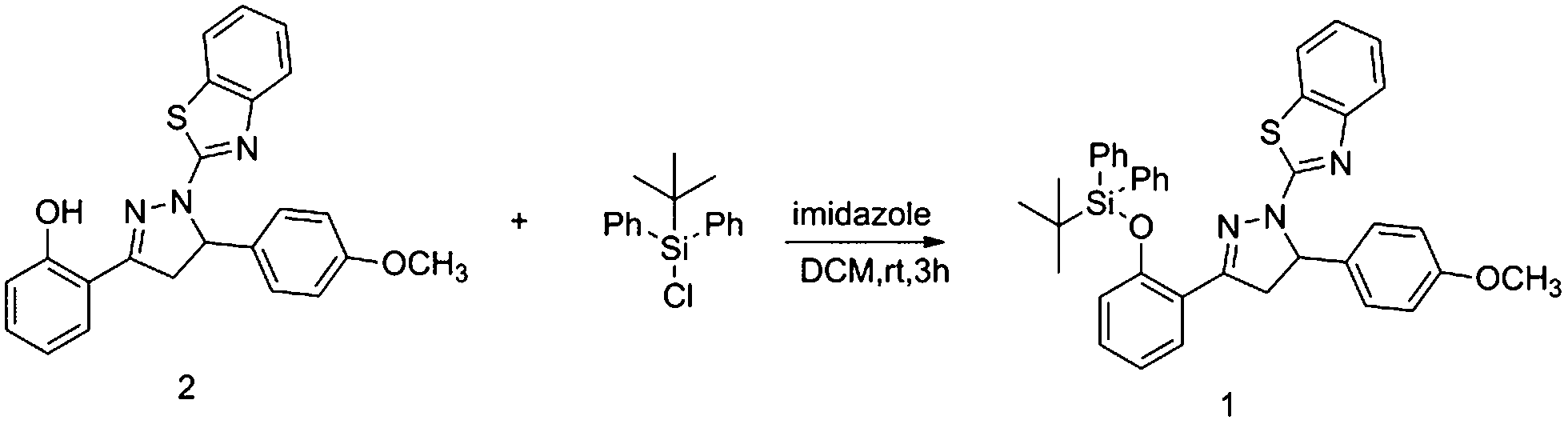
We chose tert-butylchlorodiphenylsilane (TBDPS) as a protecting group of –OH in pyrazoline moiety to form O–Si bond, because F− can easily trigger the cleavage of Si–O bond.36–59 The detection mechanism was verified by the HRMS data of the reaction products of probe 1 with NaF in this work (Fig. S1†). Probe 1 was easily synthesized from 2-(5-(4-methoxyphenyl)-1-phenyl-4,5-dihydro-1H-pyrazol-3-yl)phenol 2 and tert-butylchlorodiphenylsilane by one step in 62% yield and the structure was characterized by IR, NMR and HRMS. The synthetic route is shown in Scheme 1.
 |
| | Scheme 1 Synthesis of probe 1. | |
Absorption and fluorescence spectra of probe 1 titrated with F−
The absorption spectra of probe 1 in HEPES solution (CH3CN![[thin space (1/6-em)]](https://www.rsc.org/images/entities/char_2009.gif) :water = 8:2, v/v, pH = 7.4) showed a peak at 330 nm, however, the addition of F− resulted in 20 nm red shift of the maximum absorbance (Fig. S2†). In the next stage, we examined the quantitative on-off responses of probe 1 (1 μM) toward F− by fluorometric titration (0–400 equiv.). The emission spectra were recorded in HEPES solution (CH3CN:water = 8:2, v/v, pH = 7.4). The free probe 1 exhibited a bright blue fluorescence (fluorescence quantum yield Φ = 0.734) with a peak at 444 nm; however, with the addition of F− to the solution, the emission intensity gradually decreased and showed no significant changes above 0.4 mM, which indicated that the probe can detect a wide range of concentration of F−. The fluorescence intensity showed a good linear relationship with the concentration of F− in the range of 1–10 μM (Fig. 1). The detection limit was found to be 0.74 μM, which is much lower than the level of drinking water standard. The fluorescence quantum yield (Φ) of the compound produced by the reaction of probe 1 and F− is 0.088. Therefore, the probe 1 is very promising for the quantitative detection of F− in aqueous media. To further verify the detection mechanism, we compared the emission spectra of compound 2 and the products of probe 1 with NaF (Fig. S3†). The fluorescence intensity of the products of probe 1 with NaF was similar to that of the compound 2.
:water = 8:2, v/v, pH = 7.4) showed a peak at 330 nm, however, the addition of F− resulted in 20 nm red shift of the maximum absorbance (Fig. S2†). In the next stage, we examined the quantitative on-off responses of probe 1 (1 μM) toward F− by fluorometric titration (0–400 equiv.). The emission spectra were recorded in HEPES solution (CH3CN:water = 8:2, v/v, pH = 7.4). The free probe 1 exhibited a bright blue fluorescence (fluorescence quantum yield Φ = 0.734) with a peak at 444 nm; however, with the addition of F− to the solution, the emission intensity gradually decreased and showed no significant changes above 0.4 mM, which indicated that the probe can detect a wide range of concentration of F−. The fluorescence intensity showed a good linear relationship with the concentration of F− in the range of 1–10 μM (Fig. 1). The detection limit was found to be 0.74 μM, which is much lower than the level of drinking water standard. The fluorescence quantum yield (Φ) of the compound produced by the reaction of probe 1 and F− is 0.088. Therefore, the probe 1 is very promising for the quantitative detection of F− in aqueous media. To further verify the detection mechanism, we compared the emission spectra of compound 2 and the products of probe 1 with NaF (Fig. S3†). The fluorescence intensity of the products of probe 1 with NaF was similar to that of the compound 2.
 |
| | Fig. 1 Fluorescence responses of probe 1 (1 μM) toward different concentrations of F− in HEPES solution (CH3CN:water = 8:2, v/v, pH = 7.4). (a) Fluorescence spectra of probe 1 in the presence of increasing concentrations of F− (0–0.4 mM); (b) fluorescence intensity of probe 1 versus increasing concentrations of F− (final concentration: 1, 2, 3, 4, 5, 6, 7, 8, 9, 10 μM). Each spectrum was obtained after F− addition at least for 12 h (λex = 340 nm). | |
Selectivity and anti-interference of probe 1 for F− detection
To explore the selectivity of probe 1 in the presence of various anions, we recorded the emission spectra of probe 1 (1 μM) in HEPES solution (CH3CN:water = 8:2, v/v, pH = 7.4) upon addition of 0.4 mM of various anions (F−, AcO−, Br−, Cl−, ClO−, HCO3−, HSO4−, SO42−, SO32−, I−, HPO42−), only F− caused significant fluorescent intensity quenching (Fig. 2a). Then, we tested the interference of various anions, subsequent addition of F− to the above solution (probe 1 + other anions) including AcO−, Br−, Cl−, ClO−, HCO3−, HPO42−, HSO4−, I−, SO42−, SO32− cannot influence the detection of F− (Fig. 2b). The results demonstrate that probe 1 has excellent selectivity for F− over other anions and great capability of resisting the disturbance of other anions.
 |
| | Fig. 2 (a) Emission spectra of the probe 1 (1 μM) in HEPES solution (CH3CN:water = 8:2, v/v, pH = 7.4) upon addition of 0.4 mM of various anions (F−, AcO−, Br−, Cl−, ClO−, HCO3−, HSO4−, SO42−, SO32−, I−, HPO42−); (b) the red bars represent the emission intensity that occurs upon the subsequent addition of 0.4 mM of F− to the above solution (probe 1 + other anions). From 2 to 12: none, AcO−, Br−, Cl−, ClO−, HCO3−, HPO42−, HSO4−, I−, SO42−, SO32− (1 represents probe 1). | |
pH-dependent
In order to determine the appropriate pH range for the probe to function, the reaction of probe 1 with F− at different pH was examined (Fig. 3). The free probe 1 showed high fluorescent intensity at pH 3.0–11.0, so the probe is stable at pH 3.0–11.0. However, the reaction of probe 1 with F− was sensitive to the pH in the range of 3.0–4.0. Therefore, the probe can work at a wide range of pH 5.0–11.0, which indicates that the probe can be applied to analyse a broader scope of real life samples.
 |
| | Fig. 3 Fluorescence intensity of probe 1 (1 μM) in CH3CN upon addition of 0.4 mM F− in HEPES buffer of pH range 3–11 (CH3CN:water = 8:2, v/v) at λem = 444 nm (λex = 340 nm). | |
Application of the probe on real samples
Fluoride is the essential ingredient in toothpaste to protect dental and bone by forming the enamel. To prove whether probe 1 can determine fluoride content in toothpaste, we firstly examined the response dynamics of probe 1 and chose an examination time of 12 h for studying the application of probe 1 (Fig. S4†) and then took widely used toothpaste and measured the concentration of F− in it.47 We compared the concentration of F− in tested samples (as calculated from weight of toothpaste) and detected value (as obtained from the fluorescence intensity changes, Fig.1b). As shown in Fig. 4, the two parameters have good correlation. The results demonstrate that probe 1 can be used to estimate F− in the toothpaste. According to the detected value in Fig. 4, we found that the Crest® contains 0.13% F− which is similar to that labeled in the toothpaste ingredients (0.14%). To test the capacity of the present system in analyzing diverse water samples, distilled water, tap water (Jinan China), and Yellow River were chosen as the water samples. Replacing the deionized water with natural water and added F− in known concentration.18,59,63 The corresponding emission spectra were recorded in solution (CH3CN:H2O = 8:2, v/v, pH = 7.4). As shown in Table 1, the tested concentrations of F− (as obtained from the fluorescence intensity changes, Fig.1b) are in great agreement with those added in natural water samples. The results indicate that we can detect F− in real water samples at the ppm level using probe 1 without any further process. Meanwhile, the results show that the concentrations of F− in real water samples can really be ignored. Therefore, probe 1 has great potential for practical application.
 |
| | Fig. 4 Plot presenting linear correlation between the concentration of F− present in tested samples and the detected concentration of F−. | |
Table 1 Measurement of F− in various water samples by probe 1
| Amount of F− added [ppm] |
Distilled water tested [ppm] |
Tap water tested [ppm] |
Yellow River tested [ppm] |
| 0.057 |
0.060 ± 0.003 |
0.054 ± 0.004 |
0.062 ± 0.006 |
| 0.095 |
0.090 ± 0.004 |
0.103 ± 0.004 |
0.100 ± 0.010 |
| 0.133 |
0.132 ± 0.007 |
0.124 ± 0.003 |
0.135 ± 0.006 |
| 0.190 |
0.185 ± 0.001 |
0.194 ± 0.001 |
0.189 ± 0.003 |
Experimental
Materials and instruments
All reagents were purchased from commercial suppliers and used without further purification. TLC was conducted on silica gel 60 F254 plates (Merck KGaA) with a fluorescent indicator for 254 nm excitation. 1H NMR and 13C NMR spectra were recorded on a Bruker Avance 300 (300 and 75 MHz) spectrometer using either tetramethylsilane (TMS) as an internal standard with CDCl3 as a solvent. Melting points were carried out on an XD-4 digital micro melting point apparatus. IR spectra were determined with an IR spectrophotometer VERTEX 70 FT-IR (Bruker Optics). UV-vis spectra were recorded on a U-4100 spectrophotometer (Hitachi). Fluorescent measurements were recorded on a Perkin-Elmer LS-55 luminescence spectrophotometer. HRMS spectra were recorded on a Q-TOF6510 spectrograph (Agilent). All pH measurements involved the use of a Model PHS-3C pH meter (Shanghai) at room temperature about 298 K. Deionized water was used throughout the experiment.
Synthesis of 2-(1-(benzo[d]thiazol-2-yl)-5-(4-methoxyphenyl)-4,5-dihydro-1H-pyrazol-3-yl)phenol (compound 2)
Compound 2 was synthesized as described in literature.64
Synthesis of 2-(3-(2-((tert-butyldiphenylsilyl)oxy)phenyl)-5-(4-methoxyphenyl)-4,5-dihydro-1H-pyrazol-1-yl)benzo[d]thiazole (probe 1)
Compound 2 (0.3 mmol), imidazole (0.5 mmol) and 30 mL DCM were added to 50 mL round-bottomed flask. After stirring for 10 min, tert-butyldiphenylchlorosilane (0.3 mmol) was added to the reaction mixture and stirred at room temperature for 3 h. The mixture was washed with water and extracted with DCM. The extract was dried over anhydrous Na2SO4. The solvent was concentrated and the crude product was purified by column chromatography on silica gel using EA:PE (1:8) to obtain pale yellow solid (62%). M.P. 142–144 °C.1H NMR (300 MHz, CDCl3) δ 7.84 (dd, J = 6.1, 3.4 Hz, 1H), 7.69 (m, 4H), 7.63 (d, J = 7.6 Hz, 1H), 7.53 (d, J = 8.0 Hz, 1H), 7.39 (m, 6H), 7.31–7.26 (m, 2H), 7.24 (s, 1H), 7.07 (t, J = 7.5 Hz, 1H), 6.94–6.87 (m, 2H), 6.85 (t, J = 5.9 Hz, 2H), 6.52–6.43 (m, 1H), 5.78 (dd, J = 11.8, 5.1 Hz, 1H), 4.19 (dd, J = 18.0, 11.8 Hz, 1H), 3.76 (s, 3H), 3.61 (dd, J = 18.0, 5.1 Hz, 1H), 1.00 (s, 9H); 13C NMR (75 MHz, CDCl3) 163.59, 159.07, 154.10, 135.33, 134.80, 133.45, 132.16, 132.04, 130.47, 130.09, 129.83, 127.98, 127.71, 127.24, 125.53, 122.43, 121.64, 121.27, 120.70, 120.58, 119.98, 114.21, 77.42, 77.00, 76.58, 63.29, 55.26, 47.22, 26.55, 19.25; IR (KBr, cm−1): 2928.7, 2855.6, 1735.3, 1598.9, 1537.6, 1513.1, 1489.3, 702.5; HRMS: calcd for [M + H]+ C39H37N3O2SSi: 640.2454; found: 640.2421.
Determination of quantum yields
The quantum yield of probe 1 was determined according to the eqn (1):| | |
ΦF = (ΦFS)(FAu)(As)(ηu2)/(FAs)(Au)(ηs2)
| (1) |
where ΦF and FA are the fluorescence quantum yield and integrated area under the corrected emission spectra, respectively; A is the absorbance at the excitation wavelength; η presents the refractive index of the solution; and subscripts u and s refer to the unknown and the standard, respectively.65
Detection limit calculation
Detection limit for F− was calculated using fluorometric titrations using equation: detection limit = 3σ/m, where σ = standard deviation of 10 blank measurements and m = slope obtained from the graph of fluorescence intensity vs. concentration of F− added.48
Preparation for the detection of fluoride ion in toothpaste
We chose Crest® as toothpaste sample, which contains 0.14% F− labeled in ingredients. Four samples with different weights (30, 40, 50, 60 mg) were prepared in small vials. The samples were dried overnight in drying oven (temperature = 75 °C). To each sample 15 mL water was added. Then the solutions were sonicated at 50 °C for 1 h and balanced for 4 h at room temperature. Each sample was centrifuged and filtered to get clear solution. The solutions were diluted 100-fold and then treated with probe 1 solution. The corresponding emission spectra were recorded in HEPES solution (CH3CN:water = 8:2, v/v, pH = 7.4).
Conclusions
In this study, we have developed a new pyrazoline-based fluorescent probe 1 for the detection of F−. Probe 1 shows several good properties involving low detection limit in aqueous solution, high selectivity and sensitivity, a wide range of pH 5.0–11.0. Furthermore, the probe can be applied to detect F− in real life sample without any further purification. Therefore, probe 1 will be an excellent candidate for highly selective and sensitive detection of F−. And this work will prove useful for developing organic dyes as chemosensors.
Acknowledgements
This study was supported by the Natural Science Foundation of Shandong Province (ZR2014BM004).
Notes and references
- R. M. Duke, E. B. Veale, F. M. Pfeffer, P. E. Kruger and T. Gunnlaugsson, Chem. Soc. Rev., 2010, 39, 3936–3953 RSC.
- P. D. Beer and P. A. Gale, Angew. Chem., Int. Ed., 2001, 40, 486–516 CrossRef CAS.
- S. Ayoob and A. K. Gupta, Crit. Rev. Environ. Sci. Technol., 2006, 36, 433–487 CrossRef CAS.
- A. Choi, G. Sun, Y. Zhang and P. Grandjean, Environ. Health Perspect., 2012, 120, 1362–1368 CrossRef CAS PubMed.
- P. P. Singh, M. K. Barjatiya, S. Dhing, R. Bhatnagar, S. Kothari and V. Dhar, Urol. Res., 2001, 29, 238–244 CrossRef CAS.
- M. L. Cittanova, B. Lelongt and M. C. Verpont, Anesthesiology, 1996, 84, 428–435 CrossRef CAS PubMed.
- http://www.epa.gov/safewater/mcl.html#mcls.
- Y. T. Yang, C. X. Yin, F. J. Huo and J. B. Chao, RSC Adv., 2013, 3, 9637–9640 RSC.
- A. K. Mahapatra, S. K. Manna, K. Maiti, R. Maji, C. D. Mukhopadhyay, D. Sarkar and T. K. Mondal, RSC Adv., 2014, 4, 36615–36622 RSC.
- Y. R. Zhang, X. P. Chen, J. Shao, J. Y. Zhang, Q. Yuan, J. Y. Miao and B. X. Zhao, Chem. Commun., 2014, 50, 14241–14244 CAS.
- Q. Zhang, D. H. Yu, S. S. Ding and G. Q. Feng, Chem. Commun., 2014, 50, 14002–14005 RSC.
- G. Liu, J. Q. Wang, M. D. Yang, X. Z. Zhang, L. N. Ye, F. Y. Hao, H. P. Zhou, X. Y. Tian, J. Y. Wu and Y. P. Tian, Sens. Actuators, B, 2014, 192, 586–593 CrossRef CAS PubMed.
- A. K. Mahapatra, S. Mondal, K. Maiti, S. K. Manna, R. Maji, D. Mandal, S. Mandal, S. Goswami, C. K. Quah and H. K. Fun, RSC Adv., 2014, 4, 56605–56614 RSC.
- J. Zhao, H. J. Li, K. Yang, S. G. Sun, A. P. Lu and Y. Q. Xu, New J. Chem., 2014, 38, 3371–3374 RSC.
- J. F. Li, F. J. Huo and C. X. Yin, RSC Adv., 2014, 4, 44610–44613 RSC.
- J. T. Hou, K. Li, K. K. Yu, M. Z. Ao, X. Wang and X. Q. Yu, Analyst, 2013, 138, 6632–6638 RSC.
- B. C. Zhu, Y. H. Xu, W. Q. Liu, C. X. Shao, H. F. Wu, H. L. Jiang, B. Du and X. L. Zhang, Sens. Actuators, B, 2014, 191, 473–478 CrossRef CAS PubMed.
- P. Ashokkumar, H. Weißhoff, W. Kraus and K. Rurack, Angew. Chem., Int. Ed., 2014, 53, 2225–2229 CrossRef CAS PubMed.
- Z. Q. Hu, C. L. Cui, H. Y. Lu, L. Ding and X. D. Yang, Sens. Actuators, B, 2009, 141, 200–204 CrossRef CAS PubMed.
- M. M. M. Raposo, B. García-Acosta, T. Ábalos, P. Calero, R. Martínez-Máňez, J. V. Ros-Lis and J. Soto, J. Org. Chem., 2010, 75, 2922–2933 CrossRef CAS PubMed.
- M. Boiocchi, L. D. Boca, D. E. Gomez, L. Fabbrizzi, M. Licchelli and E. Monzani, J. Am. Chem. Soc., 2004, 126, 16507–16514 CrossRef CAS PubMed.
- M. Cametti and K. Rissanen, Chem. Commun., 2009, 20, 2809–2829 RSC.
- C. R. Wade, A. E. J. Broomsgrove, S. Aldridge and F. P. Gabbaï, Chem. Rev., 2010, 110, 3958–3984 CrossRef CAS PubMed.
- M. Mascal, I. Yakovlev, E. B. Nikitin and J. C. Fettinger, Angew. Chem., Int. Ed., 2007, 46, 8782–8784 CrossRef CAS PubMed.
- Z. C. Xu, N. J. Singh, S. K. Kim, D. R. Spring, K. S. Kim and J. Yoon, Chem.–Eur. J., 2011, 17, 1163–1170 CrossRef CAS PubMed.
- S. Guha, F. S. Goodson, L. J. Corson and S. Saha, J. Am. Chem. Soc., 2012, 134, 13679–13691 CrossRef CAS PubMed.
- K. C. Song, H. Kim, K. M. Lee, Y. S. Lee, Y. Do and M. H. Lee, Sens. Actuators, B, 2013, 176, 850–857 CrossRef CAS PubMed.
- V. Kumar, M. P. Kaushik, A. K. Srivastava, A. Pratap, V. Thiruvenkatam and T. N. G. Row, Anal. Chim. Acta, 2010, 663, 77–84 CrossRef CAS PubMed.
- B. Chandra, S. P. Mahanta, N. N. Pati and S. Baskaran, Org. Lett., 2013, 15, 306–309 CrossRef CAS PubMed.
- A. K. Mahapatra, R. Maj, K. Maiti, S. S. Adhikari, C. D. Mukhopadhyay and D. Mandal, Analyst, 2014, 139, 309–317 RSC.
- X. Yong, M. J. Su, W. Wan, W. W. You, X. W. Lu, J. Q. Qu and R. Y. Liu, New J. Chem., 2013, 37, 1591–1594 RSC.
- M. R. Rao, S. M. Mobin and M. Ravikanth, Tetrahedron, 2010, 66, 1728–1734 CrossRef CAS PubMed.
- L. Fu, F. L. Jiang, D. Fortin, P. D. Harvey and Y. Liu, Chem. Commun., 2011, 47, 5503–5505 RSC.
- D. Buckland, S. V. Bhosale and S. J. Langford, Tetrahedron Lett., 2011, 52, 1990–1992 CrossRef CAS PubMed.
- H. Lu, Q. H. Wang, Z. F. Li, G. Q. Lai, J. X. Jiang and Z. Shen, Org. Biomol. Chem., 2011, 9, 4558–4562 CAS.
- S. Goswami, A. K. Das, A. Manna, A. K. Maity, H. K. Fun, C. K. Quah and P. Saha, Tetrahedron Lett., 2014, 55, 2633–2638 CrossRef CAS PubMed.
- S. Y. Kim, J. Park, M. Koh, S. B. Park and J. I. Hong, Chem. Commun., 2009, 31, 4735–4737 RSC.
- J. Cao, C. C. Zhao and W. H. Zhu, Tetrahedron Lett., 2012, 53, 2107–2110 CrossRef CAS PubMed.
- R. Hu, J. Feng, D. H. Hu, S. Q. Wang, S. Y. Li, Y. Li and G. Q. Yang, Angew. Chem., Int. Ed., 2010, 49, 4915–4918 CrossRef CAS PubMed.
- B. C. Zhu, F. Yuan, R. X. Li, Y. M. Li, Q. Wei, Z. M. Ma, B. Du and X. L. Zhang, Chem. Commun., 2011, 47, 7098–7100 RSC.
- G. H. Wei, J. X. Yin, X. Ma, S. Y. Yu, D. B. Wei and Y. G. Du, Anal. Chim. Acta, 2011, 703, 219–225 CrossRef CAS PubMed.
- J. F. Zhang, C. S. Lim, S. Bhuniya, B. R. Cho and J. S. Kim, Org. Lett., 2011, 13, 1190–1193 CrossRef CAS PubMed.
- P. Sokkalingam and C. H. Lee, J. Org. Chem., 2011, 76, 3820–3828 CrossRef CAS PubMed.
- O. A. Bozdemir, F. Sozmen, O. Buyukcakir, R. Guliyev, Y. Cakmak and E. U. Akkaya, Org. Lett., 2010, 12, 1400–1403 CrossRef CAS PubMed.
- X. W. Cao, W. Y. Lin, Q. X. Yu and J. L. Wang, Org. Lett., 2011, 13, 6098–6101 CrossRef CAS PubMed.
- M. Dong, Y. Peng, Y. M. Dong, N. Tang and Y. W. Wang, Org. Lett., 2012, 14, 130–133 CrossRef CAS PubMed.
- N. Kumari, N. Dey and S. Bhattacharya, Analyst, 2014, 139, 2370–2378 RSC.
- A. Roy, D. Kand, T. Saha and P. Talukdar, Chem. Commun., 2014, 50, 5510–5513 RSC.
- Y. Y. Bao, B. Liu, F. F. Du, J. Tian, H. Wang and R. K. Bai, J. Mater. Chem., 2012, 22, 5291–5294 RSC.
- J. Cao, C. C. Zhao, P. Feng, Y. L. Zhang and W. H. Zhu, RSC Adv., 2012, 2, 418–420 RSC.
- P. Hou, S. Chen, H. B. Wang, J. X. Wang, K. Voitchovsky and X. Z. Song, Chem. Commun., 2014, 50, 320–322 RSC.
- J. Ren, Z. Wu, Y. Zhou, Y. Li and Z. X. Xu, Dyes Pigm., 2011, 91, 442–445 CrossRef CAS PubMed.
- Y. Y. Bao, B. Liu, H. Wang, J. Tian and R. K. Bai, Chem. Commun., 2011, 47, 3957–3959 RSC.
- L. K. Calderón-Ortiz, E. Täuscher, E. L. Bastos, H. Görls, D. Weiß and R. Beckert, Eur. J. Org. Chem., 2012, 13, 2535–2541 CrossRef.
- L. Z. Gai, H. C. Chen, B. Zou, H. Lu, G. Q. Lai, Z. F. Li and Z. Shen, Chem. Commun., 2012, 48, 10721–10723 RSC.
- B. W. Ke, W. X. Chen, N. T. Ni, Y. F. Cheng, C. F. Dai, H. Dinh and B. H. Wang, Chem. Commun., 2013, 49, 2494–2496 RSC.
- P. Hou, S. Chen and X. Z. Song, Luminescence, 2014, 29, 423–426 CrossRef CAS PubMed.
- S. J. Yang, Y. Liu and G. Q. Feng, RSC Adv., 2013, 3, 20171–20178 RSC.
- F. Y. Zheng, F. Zeng, C. M. Yu, X. F. Hou and S. Z. Wu, Chem.–Eur. J., 2013, 19, 936–942 CrossRef CAS PubMed.
- C. J. Fahrni, L. C. Yang and D. G. Van-Derveer, J. Am. Chem. Soc., 2003, 125, 3799–3812 CrossRef CAS PubMed.
- J. F. Li, D. X. Li, Y. Y. Han, S. M. Shuang and C. Dong, Spectrochim. Acta, Part A, 2009, 73, 221–225 CrossRef PubMed.
- S. Q. Wang, Q. H. Wu, H. Y. Wang, X. X. Zheng, S. L. Shen, Y. R. Zhang, J. Y. Miao and B. X. Zhao, Analyst, 2013, 138, 7169–7174 RSC.
- Y. Xu, X. Dai and B. X. Zhao, Spectrochim. Acta, Part A, 2015, 138, 164–168 CrossRef CAS PubMed.
- V. Sharma and K. V. Sharma, Chem.–Eur. J., 2009, 6, 348–356 CAS.
- R. A. Velapoldi and K. D. Mielenz, A fluorescence standard reference material: quinine sulfate dehydrate, National Bureau of Standards, Washington DC, USA, 1980, pp. 260 Search PubMed.
Footnote |
| † Electronic supplementary information (ESI) available: Supplementary Fig. S1–S5. See DOI: 10.1039/c5ra04933d |
|
| This journal is © The Royal Society of Chemistry 2015 |
Click here to see how this site uses Cookies. View our privacy policy here.