DOI:
10.1039/C5RA04894J
(Paper)
RSC Adv., 2015,
5, 51364-51370
Preparation of a novel anti-fouling β-cyclodextrin–PVDF membrane
Received
20th March 2015
, Accepted 2nd June 2015
First published on 3rd June 2015
Abstract
As part of this work, polyvinylidene fluoride (PVDF) membranes were prepared via a phase inversion method. A novel β-cyclodextrin (β-CD)–PVDF membrane was prepared via an interfacial reaction, using Trimesoyl Chloride (TMC) and β-CD as cross-linking and modification agents, respectively. The membranes were modified by a simple dip-coating method. The results of comparison between the modified and unmodified membranes by attenuated total reflectance Fourier transform infrared spectroscopy (FTIR-ATR) and X-ray photoelectron spectroscopy (XPS) methods confirmed successful grafting of β-CD on the membrane surfaces. The morphology of the membrane was investigated by scanning electron microscopy (SEM) and atomic-force microscopy (AFM). The roughness of PVDF membranes increased after their surfaces were grafted with β-CD. It was also found that β-CD–PVDF membranes had higher permeate flux and hydrophilicity than those of the pristine PVDF membranes. Furthermore, in protein fouling experiments, bovine serum albumin (BSA) was used as a protein model solution. Modified membranes exhibited a higher flux recovery ratio. The experiment showed that the anti-fouling performance of β-CD–PVDF membranes was more effective in reducing the irreversible membrane fouling. The results of this study hold promise for useful applications of β-CD–PVDF membranes in membrane separation areas.
1. Introduction
Over the past few decades, membrane separation technology has been attracting greater attention than conventional separation technology, because of its high efficiency, high selectivity, reliability in separation, ease of operation, and low energy consumption.1 Membrane technology is widely believed to be an effective method for water treatment and environmental protection.2
Polyvinylidene fluoride (PVDF), as one of the most extensively used substances in membrane preparation, has been attracting the attention of several researchers, because of its special mechanical properties, chemical and thermal stability, and resistance to radiation.3 PVDF membrane can perform well in water treatment, but that leads to fouling, which is now the foremost issue hindering its successful application. The hydrophobic PVDF membrane, while treating an aqueous solution containing natural organic matters, e.g. proteins, tends to foul the solution. Proteins are prone to be easily absorbed onto the membrane surface or they may block the surface pores, reducing the membrane flux. It is generally accepted that hydrophilic membranes have better fouling resistance and can significantly reduce the deposition and adsorption of proteins.4–6 Numerous studies have been carried out to increase the hydrophilic of PVDF membranes. Permeation flux and anti-fouling capability of such modified membranes should be significantly higher than those of pristine membranes for filtration. In recent years, different methods have been explored to enhance the hydrophilic of PVDF membranes, such as blending modification, surface coating and surface grafting. Among these, blending inorganic nanoparticles is the most effective way in practical applications. TiO2,7–9 SiO2,10,11 Al2O3,12,13 ZnO,5 ZrO2,14 Ag15 and MWCNT16 are blended in membranes, but one of the biggest issues is the agglomeration phenomenon.2 The main problem of the surface coated layer is its instability, which can be washed away during operation and cleaning process, because of the rather weak physical adsorption interaction between PVDF membrane and the coated layer.3 Surface grafting can solve the problem of instability completely. The covalent attachment of modifiers on membrane surface offers a long-term chemical stability, because the covalent bonding interaction between the grafted chains and the membrane. Surface grafting can be done in different ways, such as UV irradiation, low-temperature plasma, high energy irradiation and chemical method. For grafting by UV irradiation, low-temperature gamma ray or high energy irradiation is not easy to apply on a large industrial scale; therefore, chemical method is a promising way to achieve hydrophilic modification of PVDF membranes.
β-Cyclodextrin (β-CD) is composed of seven α-(1,4) linked glycosyl units, among which β-CD is the most easily accessible, the least expensive and generally the most useful one. Each cyclodextrin is a torus (doughnut-shaped) molecule. The internal cavity of the doughnut is hydrophobic, whereas the external surface is hydrophilic.17–19 The multi-hydroxyl of β-CD can be replaced by other functional groups also for better performance or other applications. Besides, the functional groups of β-CD can also contribute to membrane's hydrophilicity. However, to the authors' best knowledge, there has been little research on the grafting of β-CD on the surface of the PVDF membrane and its performance evaluation.
Our focus in this work was on preparing hydrophilic and antifouling PVDF membranes by using a remarkably simple and effective dip-coating method (Fig. 1).
 |
| | Fig. 1 Schematic diagram illustrating the preparation of β-CD–PVDF membrane. | |
2. Experiment
2.1. Materials
PVDF (FR904) was purchased from Shanghai 3F New Materials Ltd. China, (Shanghai, China), dimethylacetamide (DMAc, ≥99.5%, reagent), n-hexane, polyvinyl pyrrolidone (PVP, K30, Mw = 35![[thin space (1/6-em)]](https://www.rsc.org/images/entities/char_2009.gif) 000) and β-cyclodextrin (β-CD) from Chengdu Kelong Chemical Co. Ltd. (Sichuan, China), Trimesoyl Chloride (TMC) from Aladdin Industrial Corporation (Shanghai, China), and BSA (Mw = 67000) from Shanghai Ruji Bio Co. Ltd.
000) and β-cyclodextrin (β-CD) from Chengdu Kelong Chemical Co. Ltd. (Sichuan, China), Trimesoyl Chloride (TMC) from Aladdin Industrial Corporation (Shanghai, China), and BSA (Mw = 67000) from Shanghai Ruji Bio Co. Ltd.
2.2. Preparation of PVDF membrane
PVDF membranes were prepared via the phase inversion method. PVDF (18 wt%) and PVP (4 wt%) were dissolved in DMAc in a 200 mL beaker and the polymer solution was vigorously stirred at 60 °C for 8 h. Then, the membranes were dried in vacuum oven at 30 °C for 12 h to allow complete release of air bubbles and then casted the solutions with a casting knife of 200 μm onto a glass plate. After 30 s evaporation time in air, the glass plate was immersed in a coagulation bath (distilled water) to induce phase inversion. After peeling the membranes from the glass plate, they were placed in a water bath for 48 h. Finally, the membranes were allowed to dry under a temperature of 60 °C for 30 min.
2.3. Modification of PVDF membrane
The pristine PVDF membrane was pre-wetted with ethanol and then immersed in deionized water for 30 min. The deionized water, packed into the pores of the membranes, replaced ethanol. The membrane was then immersed in an aqueous solution of 3 mol L−1 KOH and 5 wt% KMnO4. The reaction between the membrane and the aqueous solution lasted 5 min and was kept at 80 °C. After the original white color of the membrane surface changed to brown, the PVDF membrane was immersed in the aqueous solution of 5 wt% NaHSO3 and 5 wt% H2SO4. After 30 minutes, the membrane regained its original white color. After washing with deionized water, the hydroxyl modified PVDF membranes were immersed in hexane (150 mL TMC/0.6%, w/v) oil solution for 30 minutes. Then, the PVDF membranes were soaked in a solution of β-CD (0.9%, 1.8% and 2.7%, w/v) at pH = 9 for 90 min (the pH of the solution was adjusted by NaOH and Na2CO3). Finally, the modified membranes were washed repeatedly with deionized water and dried under a temperature of 60 °C for 30 min.
2.4. Membrane characterization
2.4.1. FTIR-ATR. The chemical composition of the modified membrane surfaces was determined by using the attenuated total reflectance Fourier transform infrared spectroscopy (FTIR-ATR) with a Bruker TENSOR27 FT-IR spectroscope.
2.4.2. XPS. The surface composition of the membranes was determined by X-ray photoelectron spectroscopy (Kratos, XSAM800), using Al Kα as the excitation source (hν = 1486.6 eV).
2.4.3. Contact angle measurement. The contact angle of water on the membrane was determined at 25 °C with a contact-angle measuring instrument (Beijing Hake, XED-SPJ), following the sessile-drop method. For this, 2.0 μL distilled water was dropped on the top surface of the membrane, and the advancing angle measured as the water started spreading over the membrane surface. At least five angles were measured and their average taken for every sample.
2.4.4. Water absorption measurement. All the membranes were dried in vacuum at 50 °C for 1 h before testing and then immersed in deionized water at room temperature for 24 h. Before weighing, the water remaining on the surface of the wetted membranes was removed using filter paper. The water absorption (η) was calculated by the following equation:| |
 | (1) |
where mwet and mdry were the masses of dried and wet samples respectively.
2.4.5. Permeation performance. The pure water flux of the membranes was measured by ultrafiltration experimental equipment (SINAP, SCM-300). The experiments were performed at 0.1 MPa. To obtain a stable flux before measurement, the newly prepared membranes were pre-pressured at 0.1 MPa using pure water for 30 min. The pure water flux (Jw) is defined as follows:| |
 | (2) |
where V is the volume of penetrative water (L), A the effective membrane area (m2), and t the running time (h).
2.4.6. The multi-cycle filtration experiment. The antifouling properties of membranes were evaluated through using cycle filtration of 0.1 g L−1 BSA solution as a model protein. First, the BSA solution was filtered by the membranes under the operation pressure of 0.1 MPa for 30 min and the resulting flux recorded as Jp. After washing the membranes with deionized water for 30 min, the operation pressure was kept at 0.1 Mpa for 30 min and the resulting flux recorded as Jr. After that, the experiment was repeated twice.The relative flux reduction (RFR) and the flux recovery ratio (FRR) were calculated using the following equations:
| |
 | (3) |
| |
 | (4) |
2.4.7. Morphology. The morphology of the surface of membranes was studied by a scanning electron microscope (SEM, JSM-7500F, JEOL, Tokyo, Japan). The membrane samples were pre-treated by spraying gold on them before scanning.The topography of membranes was characterized using an atomic force microscopy (AFM, SPA300HV). The membrane surfaces were scanned and imaged in a scan size of 20 μm × 20 μm. The surface roughness was reported in terms of average plane roughness (Ra) and root mean square roughness (Rms).
3. Results and discussion
3.1. FTIR-ATR analysis
To ascertain if surface functionalization of β-CD on PVDF membranes is successful, the attenuated total reflectance Fourier transform infrared spectroscopy (FTIR-ATR) was performed and the result shown in Fig. 2.
 |
| | Fig. 2 FTIR-ATR spectrum of pristine PVDF membrane, hydroxylated membrane, TMC-modified membrane and β-CD–PVDF membrane. | |
The characteristic peaks at 1405 cm−1 and at 1168 cm−1 are ascribed to CH2 and CF2 stretching vibrations. The skeletal vibration of C–C bond is shown at 877 cm−1.20 An obvious characteristic peak at 1660 cm−1 is attributed to the bending vibration of carbonyl group (C![[double bond, length as m-dash]](https://www.rsc.org/images/entities/char_e001.gif) O), which originated from PVP because of the residue trapped in the membrane matrix.5 Compared to the pristine PVDF membrane, a new peak showed up in the curve of hydroxylated membrane. The broad and strong band, ranging from 3200 to 3600 cm−1, indicates the presence of –OH groups. For the FTIR spectra of TMC-modified membranes, the bending vibration of CO in TMC is shown at 1727 cm−1. In addition, the absorption peak of –OH groups disappeared. These results demonstrate that the TMC was successfully grafted onto PVDF membrane. For the FTIR spectra of β-CD–PVDF membrane, the band ranging from 3200 to 3600 cm−1, reveals the presence of –OH groups in β-CD, and C–O–C stretching vibration at around 1233 cm−1 and 1031 cm−1.21 It indicates that β-CD was grafted onto the outer surface of the PVDF membrane by TMC's linking.
O), which originated from PVP because of the residue trapped in the membrane matrix.5 Compared to the pristine PVDF membrane, a new peak showed up in the curve of hydroxylated membrane. The broad and strong band, ranging from 3200 to 3600 cm−1, indicates the presence of –OH groups. For the FTIR spectra of TMC-modified membranes, the bending vibration of CO in TMC is shown at 1727 cm−1. In addition, the absorption peak of –OH groups disappeared. These results demonstrate that the TMC was successfully grafted onto PVDF membrane. For the FTIR spectra of β-CD–PVDF membrane, the band ranging from 3200 to 3600 cm−1, reveals the presence of –OH groups in β-CD, and C–O–C stretching vibration at around 1233 cm−1 and 1031 cm−1.21 It indicates that β-CD was grafted onto the outer surface of the PVDF membrane by TMC's linking.
3.2. XPS analysis
The surface compositions of membranes are investigated by XPS analysis and the results are shown in Table 1. Fig. 3 shows the high-resolution XPS spectra of C 1s region of the prepared membranes, comprising the pristine PVDF membrane, hydroxylated membrane, TMC-modified membrane, β-CD–PVDF membrane (0.9%, w/v), β-CD–PVDF membrane (1.8%, w/v). The C 1s spectrum of pristine PVDF membrane is curve-fitted with four peak components for C–H/C–C, CH2/C–N, CO, CF2 species, at the binding energies of about 285, 286, 288 and 290.5 eV, respectively (Fig. 3a). The peak at 286 eV and 288 eV is the contribution of C–N and CO species from PVP. The surface elemental mole percentage of fluorine in the hydroxylated membranes decreased from 44.1 to 43.2% compared with the pristine PVDF membranes. This is caused by the elimination of H–F during surface treatment.22 As seen in Fig. 3b, a new peak with the binding energy locating at around 286.5 eV was for C–OH. Besides, the surface elemental mole percentage of oxygen in the hydroxylated membranes (3.9%) is higher than the pristine PVDF membrane (2.2%), indicating that there are hydroxyl groups on the surface of membranes. As seen in Fig. 3c, the binding energy locating at around 289 eV was for CO, which is attributed to the TMC on the surface of membrane. In addition, the peak area percentage of CO is about 8.5% for the TMC-modified membrane, which is significantly higher than the pristine PVDF membrane (3.1%) and hydroxylated membrane (2.8%). These results demonstrate that the TMC was successfully grafted onto PVDF membrane. As shown in Fig. 3d and e, a peak at around 286.5 eV is assigned to the C–O–C/C–OH species of β-CD.23 This peak component becomes more prominent with increasing β-CD concentration. Moreover, the peak area percentage of C–O–C/C–OH and the surface elemental mole percentage of oxygen increased which is attributed to the β-CD grafted onto the PVDF membranes.
Table 1 Changes in the chemical structure of the membrane surface
| Sample |
C 1s (at%) |
C–H/C–C |
CH2/C–N |
C–OH/C–O–C |
CO |
CF2 |
O 1s (at%) |
N 1s (at%) |
F 1s (at%) |
| 285.0 eV |
286.0 eV |
286.5 eV |
288.5 eV |
290.5 eV |
| Pristine |
52.4 |
10.2 |
52.4 |
— |
3.1 |
34.3 |
2.2 |
1.3 |
44.1 |
| Hydroxylated |
51.1 |
32.8 |
9.5 |
18.6 |
2.8 |
36.3 |
3.9 |
1.8 |
43.2 |
| TMC-modified |
54.0 |
29.7 |
5.9 |
27.2 |
8.5 |
28.7 |
13.0 |
1.1 |
31.8 |
| 0.9% β-CD |
57.4 |
41.9 |
12.3 |
23.4 |
4.9 |
17.6 |
12.8 |
2.3 |
27.5 |
| 1.8% β-CD |
54.4 |
37.2 |
9.8 |
29.5 |
4.8 |
18.7 |
16.7 |
1.9 |
27.0 |
 |
| | Fig. 3 XPS curve-fitted spectra of C 1s of sample membranes: (a) pristine PVDF membrane; (b) hydroxylated membrane; (c) TMC-modified membrane; (d) β-CD–PVDF membrane (0.9%, w/v) and (e) β-CD–PVDF membrane (1.8%, w/v). | |
3.3. Water contact angle and absorption
Water contact angle can be used to evaluate the hydrophilicity of the substance surface. As can be seen in Fig. 4, the water contact angle of pristine, hydroxylated, TMC-modified, β-CD–PVDF (0.9%, w/v), β-CD–PVDF (1.8%, w/v) and β-CD–PVDF (2.7%, w/v) membranes were 85.9°, 61.3°, 66.0°, 64.9°, 48.5° and 45.9°, respectively. The water contact angle of β-CD–PVDF membranes is obviously lower than that of pristine PVDF membranes. Additionally, as the β-CD content increased from 0.9% to 2.7%, the water contact angle decreased from 64.9° to 45.9°. This clearly implies that β-CD can contribute to increase the hydrophilicity of the membrane. The possible explanation for this increase is as follows. On the one hand, when β-CD was grafted on to the surface of the PVDF membrane, the hydroxyl groups at the outer surface of β-CD could have improved the hydration effect between the membrane and water. On the other hand, when the membrane surface was rough, the hydrophilic surface (θ < 90°) could have become more hydrophilic,24 whereas the contact angle of pristine PVDF membrane was 85.9°. Consequently, the decrease of contact angles, which can be observed in the AFM results, can also be attributed to the increase of surface roughness after modification of the membrane by β-CD.25
 |
| | Fig. 4 Typical images of water droplets on the pristine PVDF membrane, hydroxylated membrane, TMC-modified membrane and β-CD–PVDF membranes, illustrating the contact angle. All measurements were performed at 25 °C. | |
The water absorption results of the membranes are presented in Table 2. The water absorption of pristine, hydroxylated, TMC-modified, β-CD–PVDF (0.9%, w/v), β-CD–PVDF (1.8%, w/v) and β-CD–PVDF (2.7%, w/v) membranes were 17.1%, 50.9%, 42.3%, 52.6%, 55.6% and 56.9%. On the whole, the trend of changes in water absorption conforms to that in water contact angle. The water absorption of β-CD–PVDF membranes is conspicuously higher than that of pristine PVDF membranes. Additionally, water absorption increased when the β-CD content increased from 0.9% to 2.7%, thus confirming that β-CD–PVDF membrane has good wettability for water. Overall, the measurement results of contact angle and water absorption demonstrate that the surface of β-CD–PVDF membranes have higher hydrophilicity.26 Higher hydrophilicity can induce an increase in the pure water flux, as can be observed in the following permeation flux experiments.
Table 2 Water absorption and contact angle of pristine and modified membranes
| Sample |
Water absorption (%) |
Contact angle (°) |
| Pristine |
17.1 |
85.9 |
| Hydroxylated |
50.9 |
61.3 |
| TMC-modified |
42.3 |
66.0 |
| 0.9% β-CD |
52.6 |
64.9 |
| 1.8% β-CD |
55.6 |
48.5 |
| 2.7% β-CD |
56.9 |
45.9 |
3.4. Permeability property of membranes
Measurements of the flux of pure water (Jw) and that of BSA solution (Jp) (see Fig. 5) were performed at 0.1 MPa. The water flux and the BSA solution flux of the β-CD–PVDF membranes increased significantly as compared with those of the pristine PVDF membrane. Moreover, the permeability increased with increase in β-CD content, thus indicating that multiple hydroxyl groups of β-CD play a major role in enhancing permeation flux. The reasons for this increase could be that β-CD–PVDF membranes had higher hydrophilicity and surface roughness.27 The increase in flux with increase in surface roughness can be attributed to the increase in the area available for membrane transport.4 Besides, it could also be due to the increase of pore sizes, which can be observed in the SEM results.
 |
| | Fig. 5 The flux of pure water and BSA solution of membranes. | |
3.5. Microstructures of membranes
SEM and AFM analyses were performed to compare the morphological changes between pristine and modified membranes. Fig. 6 shows SEM micrographs of surface and cross-section morphology of PVDF and β-CD–PVDF membranes. The membranes exhibit a typical asymmetric cross-section, consisting of a dense layer of skin with finger-like structure. In Fig. 6b1, a slight increase can be seen in the pore sizes of PVDF membrane when the membrane was defluorinated. However, there is no significant change in the cross-sectional structure of the modified membrane compared with the pristine membrane. One main reason for the increase of the pore size could be the corrosion caused by defluorination on the membrane surface. The images further demonstrate that the increase in membranes' hydrophilicity and efficient filtration area is the main reason for the increase in water flux. In addition, β-CD content shows no effect on morphology. Fig. 7 shows the three-dimensional AFM images of the membrane surfaces. The results in respect of Ra and Rms are presented in Table 3. The surface roughness of the β-CD–PVDF membrane is apparently higher than that of the pristine PVDF membrane. Within the range of 20 μm × 20 μm scan areas, the Ra values of the β-CD–PVDF membranes and the pristine membranes are 71.5 and 40.6 nm, respectively, the corresponding values of Rms being 90.6 and 53.6 nm, respectively. Increase in the roughness of surface usually results in increasing the effective filtration area and decreasing the anti-fouling performance. The enlarged effective filtration area increases the membrane flux directly. β-CD on the membrane surface can increase the effective filtration area of membrane, which would, in turn, increase the permeation flux. The membrane-fouling trend increases with the roughness of the membrane's surface, because the “valleys” of the rough membrane surface facilitate accumulation of contaminants.27 On the other hand, the increase in surface roughness of the membranes leads to more effective contact between BSA and the membrane surface,28 decreasing thereby the anti-fouling performance of the membranes. However, grafting hydrophilic β-CD onto the membrane surface results in increasing the surface roughness, which, in turn, improves hydrophilicity of the membrane surface, besides reducing the interaction between the contaminants and the membrane surface, and thus enhancing the anti-fouling performance and the permeation flux.
 |
| | Fig. 6 SEM images of surface and cross-section morphology of the membranes: (a1 and a2) pristine PVDF membranes; (b1 and b2) defluorinated PVDF membrane; (c1 and c2) β-CD–PVDF membranes (0.9%, w/v); (d1 and d2) β-CD–PVDF membranes (1.8%, w/v); and (e1 and e2) β-CD–PVDF membranes (2.7%, w/v). | |
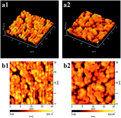 |
| | Fig. 7 2D and 3D AFM images of pristine PVDF membrane (a1 and b1) and β-CD–PVDF membrane (1.8%, w/v) (a2 and b2). | |
Table 3 Surface roughness data
| Sample |
Roughness |
| Ra (nm) |
Rms (nm) |
| Pristine PVDF membrane |
40.6 |
53.6 |
| β-CD–PVDF membrane |
71.5 |
90.6 |
3.6. Antifouling property of membranes
The antifouling performance of membrane plays a crucial role in the application of membranes. The antifouling properties of pristine PVDF membrane and those of β-CD–PVDF membrane (1.8%), during two cycles of flux recovery, are shown in Fig. 8. The results of relative flux reduction (RFR) and the flux recovery ratio (FRR) are summarized in Table 4. In the first filtration cycle, the initial water flux of β-CD–PVDF membrane is significantly higher than that of pristine membrane. When 0.1 g L−1 BSA solution was filtered through the membrane, it was found that the BSA flux for pristine membrane and β-CD–PVDF membrane suffered a rapid flux decline due to deposition and adsorption of BSA. Although the RFR of the two membranes is 75.0%, the β-CD–PVDF membrane has higher BSA flux than that of pristine membrane. In the last stage, after the membrane was cleaned by water, the pristine PVDF membrane water flux recovered up to only 40.0% (FRR) of the initial flux, while β-CD–PVDF membrane water flux recovered up to 47.4% (FRR). Furthermore, in the subsequent filtration cycle, FRR of the pristine membrane (49.1%) is significantly higher than that of pristine PVDF membrane (31.0%). The results indicate that β-CD–PVDF membrane has remarkable antifouling properties, especially after repeated filtration cycles. RFR changed a little, but FRR increased, indicating that part of irreversible fouling changed to reversible fouling. This is because the multi-hydroxyl molecules of β-CD can improve hydrophilicity of the membrane surface, besides effectively reducing the deposition and adsorption of BSA. β-CD–PVDF membranes display a better antifouling property than does the pristine PVDF membrane, thus demonstrating that the antifouling property of membranes can be effectively improved after grafting the membranes by β-CD.
 |
| | Fig. 8 Water flux recovery of pristine PVDF membrane and β-CD–PVDF membrane (1.8%) during two cycles. | |
Table 4 The relative flux reduction (RFR) and the flux recovery ratio (FRR) of the membranes in BSA filtration experiments
| Sample |
First cycle |
Second cycle |
| RFR (%) |
FRR (%) |
RFR (%) |
FRR (%) |
| Pristine PVDF membrane |
75.0% |
40.0% |
80.0% |
31.0% |
| β-CD–PVDF membrane |
75.0% |
47.4% |
82.3% |
49.1% |
4. Conclusion
Novel β-CD–PVDF membranes were prepared via interfacial reaction using TMC and β-CD as cross-linking and modification agents, respectively. β-CD was bound to the PVDF membrane surface via a simple dip-coating technique. The roughness of PVDF membranes increased after their surfaces were grafted with β-CD. β-CD–PVDF membranes had lower water contact angle and higher absorption than the pristine PVDF membranes. The results indicate that β-CD–PVDF has higher hydrophilicity than the pristine PVDF membranes. And, the modification of PVDF membranes led to significant increase in their antifouling property. The flux recovery ratio of modified membrane is better than that of the pristine membrane. Furthermore, the modification increased pure water flux of PVDF membrane. The superior performance of our β-CD–PVDF membranes holds promise for their practical application.
Acknowledgements
This work was financially supported by The Applied Basic Research Project in Sichuan Province (2013JY0099).
References
- M. M. Pendergast and E. M. V. Hoek, Energy Environ. Sci., 2011, 4, 1946 CAS.
- G.-D. Kang and Y.-M. Cao, J. Membr. Sci., 2014, 463, 145–165 CrossRef CAS PubMed.
- F. Liu, N. A. Hashim, Y. Liu, M. R. M. Abed and K. Li, J. Membr. Sci., 2011, 375, 1–27 CrossRef CAS PubMed.
- D. Rana and T. Matsuura, Chem. Rev., 2010, 110, 2448–2471 CrossRef CAS PubMed.
- S. Liang, K. Xiao, Y. Mo and X. Huang, J. Membr. Sci., 2012, 394–395, 184–192 CrossRef CAS PubMed.
- X.-M. Wang, X.-Y. Li and K. Shih, J. Membr. Sci., 2011, 368, 134–143 CrossRef CAS PubMed.
- X. Cao, J. Ma, X. Shi and Z. Ren, Appl. Surf. Sci., 2006, 253, 2003–2010 CrossRef CAS PubMed.
- T.-H. Bae and T.-M. Tak, J. Membr. Sci., 2005, 249, 1–8 CrossRef CAS PubMed.
- Q. Y. Wang, Z. W. Wang, J. Zhang, J. Wang and Z. C. Wu, RSC Adv., 2014, 4, 43590–43598 RSC.
- L.-Y. Yu, Z.-L. Xu, H.-M. Shen and H. Yang, J. Membr. Sci., 2009, 337, 257–265 CrossRef CAS PubMed.
- A. Qin, X. Wu, B. Ma, X. Zhao and C. He, J. Mater. Sci., 2014, 49, 7797–7808 CrossRef CAS.
- L. Yan, Y. S. Li and C. B. Xiang, Polymer, 2005, 46, 7701–7706 CrossRef CAS PubMed.
- H. Dong, K. J. Xiao, X. L. Li, Y. Ren and S. Y. Guo, Desalin. Water Treat., 2013, 51, 3685–3690 CrossRef CAS PubMed.
- R. Z. Pang, X. Li, J. S. Li, Z. Y. Lu, C. Huang, X. Y. Sun and L. J. Wang, Acta Phys.-Chim. Sin., 2013, 29, 2592–2598 CAS.
- J.-H. Li, X.-S. Shao, Q. Zhou, M.-Z. Li and Q.-Q. Zhang, Appl. Surf. Sci., 2013, 265, 663–670 CrossRef CAS PubMed.
- T. L. S. Silva, S. Morales-Torres, J. L. Figueiredo and A. M. T. Silva, Desalination, 2015, 357, 233–245 CrossRef CAS PubMed.
- M. Singh, R. Sharma and U. C. Banerjee, Biotechnol. Adv., 2002, 20, 341–359 CrossRef CAS.
- T. Loftsson and M. E. Brewster, J. Pharmaceut. Sci., 1996, 85, 1017–1025 CrossRef CAS PubMed.
- T. Uyar, R. Havelund, J. Hacaloglu, F. Besenbacher and P. Kingshott, ACS Nano, 2010, 4, 5121–5130 CrossRef CAS PubMed.
- F. Zhang, W. Zhang, Y. Yu, B. Deng, J. Li and J. Jin, J. Membr. Sci., 2013, 432, 25–32 CrossRef CAS PubMed.
- J. Hu, D. D. Shao, C. L. Chen, G. D. Sheng, J. X. Li, X. K. Wang and M. Nagatsu, J. Phys. Chem. B, 2010, 114, 6779–6785 CrossRef CAS PubMed.
- Z. Xu, L. Li, F. Wu, S. Tan and Z. Zhang, J. Membr. Sci., 2005, 255, 125–131 CrossRef CAS PubMed.
- T. Uyar, R. Havelund, Y. Nur, J. Hacaloglu, F. Besenbacher and P. Kingshott, J. Membr. Sci., 2009, 332, 129–137 CrossRef CAS PubMed.
- D. Quéré, Annu. Rev. Mater. Res., 2008, 38, 71–99 CrossRef.
- G. Li, L. Shen, Y. Luo and S. Zhang, Desalination, 2014, 338, 115–120 CrossRef CAS PubMed.
- H. Fan, C. Wang, Y. Li and Y. Wei, J. Membr. Sci., 2012, 415–416, 161–167 CrossRef CAS PubMed.
- L. Yan, Y. Li, C. Xiang and S. Xianda, J. Membr. Sci., 2006, 276, 162–167 CrossRef CAS PubMed.
- Y. Liu, Y. Su, X. Zhao, Y. Li, R. Zhang and Z. Jiang, J. Membr. Sci., 2015, 486, 195–206 CrossRef CAS PubMed.
|
| This journal is © The Royal Society of Chemistry 2015 |
Click here to see how this site uses Cookies. View our privacy policy here. 


