Low temperature synthesis of rutile TiO2 single crystal nanorods with exposed (002) facets and their decoration with gold nanoparticles for photocatalytic applications†
Lijuan Bua,
Wenjing Yangc and
Hai Ming*b
aKey Laboratory of Chemical Biology and Traditional Chinese Medicine Research (Ministry of Education), College of Chemistry and Chemical Engineering, Hunan Normal University, Changsha 410081, P. R. China
bCollege of Chemistry, Chemical Engineering and Materials Science, Soochow University, Suzhou 215123, P. R. China. E-mail: lunaticmh@163.com
cReliability Research and Analysis Center, CEPREI (East China) Laboratories, The Fifth Research Institute of MIIT East China, P. R. China
First published on 5th May 2015
Abstract
Uniform rutile TiO2 single crystal nanorods (TNRs) enclosed by a high amount of active (002) facets were synthesized for the first time by treating anatase TiO2 with concentrated HNO3 under hydrothermal conditions. It was found that these TNRs exhibited considerably enhanced photocatalytic activity compared to bulk rutile TiO2 owing to the highly exposed active (002) facets. In addition, gold nanoparticles with diameters of 1–5 nm were successfully deposited on the TNRs (Au/TNRs) using a simple photocatalytic reduction of HAuCl4 by the TNRs in the presence of 2-propanol. The plasmon-induced photocatalytic chemistry of the Au/TNRs under ultraviolet and visible light was investigated. The photocatalytic ability of TNRs was clearly enhanced under ultraviolet light with the decoration of Au nanoparticles. In particular, in our experimental conditions, the Au/TNRs nanocomposite demonstrated much better photocatalytic ability under visible-light than under ultraviolet light. This phenomenon may be attributed to the intense localization of plasmonic near-fields close to the Au/TiO2 interface, which brings about enhanced optical absorption and is good for generating electron–hole pairs for photocatalysis.
Introduction
The synthesis of micro-particles and nanocrystals with exposed high-energy or active facets (e.g., Pt, Au, TiO2, V2O5, ZnO, Fe3O4, WO3) has attracted considerable attention because they usually exhibit fascinating interfacial behaviour and have been applied in many fields including catalysis, sensors, photovoltaics, environmental and energy storage applications.1–5 To date, TiO2 has been proven to be one of the most versatile materials among various oxides used in photo-catalysis, paints, sensor devices and rechargeable battery applications, benefiting from its impressive semiconductor properties: low cost, robust crystal structure and excellent stability. As a result, studies concerning the morphology, size, surface and crystal structures of TiO2 are being intensively explored.6 For example, the order of the average surface energies of anatase TiO2 was proved to be 0.90 J m−2 for (001) > 0.53 J m−2 for (100) > 0.44 J m−2 for (101),7 and the meta-stable (001) facets of anatase have a positive effect on the photocatalytic ability under ultraviolet (UV) or visible (vis) light. In particular, numerous studies aiming to produce TiO2 anatase with dominant facets have been developed worldwide since the pioneering work by Yang et al.8,9In addition, the decoration of noble metal particles (e.g., Au, Pt, Pd, and Ru) on certain substrates of metal oxides, carbon, or polymer has been beneficial for pursuing higher performance in many photo-induced reactions.10 In particular, the use of Au nanoparticles (AuNPs) was confirmed to be extremely effective in promoting photocatalytic reactions within a wide range of light spectra because the surface plasmon resonance (SPR) effect from AuNPs can be excited by visible light illumination.10 Moreover, the interfacial loading of AuNPs on TiO2 could largely increase the migration of photoelectrons, which can promote the separation of electrons and holes, and thus play an important role to enhance the photocatalytic activity.10,11 Therefore, the composite of Au/TiO2 has great potential application in photocatalytic reactions. As mentioned, the interfacial properties of TiO2 single crystals are crucial to determine its stability and reactivity and also affect the loading of AuNPs. High-energy surface atoms exhibit high activity and are easy to combine with the foreign atoms such as the Au atoms to form a stable structure.12 Therefore, preparing TiO2 single crystals with exposed high-energy facets loaded with AuNPs is always interesting to obtain certain specific photocatalytic behaviors, as proved by Gong et al. They found that the exposed active (001) and (110) facets of TiO2 loaded with AuNPs dramatically enhanced the UV light photocatalytic performance.13 Considering the greater thermodynamic stability of rutile TiO2 as compared to anatase, with only a slightly lower direct band gap (∼3.0 eV vs. 3.2 eV of anatase),14 it would be considerably attractive to load AuNPs on rutile TiO2 single crystals with highly exposed facets and then investigate its photocatalytic ability for enriching the fundamental study and obtaining a better understanding of the synergetic behaviour between Au and TiO2.
Although considerable efforts have been devoted to the synthesis and applications of hierarchical structures consisting of anatase or rutile nanocrystals with dominant facets, scarcely any successful method achieved the preparation of rutile crystals with a large percentage of (002) facets in scalable production. In this study, a new type of rutile TiO2 single crystal nanorod with highly exposed (002) facets was introduced and readily prepared for the first time by treating anatase TiO2 with concentrated HNO3 under mild hydrothermal conditions. The TNRs exhibited considerably enhanced photocatalytic activity compared to bulk rutile TiO2 owing to their highly exposed active (002) facets. Furthermore, ultra-small AuNPs (1–5 nm) were successfully deposited on TNRs (Au/TNRs) and the plasmon-induced photocatalytic chemistry of Au/TNRs in the UV and vis region was studied. Compared to bare TNRs, the photocatalytic ability of Au/TNRs was obviously enhanced under UV light. More interestingly, Au/TNRs hybrids exhibited a much better visible-light photocatalytic performance than that under UV light in our experimental conditions. This phenomenon may be due to the intense localization of plasmonic near-fields close to the Au/TiO2 interface, which could bring about enhanced optical absorption and facilitate the generation of electron–hole pairs.
Experimental section
Preparation of anatase TiO2 nanoparticles
All the chemicals were used as received without further purification. Typically, 4 mL tetra-n-butyl titanate (purchased from Alfa Aesar Co. Ltd., named as TBT) was dissolved in 100 mL ultra-pure ethanol. On the other hand, a certain amount of HCl was added into 200 mL H2O to adjust the pH value to around 1, and it was then transferred to a 500 mL three-neck flask and heated to 343 K in an oil bath. The abovementioned ethanol solution of TBT was dropped into the solution of HCl–H2O at a rate of 1 mL min−1 with vigorous stirring to promote the hydrolysis of TBT because of which a white colloid solution was formed. The solution was filtered and the precipitate was washed by ethanol and water several times. Finally, the white powder obtained was dried at 343 K for 48 h.Formation of the rutile TNRs
The prepared TiO2 nanoparticles were dispersed in concentrated HNO3 (69 wt%) with a concentration of 10 mg mL−1, and the colloid was then transferred into Teflon-lined stainless steel autoclaves maintained at 453 K for 24 h. After the autoclaves cooled down to room temperature, a white suspension was obtained. The products were collected by centrifugation at 12![[thin space (1/6-em)]](https://www.rsc.org/images/entities/char_2009.gif) 000 rpm for 20 min and washed by ethanol and water several times, giving rise to highly crystallized TNRs.
000 rpm for 20 min and washed by ethanol and water several times, giving rise to highly crystallized TNRs.
Preparation of Au/TNRs nanocomposite
Typically, 0.18 g of the as-prepared TNRs or P25, modified with 3-mercaptopropionic acid and then dispersed in the solution of H2O/2-propanol (IPA) (20 mL/5 mL), and 4.5 mL of an aqueous solution of 25 mM HAuCl4 were mixed. Subsequently, the solution was transferred into a one-neck quartz flask and stirred vigorously for 4 h. Finally, this suspension was exposed to UV light and stirred for 12 h, and Au/TNRs or Au/P25 nanohybrids were obtained after centrifugation.Photocatalytic activities and photoelectrochemical tests
The photocatalytic activity of bulk rutile TiO2, P25, TNRs, Au/P25, and Au/TNRs was evaluated by the degradation of rhodamine B (RhB) in an aqueous solution. In a typical procedure, 50 mg TNRs, P25 or Au/TNRs was dispersed first into 100 mL RhB (10 ppm), and then the suspension was stirred and irradiated by UV lamp (Spectroline SB-100P/F; 100 W, λ: 50–400 nm) or a tungsten–halogen (100 W, λ: 400–2500 nm) with a UV cut-off filter (λ ≥ 400 nm). A cooling fan was used to avoid the increase in temperature of the reaction system during irradiation. Before irradiation, the aqueous solution was magnetically stirred in the dark for 12 h to reach the adsorption equilibrium of dye on the catalyst surface; the absorption plots (Fig. S1†) of RhB on different photocatalysts showed that the adsorption equilibrium is nearly stable after 12 h. Compared to the starting concentration of RhB (10 ppm), the absorption amount of these photocatalysts are about 10.2% to 21.5% of the total pollutant (RhB). The adsorption capability of P25, TNRs, and bulk rutile TiO2 decreased successively, which might agree with their decreasing specific surface areas. Moreover, the adsorption ability of Au/P25 and Au/TNRs were a little lower than pristine TNRs due to the decoration of Au particles. After the initiation of irradiation, 5 mL suspensions were withdrawn at regular intervals of 10 or 30 min and centrifuged to remove the catalyst completely. The UV-vis absorption spectra of the centrifuged solution were recorded using a spectrophotometer.Measurement and characterization
X-ray powder diffraction (XRD) was obtained using an X'Pert-ProMPD (Holland) D/max-γA diffractometer with Cu-Kα radiation (λ = 0.154178 nm). Transmission electron micrographs (TEM) and high-resolution TEM (HRTEM) images were obtained on an FEI-Tecnai F20 (200 kV) transmission electron microscope (FEI). Room temperature solid UV-vis diffuse reflectance absorption spectra (UVDRS) were recorded on a Lambda 750 (Perkin Elmer) spectrophotometer in the wavelength range of 200–800 nm.Results and discussion
Nanostructure of TNRs
As shown in XRD patterns (Fig. 1a), the crystalline structure of the TNRs (red curve) sample was classified as the rutile phase (JCPDS: 87-0710). The intensity of characteristic peak at 62.79°, corresponding to the (002) crystal facets, is much higher than that of normal rutile TiO2, indicating the highly exposed (002) facets. Even after the thermal treatment of the TNRs at 973 K, their crystal phase was well maintained and there was no obvious change, confirming the good stability and highly crystallinity of the pristine TNRs synthesized under mild hydrothermal conditions. Before the hydrothermal treatment, raw materials were mainly composed of anatase (black curve), and XRD results show that crystalline anatase TiO2 can transform to rutile TiO2 through a simple acid hydrothermal strategy at a low temperature of 453 K.The high magnification SEM image of TNRs shows that their morphology consisted of uniform nanorods with a diameter of 20–50 nm and length of 100–1000 nm (Fig. 2a), as further characterized by the TEM (Fig. 2b). They have very thin structure and smooth surface. The lattice fringes with lattice space of 0.322 nm along the [001] direction were clearly observed under HRTEM (Fig. 2c). Furthermore, the well-separated diffraction spots in the selected area electron diffraction (SAED) pattern fully reveal the single-crystalline nature of TNRs, and the spacing of diffraction spots are 0.322 nm and 0.148 nm, corresponding to the facets of (110) and (310) (Fig. 2d); moreover, the angle between the (110) and (310) is about 28.3°, which is identical to the theoretical value between the {110} and {310} facets. This information confirms that the TNRs nanocrystals mainly expose {002} surfaces.15
Based on the crystalline structure of TNRs, a possible conversion mechanism was presented (Fig. 3). As is well known, the different polymorphs of anatase, rutile, and/or brookite can be induced by local pH variations or the relative complexation effect. In TiO2, the rutile crystalline structure is based on linear chains of TiO6 octahedra by sharing equatorial edges, whereas anatase is based on a spiral chain of apical edge sharing TiO6 octahedra.16 First, in the hydrothermal process at 453 K, the anatase precursor may bond with the anionic ions of NO3− to form a anionic matrix TiO2−x/2[NO3−]x[OH2]x/2. Next, with increasing temperature and pressure, the value of x will increase and the TiO2−x/2[NO3−]x[OH2]x/2 will further dehydrate due to the strongly acidic medium. In addition, the anion of NO3− will decompose under these critical conditions, while TiO2−x/2[NO3−]x[OH2]x/2 transfers to an octahedral monomer ([TiO(OH2)5]2) in a strongly acidic solution.17 In the interfacial region of the confined mesostructure, a linear chain arrangement of [TiO(OH2)5]2 octahedral monomers (i.e., rutile nuclei) sharing equatorial edges may be more favorable rather than an apical edge-sharing spiral structure (i.e., anatase nuclei) due to the geometric confinement.18 Thus, the lamellar rutile nanocrystals grow along the [001] direction. The interfacial charge density of TiO2 decreases due to the elimination of positive charges in the TiO2 framework, and then the two-dimensional lamellar rutile nanocrystals transform into rutile nanorods. In particular, a high pressure in thermal treatment and a supercritical state were beneficial for the formation of highly crystallized TiO2.
Nanostructure of Au/TNRs
Because TNRs have a special single crystalline structure similar to the monocrystal Si wafer and mica plate, they could be a good processing substrate for studying photocatalytic mechanisms.To verify this, Au/TNRs nanocomposites were prepared and their structural, physical and photocatalytic properties were investigated in detail to enrich the knowledge of the effect of AuNPs on the photocatalytic properties of TiO2. Except the XRD peaks of TiO2, four other peaks corresponding to Au (111), (200), (220) and (311) appeared (Fig. 4a), directly demonstrating the existence of Au. This was further confirmed by the EDX spectrum of Au/TNRs, which contained Ti, Au and O elements (Fig. 4b). Furthermore, as demonstrated by SEM image and EDS-mapping, the mass ratio of Au is about 10.1 wt% with a very uniform distribution (Fig. 4c–f).
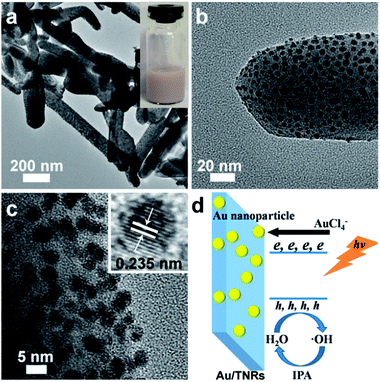
The distribution of AuNPs on TNRs was further demonstrated by TEM images. As shown in Fig. 5a and b, TNRs were uniformly decorated with plenty of AuNPs with sizes of 1–5 nm. The close-knitting of AuNPs and one dimensional TNRs in junction structures indicate their good combination after UV irradiation treatment. The fine and well-resolved lattice fringe of black dots exhibit an inter-planar distance of 0.235 nm, which corresponds well to the Au (111) planes. The digital image of the Au/TNRs solution exhibits a typical white-purple color (inset of Fig. 5a). The reason for the close-knitting of AuNPs and TNRs can be ascribed to the ingenious design of the experiment (Fig. 5d). First, the irradiation of TNRs by UV light can produce a lot of electrons and holes due to the narrow band gap of 3.0 eV, which is sensitive to the UV light. Once the AuCl4− comes into contact with the surface of TNRs in the solution, Au(III) could be reduced in situ to Au(0) atoms, which can closely link with the TNRs due to the low polarization and surface tension of the IPA/H2O solution. Accompanying the deposition of Au(0) on TNRs, the site of Au(0)/TNRs could produce more electrons around the surface of the Au(0) nuclei and then further facilitate its growth. In addition, the IPA is a hole-trapping agent, and it could sufficiently separate electrons and holes in Au(0)/TNRs, which could continually reduce Au(III) and maintain the regular growth of AuNPs.19 Finally, the AuNPs can tightly combine with TNRs. However, for the sample P25, the particle size of loaded Au on P25 was as large as 10–30 nm and not uniform (Fig. S2†). The reason can be ascribed to the shortage of high energy crystal surface of P25 for loading the Au nuclei and a fast growth of Au particles on P25 under the UV irradiation.13
 | ||
| Fig. 5 (a and b) TEM and (c) HRTEM images of Au/TNRs; (d) scheme for the formation of Au/TNRs. Insert: (a) digital photo of Au/TNRs suspension and (c) HRTEM image of single AuNP. | ||
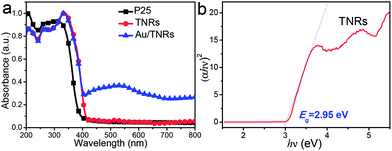
UV-vis diffuse reflectance spectroscopy (UVDRS) was used to characterize the electronic states (Fig. 6a), and it indicated that the band gap absorption onset of P25 and TNRs are located around 380 nm (black curve) and 420 nm (red curve), respectively, while Au/TNRs show an efficient absorption in the visible-range of 420–800 nm, implying a good photocatalytic ability of Au/TNRs under visible light. The UVDRS of pristine TNRs exhibits a broad absorption band from 200 to 420 nm; thus, the band gap energy of this sample can be calculated using the equation (αhν)n = k(hν − Eg), where α is the absorption coefficient, k is the parameter that relates to the effective masses associated with the valence and conduction bands, n is 2 for a direct transition, hν is the absorption energy, and Eg is the band gap energy. Plotting (αhν)2 vs. hν based on the spectral response gives the extrapolated intercept corresponding to the Eg value (Fig. 6b). The optical band energy of the rutile TNRs is 2.95 eV, which exhibits a slight red-shift of 0.05 eV with respect to 3.0 eV of normal rutile TiO2. This phenomenon is acceptable because changes of crystal structure and morphology could affect the optical properties.14,20,21
 | ||
| Fig. 6 (a) UVDRS spectra of the TNRs, Au/TNRs, and P25; (b) indirect inter band transition energy of the TNRs. | ||
Photocatalytic properties of TNRs and Au/TNRs
To investigate the photocatalytic activity of Au/TNRs, the photo-degradation of RhB under UV (λ: 50–400 nm) and visible light (λ: 400–2500 nm) was performed in comparison to the TNRs, P25, and Au/P25 catalysts. As shown in Fig. 7a, when irradiated with UV light, 10 ppm of RhB can be degraded completely by P25 and Au/P25 within 80 min because of their double crystalline phase of anatase/rutile, while the degradative amount of RhB were only 28%, 45% and 58% for the bulk rutile TiO2, TNRs and Au/TNRs, respectively, even by prolonging the irradiation time to 120 min. The TNRs exhibited improved catalytic ability compared to bulk rutile TiO2 owing to the dominant active (002) facets; moreover, the introduction of AuNPs on TNRs gives rise to higher catalytic activity than with pristine TNRs.This phenomena could be interpreted from the reaction mechanism, and generally the photocatalytic pathway in Au/semiconductors could be summarized as follows: the semiconductor absorbs photons and excites electrons from the valence band to the conduction band, creating highly reactive electron–hole pairs. Some of the pairs recombine, while some migrate towards the semiconductor surface to be involved in the photocatalytic reaction. The electrons may be trapped by oxygen to form oxygen species (˙O2−).22 The photo-generated holes may be trapped by hydroxyl groups attached on the surface to form hydroxyl radicals (˙OH).23 These hydroxyl radicals and oxygen species oxidize the RhB to carbon dioxide, water and some simple mineral acids in an aqueous solution. However, for the sample of Au/TNRs, the photoelectrons can be captured by gold particles and subsequently be transferred to the adsorbed O2, leading to the effective separation of electrons and holes and they are then available to increase the photocatalytic activity.24
More interesting and entirely different results were collected in the reaction system irradiated by a tungsten–halogen lamp with a UV cut-off filter (λ ≥ 400 nm) (Fig. 7b). The Au/TNRs are able to degrade RhB completely within 3 hours, while the residual amount of RhB for bulk rutile TiO2, P25, TNRs, and Au/P25 are as high as 98.5%, 97.4%, 87.6% and 55.8%, respectively. Moreover, the catalyst of Au/TNRs demonstrate a high cycle ability in the initial 10 cycles and was available to fully degrade RhB within 3 hours (Fig. S3†). It has a better catalytic ability under the visible light than under the UV light when both light powers are the same, while this phenomenon is completely inverse for the sample of Au/P25. The impressive catalytic behaviour of Au/TNRs may be relative to their special electronic system, and various enhancement mechanisms could be proposed such as SPR-mediated charge injection from metal to semiconductor and plasmonic heating. In the SPR-mediated charge injection mechanism, the plasmonic AuNPs behave like a light absorber, and charge carriers generated from the excited AuNPs will be directly injected into the adjacent TiO2 in a manner analogous to dye sensitization.25 In this mechanism, the photoreactions take place at the phase boundaries of metal–semiconductor–liquid. Under this condition, once excited by SPR, the electrons would move from AuNPs to the conduction band of TiO2 and then drive the reduction reaction of RhB. Another mechanism was also put forward to explain the plasmonic effect when the plasmonic AuNPs are in direct contact with TiO2. SPR is able to enhance the local electric fields in the vicinity of the AuNPs, and the interaction of local electric fields with the proximate TiO2 allows the facile generation of electron–hole pairs in the interfacial area of the Au/TiO2.26 In this case, the photoreaction centres are at metal–semiconductor–liquid three phase boundaries, and they spread away from the three phase boundaries along the semiconductor–liquid interface. However, the UVDRS spectra of TNRs and Au/TNRs indicate that the TNRs are not sensitive to the visible light; thus, this plasmonic heating mechanism should not be the main factor contributing to the enhanced photocatalytic performance of the Au/TNRs.
Irrespective of the reaction mechanism, all the photoreactions occur between or near the interface of TiO2 and AuNPs, which are responsible for the better photocatalytic ability under the visible light than under UV. Once the plasmonic AuNPs were excited by the visible light and performed the role of light harvester, AuNPs can absorb photons immediately and the charge carriers could be directly injected into the adjacent TiO2 conduction band, thus rapidly separating the positive holes and negative electrons on AuNPs and the conduction band of TiO2, respectively. This promotion effect could be further strengthened due to the close interaction of AuNPs and (002) facets of TNRs, where a strong thermoelectric effect could be induced by the SPR effect on high energy crystal facets, which can further improve the transfer ability of photons. Moreover, AuNPs can introduce a very strong local electric field to the proximate TiO2 matrix because of SPR and/or plasmon assisted Forster resonance energy transfer. Electron–hole pairs could be formed in the strong local electric field even though the excitation energy of the visible light is lower than the band gap of TiO2, and the strong electric field of plasmonic AuNPs could effectively suppress the recombination of hole and electron. After separation, the electron will move to the surface of AuNPs to drive the generation of ˙O2−, and the hole will migrate to the semiconductor–liquid surface and be captured by OH in the liquid phase to form ˙OH. Therefore, all these active species mainly distributed on the interface of TNRs and AuNPs will then sufficiently come into contact with the pollutant towards an efficient degradation. Moreover, according to the literature, with regards to the effects of Au particle size (Table S1†), an appropriate size below 12 nm is good for high performance; therefore, it is convincing that the AuNPs around 1–5 nm on Au/TNRs could play a positive role in improving the catalytic activity. However, in the case of Au/P25 irradiated by the visible light, it showed a better UV photocatalytic ability, and the reasons can be ascribed to the lack of (002) facets and larger Au particles with a wide distribution (Fig. S1†), on which insufficient interfaces (metal–semiconductor–liquid) could be used to accelerate the photoreactions.27,28 Besides, the photodeposition of large Au particles can mask or block TiO2 active sites and then lead to a decreased catalytic activity.29–31
In addition, possible reasons interpreting the low photocatalytic efficiency of Au/TNRs under UV irradiation was also discussed. First, under the irradiation of Au/TNRs by UV light, there is no SPR effect on the nanostructured noble metals (e.g., AuNPs) because SPR effect was mainly excited by the visible light illumination.32,33 Second, the higher loading of AuNPs (around 10.1%) is considerably higher than the optimized amount (below 2%) that is mentioned in previous reports.34,35 Excessive amount of AuNPs covered on TiO2 surface may act as recombination centres for electrons and holes, thus leading to the decrease of photocatalytic activity.30,36 Third, according to the formula E = Wt = nh/λ (where W, t, n, h and λ are the light power, irradiation time, number of photons, Planck constant and wavelength, respectively), a higher number of photons could be obtained per unit time for the visible light (λ ≥ 400 nm) compared to the UV light (λ < 400 nm) under the same light power, and this demonstrated that a higher density of photons was introduced per unit volume in the photoreaction system under the visible irradiation. In other words, more amount of visible photons could be supplied by the visible irradiation, even if the trapping efficiency of visible photons was little lower than that of UV, and thus give rise to higher catalytic ability.
Conclusions
In summary, new uniform rutile TiO2 single crystal nanorods (TNRs) enclosed by a high amount of active (002) facets were synthesized simply through treating anatase TiO2 with concentrated HNO3 under mild hydrothermal conditions, and a possible conversion mechanism was presented preliminarily. As-prepared TNRs demonstrated considerably enhanced photocatalytic activity over bulk rutile TiO2 and it may benefit from the dominant active (002) facets. Furthermore, very uniform Au nanoparticles with sizes of 1–5 nm were successfully deposited on the TNRs (i.e., Au/TNRs) by the photocatalytic reduction of HAuCl4 on TNRs in the presence of 2-propanol. In addition, the plasmon-induced photocatalytic performance of Au/TNRs in the UV and visible region was studied and compared to that of the pristine TNRs, P25 and Au/P25 catalysts. With the decoration of AuNPs, not only the catalytic ability of Au/TNRs is obviously enhanced in the photo-degradation of RhB, but also interesting results of better photocatalytic performance under visible light than that under UV light were found and investigated. This phenomenon was discussed in terms of the reaction mechanism and it may be related to the intense localization of plasmonic near-fields close to the interface of Au/TiO2, which would bring enhanced optical absorption and facilitate the generation of electron–hole pairs for photocatalysis.Acknowledgements
Financial supports from the Nature Science Foundation of China (no. 20873089, 20975073), Nature Science Foundation of Jiangsu Province (no. BK2011272), Industry-Academia Cooperation Innovation Fund Projects of Jiangsu Province (no. BY2011130), Project of Scientific and Technologic Infrastructure of Suzhou (SZS201207), Graduate Research and Innovation Projects in Jiangsu Province (CXZZ13_0802) and Hunan Provincial Innovation Foundation For Postgraduate (CX2012B206) are gratefully acknowledged.Notes and references
- T. L. Thompson and J. T. Yates, Chem. Rev., 2006, 106, 4428–4453 CrossRef CAS PubMed.
- H. Tada, T. Kiyonaga and S. Naya, Chem. Soc. Rev., 2009, 38, 1849–1858 RSC.
- M. D'Arienzo, J. Carbajo, A. Bahamonde, M. Crippa, S. Polizzi, R. Scotti, L. Wahba and F. Morazzoni, J. Am. Chem. Soc., 2011, 133, 17652–17661 CrossRef PubMed.
- C. J. Jia, L. D. Sun, F. Luo, X. D. Han, L. J. Heyderman, Z. G. Yan, C. H. Yan, K. Zheng, Z. Zhang, M. Takano, N. Hayashi, M. Eltschka, M. Kläui, U. Rüdiger, T. Kasama, L. Cervera-Gontard, R. E. Dunin-Borkowski, G. Tzvetkov and J. Raabe, J. Am. Chem. Soc., 2008, 130, 16968–16977 CrossRef CAS PubMed.
- G. Liu, H. G. Yang, X. W. Wang, L. N. Cheng, J. Pan, G. Q. Lu and H. M. Cheng, J. Am. Chem. Soc., 2009, 131, 12868–12869 CrossRef CAS PubMed.
- M. Lazzeri, A. Vittadini and A. Selloni, Phys. Rev. B: Condens. Matter Mater. Phys., 2001, 63, 155409 CrossRef.
- U. Diebold, Surf. Sci. Rep., 2003, 48, 53 CrossRef CAS.
- H. G. Yang, C. H. Sun, S. Z. Qiao, J. Zou, G. Liu, S. C. Smith, H. M. Cheng and G. Q. Lu, Nature, 2008, 453, 638–641 CrossRef CAS PubMed.
- X. W. Zhao, W. Z. Jin, J. G. Cai, J. F. Ye, Z. H. Li, Y. R. Ma, J. L. Xie and L. M. Qi, Adv. Funct. Mater., 2011, 21, 3554–3563 CrossRef CAS PubMed.
- S. Y. Zhu, S. J. Liang, Q. Gu, L. Y. Xie, J. X. Wang, Z. X. Ding and P. Liu, Appl. Catal., B, 2012, 119–120, 146–155 CrossRef CAS PubMed; M. Murdoch, G. I. N. Waterhouse, M. A. Nadeem, J. B. Metson, M. A. Keane, R. F. Howe, J. Llorca and H. Idriss, Nat. Chem., 2011, 3, 489–492 Search PubMed; X. M. Zhang, Y. L. Chen, R.-S. Liu and D. P. Tsai, Rep. Prog. Phys., 2013, 76, 046401 CrossRef PubMed.
- X. Zhang, Y. Liu, S. T. Lee, S. H. Yang and Z. H. Kang, Energy Environ. Sci., 2014, 7, 1409–1419 CAS.
- X. Q. Gong, A. Selloni, O. Dulub, P. Jacobson and U. Diebold, J. Am. Chem. Soc., 2008, 130, 370–381 CrossRef CAS PubMed.
- M. Y. Xing, B. X. Yang, H. Yu, B. Z. Tian, S. Bagwasi, J. L. Zhang and X. Q. Gong, J. Phys. Chem. Lett., 2013, 4, 3910–3917 CrossRef CAS; R. G. Li, H. X. Han, F. X. Zhang, D. G. Wang and C. Li, Energy Environ. Sci., 2014, 7, 1369–1376 Search PubMed.
- E. Bae and T. Ohno, Appl. Catal., B, 2009, 91, 634–639 CrossRef CAS PubMed.
- S. W. Liu, J. G. Yu and M. Jaroniec, Chem. Mater., 2011, 23, 4085–4093 CrossRef CAS.
- Y. Q. Zheng, E. R. Shi, Z. Z. Chen, W. J. Li and X. F. Hu, J. Mater. Chem., 2001, 11, 1547–1551 RSC; S. Cassaignon, M. Koelsch and J. P. Jolivet, J. Phys. Chem. Solids, 2007, 68, 695–700 CrossRef CAS PubMed; A. Pottier, C. Chaneac, E. Tronc, L. Mazerolles and J. P. Jolivet, J. Mater. Chem., 2001, 11, 1116–1121 RSC.
- F. P. Rotzinger and M. Graetzel, Inorg. Chem., 1987, 26, 3704–3708 CrossRef CAS.
- Y. Z. Li, N. H. Lee, D. S. Hwang, J. S. Song, E. G. Lee and S. J. Kim, Langmuir, 2004, 20, 10838–10844 CrossRef CAS PubMed.
- H. Ming, H. Huang, K. M. Pan, H. T. Li, Y. Liu and Z. H. Kang, J. Solid State Chem., 2012, 192, 305–311 CrossRef CAS PubMed.
- D. Q. Zhang, G. S. Li, F. Wang and J. C. Yu, CrystEngComm, 2010, 12, 1759–1763 RSC.
- K. S. Lee and M. A. EI-Sayed, J. Phys. Chem. B, 2005, 109, 20331–20338 CrossRef CAS PubMed.
- A. Furube, L. C. Du, K. Hara, R. Katoh and M. Tachiya, J. Am. Chem. Soc., 2007, 129, 14852–14853 CrossRef CAS PubMed.
- M. Teranishi, S. Naya and H. Tada, J. Am. Chem. Soc., 2010, 132, 7850–7851 CrossRef CAS PubMed.
- J. D. Stiehl, T. S. Kim, S. M. McClure and C. B. Mullins, J. Am. Chem. Soc., 2004, 126, 1606–1607 CrossRef CAS PubMed.
- Y. Tian and T. Tatsuma, J. Am. Chem. Soc., 2005, 127, 7632–7637 CrossRef CAS PubMed.
- I. Thomann, B. A. Pinaud, Z. B. Chen, B. M. Clemens, T. F. Jaramillo and M. L. Brongersma, Nano Lett., 2011, 11, 3440–3446 CrossRef CAS PubMed.
- J. Li and H. C. Zeng, Chem. Mater., 2006, 18, 4270–4277 CrossRef CAS.
- S. H. Overbury, V. Schwartz, D. R. Mullins, W. F. Yan and S. Dai, J. Catal., 2006, 241, 56–65 CrossRef CAS PubMed.
- B. Z. Tian, J. L. Zhang, T. Z. Tong and F. Chen, Appl. Catal., B, 2008, 79, 394–401 CrossRef CAS PubMed.
- M. C. Hidalgo, J. J. Murcia, J. A. Navío and G. Colón, Appl. Catal., A, 2011, 397, 112–120 CrossRef CAS PubMed.
- B. Cojocaru, Ş. Neaţu, E. Sacaliuc-Pârvulescu, F. Lévy, V. I. Pârvulescu and H. Garci, Appl. Catal., B, 2011, 107, 140–149 CrossRef CAS PubMed.
- Z. Y. Zhan, J. N. An, H. C. Zhang, R. V. Hansen and L. X. Zheng, ACS Appl. Mater. Interfaces, 2014, 6, 1139–1144 CAS.
- S. Sarina, E. R. Waclawik and H. Y. Zhu, Green Chem., 2013, 15, 1814–1833 RSC.
- A. Ayati, A. Ahmadpour, F. F. Bamoharram, B. Tanhaei, M. Mänttäri and M. Sillanpää, Chemosphere, 2014, 107, 163–174 CrossRef CAS PubMed.
- R. Kaur and B. Pal, J. Mol. Catal. A: Chem., 2012, 355, 39–43 CrossRef CAS PubMed.
- B. Tian, C. Li, F. Gu and H. Jiang, Catal. Commun., 2009, 10, 925–929 CrossRef CAS PubMed.
Footnote |
| † Electronic supplementary information (ESI) available. See DOI: 10.1039/c5ra04802h |
| This journal is © The Royal Society of Chemistry 2015 |


