DOI:
10.1039/C5RA04780C
(Paper)
RSC Adv., 2015,
5, 37814-37817
Insulin templated synthesis of single-crystalline silver nanocables with ultrathin Ag cores†
Received
18th March 2015
, Accepted 20th April 2015
First published on 20th April 2015
Abstract
The synthesis of ultrathin single-crystal 1D (one dimensional) nanostructures is highly desirable for potential applications as nanoconnectors and nanoscale devices. In this work, we demonstrate the preparation of straight single-crystalline coaxial silver nanocables (13 nm core diameter, 1.5 nm sheath thickness) in large amounts with excellent dispersibility for the first time through a self assembly approach via insulin fibril templates. This outstanding method for obtaining silver nanocables with ultrathin diameters will be attractive for the fabrication of other metallic 1D nanostructures, and this study may offer an effective strategy to design a novel silver nanostructure with diverse functionalities and potential applications in conductive materials, sensors and catalysts.
Introduction
Silver has the highest electrical and thermal conductivity characteristics among all the metals, which is ideal for electrical connections. Along with the trend of miniaturizing for components, the fabrication of silver 1D nanostructures is highly desirable for numerous potential applications. For instance, silver nanowires have been adopted to fabricate transparent conductive electrodes in recent years,1 and they have already become a superb candidate for the plasmonic nanoantenna, which can transmit and receive optical signals at multiple wavelengths.2 In addition, the potential of silver nanowires in metal-enhanced fluorescence-based applications of biosensing has emerged gradually.3 Furthermore, silver coaxial nanocables have been prepared4–10 as an updated version of silver nanowires, which have much more diverse functionalities and potential applications in conductive materials,11 sensor,12 catalysts.13 In the study of preparation methods for silver nanocables, Yin et al. synthesized silver nanowires with 30–40 nm diameters by polyol method at first, and silver nanowires were coated with silica shell in the range of 2–100 nm through a sol–gel process.4 But the resulted products tended to bind together and could not serve as individual nanocable with well dispersibility. Recently, Chen et al. demonstrated a facile hydrothermal method for the direct synthesis of carbonaceous silver nanocables with an Ag core of 25 nm in diameter and a sheath thickness of 20 nm,13 but their nanocables were not pretty thin enough. According to the kubo theory, the critical parameter of core silver diameter for the appearance of the quantum size effect is below 14 nm, where they can manifest a series of specific physical and chemical characters.14,15 However, silver nanowires with diameter blow 7 nm have been prepared by Eisele and coworkers via organic nanotubular templates.10 But these silver nanowires possessed polycrystalline structures with multi stacking and twinning, which could cause a relatively large loss in propagation application.16 Therefore, it exists a pressing need for ultrathin single-crystal silver 1D nanostructures with well dispersibility in large scale.
In this study, we present a successful synthesis of single-crystalline silver nanocables with sub-14 nm Ag core diameter in large quantities via insulin fibril templates. This method may offer an effective strategy to design and construct a novel nanostructure with much more diverse functionalities and potential applications in conductive materials, sensors and catalysts.
Results and discussion
Here, we demonstrate the simple but efficient fabrication of copious insulin fibrils with double helix hollow structure (Fig. S1†). These double helix nanostructures provide internal amide sites and crystal growth environment which possess following advantages specifically: (1) insulin fibrils own a small homogeneous diameter and lengths of several micrometers as shown in Fig. S2 (in the ESI†); (2) the ripe yet stable insulin fibrils have many amide sites which can recognize and react with inorganic substrates;17 (3) their limited hollow space can control the diameter of Ag core and facilitate the sequential crystal growing process.
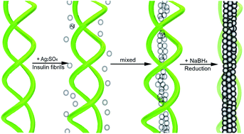
In this work, the double helix insulin amyloid fibrils are employed as scaffold templates to synthesize single-crystalline silver nanocables, which consist of a silver core about 13 nm diameter and a surrounding protein sheath about 1.5 nm thick. Detailed mechanism of silver nanocable growth using insulin fibril as templates was elaborated in Fig. 1. To begin with, the prepared insulin fibrils (Fig. 1a) were combined with Ag2SO4 precursors in solutions (Fig. 1b). After 6 h incubation, Ag ions in solution diffused to the internal amide sites of insulin fibrils and bound strongly to the fibrils at nucleation sites (Fig. 1c). At last, the mixture solution of insulin fibrils and Ag2SO4 precursors were reduced by 5 mM NaBH4 (Fig. 1d).
 |
| Fig. 1 (a) Insulin amyloid fibrils with double helix structure. (b) Mix the templates and silver precursors in solution. (c) Mix equality after 6 h. (d) Harvest silver nanocables after adding reductant. | |
In order to prove the proposed mechanism, we record the experimental process with a series of transmission electron microscopy (TEM) images in Fig. 2, and utilize UV-vis absorption spectrum as an auxiliary explanation in Fig. 3.
 |
| Fig. 2 (a) Insulin amyloid fibrils with hollow structure. (b) The obvious double helix insulin fibrils with silver precursor. (Some shapeless s insulin protein with silver precursor are scattering in the surrounding). (c) Reduction process after 3 h. (d) Entirely reduced after 6 h. | |
 |
| Fig. 3 UV-vis absorption spectra of: (a) Green: insulin fibrils solution. Red: adding silver precursors solution to insulin fibrils solution. Black: silver nanocables solution. (b) Purple red: 3 h in the reduction process. Black: 6 h in the reduction process. | |
Methods
We use phosphotungstic acid to dye the prepared insulin fibrils for making their hollow structure more clearly shown in Fig. 2a. In addition, insulin fibril presents a distinct double helix structure in Fig. 2b after combined with silver precursor. Afterwards, we tracked the silver nanocable growth during the reduction process by imaging the products after 3 h and 6 h reduction as shown in Fig. 2c and d respectively. It is certain that silver crystallize along the inner space of fibrils as demonstrated in the TEM image in Fig. 2c. As times goes on, the reduction process reach the end after 6 h, and a selected finished-nanocable is presented in Fig. 2d.
Fig. 3a displays the absorption spectrum of the neat insulin fibrils solution (green line) with one peak at 275 nm attributed to insulin fibrils. When Ag2SO4 was combined with insulin fibrils in solution, the spectrum appears a strong peak at 265 nm which obscures the templates peak (red line). While after fully reduced, dramatic changes are observed (black line); a broad silver plasmon absorption peak around 420 nm grows, which indicates the growth of silver nanostruture. The Fig. 3b absorption spectrum aims at analyzing the reduction process at two typical times such as 3 h and 6 h, the reduced 6 h sample peak (black line) is higher than the reduced 3 h sample peak (purple red line), which indicates silver crystal are increasing during this period of time.
The structure and morphology of our Ag nanocables are investigated by scanning electron microscopy (SEM). Fig. 4a–b shows the overall morphology of the obtained Ag nanocables at two different magnifications, which are about 14 nm in diameter and several micrometers in length, and some of them almost aggregate to form a large spiky ball. But they could be detached from each others after 20 min of sonication for TEM sample preparation, and the products with high dispersion are shown in Fig. S3.† The representative TEM images of multiple and single Ag nanocable are shown in Fig. 4c–d respectively.
 |
| Fig. 4 (a) SEM image of Ag nanocables in large quantities. (b) SEM image of Ag nanocables in relative high magnification. (c) TEM image of multiple Ag nanocables. (d) TEM image of single Ag nanocable. | |
Selected-area electron diffraction (SAED) patterns from different parts of single Ag nanocable (Fig. 5a) reveal several bright spots (inset in Fig. 5a), attributable to {111} and {220} crystal planes of an fcc crystalline metal Ag. The high-resolution TEM (HRTEM) image shown in Fig. 5b displays the crystallographic alignment of Ag nanocable, revealing that the entire nanocable is one single crystal with the lattice spacing between the {111} planes of 0.24 nm, which is in agreement with the value of Ag crystal. Clearly visible is that the structure is composed of an ultrathin silver nanowire as core and protein layer as sheath, similar to a coaxial cable. The overall diameter is about 16 nm and the central silver core diameter is about 13 nm. Fig. 5c shows the X-ray diffraction (XRD) pattern of the nanocables. The peaks at 2θ = 38°, 44°, 64°, 77° and 81° correspond to the diffractions from the fcc Ag structure (JCPDS PDF card 04-0783). The energy dispersive X-ray spectrum (EDS) in Fig. 5d displays that the products comprise only of silver. The Cu signals arise from Cu grid and S signal is due largely to insulin protein sheath.
 |
| Fig. 5 (a) TEM image of one single-crystalline Ag nanocable and SAED patterns of two sections (arrow). (b) HRTEM image of the white boxed area in (a). (c) XRD pattern and (d) EDS spectrum of Ag nanocables. | |
On the basis of above analysis, we confirm that the silver precursors are incorporated with internal amide sites of insulin fibrils and silver crystallize along the inner wall of templates to self assemble single crystalline nanocables. These nanocables with ultrathin core diameter will be attractive in future applications, due to their smooth surfaces and single-crystalline nature, which may lead to excellent optical performance owing to less propagation losses and higher conversion efficiency of surface plasmon polaritons (SPPs).18 The functions of the resulting nanostructures in a galvanic replacement reaction and oxygen reduction reaction (ORR) will be explored and measured in our future research.
Conclusion
In summary, we have successfully developed a simple but effective route to self assemble ultrathin silver nanocable with 13 nm core diameter by using insulin amyloid fibrils. The template controls the diameter, provides internal amide sites, and acts as ultrathin insulating sheath. Structural characterization attributes the formation of silver nanocables to silver crystallizes in situ inside insulin fibril templates which have double helix structures. This work would provide an excellent candidate route to form other metallic one-dimensional structure with ultrathin diameter for potential applications in sensors, electrocatalysts, and biomedicine.
Author contributions statement
L.T., K.Y.and F.G. wrote the main manuscript text. J.P. and X.L. prepared figures. All authors reviewed the manuscript.
Additional information
Authors are no Competing Financial Interests statement.
Acknowledgements
Financial support from the National Natural Science Foundation of China (Grants 21371149, 21101134) and Research Fund for the Doctoral Program of Higher Education of China (Grant 20131333110010) and the Natural Science Foundation of Hebei (Grant 14961107D).
References
-
(a) S. De, P. J. King, P. E. Lyons, U. Khan and J. N. Coleman, ACS Nano, 2010, 4, 7064–7072 CrossRef CAS PubMed;
(b) T. Kim, A. Canlier, C. Cho, V. Rozyyev, J. Y. Lee and S. M. Han, ACS Appl. Mater. Interfaces, 2014, 6, 13527–13534 CrossRef CAS PubMed;
(c) Y. Jin, L. Li, Y. Cheng, L. Kong, Q. Pei and F. Xiao, Adv. Funct. Mater., 2015, 25, 1581–1587 CrossRef CAS PubMed.
-
(a) Z. Fang, L. Fan, C. Lin, D. Zhang, A. J. Meixner and X. Zhu, Nano Lett., 2011, 11, 1676–1680 CrossRef CAS PubMed;
(b) T. Kang, W. Choi, I. Yoon, H. Lee, M. K. Seo, Q. H. Park and B. Kim, Nano Lett., 2012, 12, 2331–2336 CrossRef CAS PubMed;
(c) X. Wu, Y. Xiao, C. Meng, X. Zhang, S. Yu, Y. Wang, C. Yang, X. Guo, C. Z. Ning and L. Tong, Nano Lett., 2013, 13, 5654–5659 CrossRef CAS PubMed.
-
(a) B. Abel, S. Coskun, M. Mohammed, R. Williams, H. E. Unalan and K. Aslan, J. Phys. Chem. C, 2015, 119, 675–684 CrossRef CAS PubMed;
(b) M. Ahmad, H. Sun, M. Hussain, S. Karim, A. Nisar and M. Khan, Electroanalysis, 2015, 27, 1–10 CrossRef PubMed.
- Y. Yin, Y. Lu, Y. Sun and Y. Xia, Nano Lett., 2002, 2, 427–430 CrossRef CAS.
- L.-B. Luo, S.-H. Yu, H.-S. Qian and T. Zhou, J. Am. Chem. Soc., 2005, 127, 2822–2823 CrossRef CAS PubMed.
- S.-H. Yu, X. Cui, L. Li, K. Li, B. Yu, M. Antonietti and H. Cölfen, Adv. Mater., 2004, 16, 1636–1640 CrossRef CAS PubMed.
- A. Chen, H. Wang and X. Li, Chem. Commun., 2005, 14, 1863–1864 RSC.
- W. Wang, S. Xiong, L. Chen, B. Xi, H. Zhou and Z. Zhang, Cryst. Growth Des., 2006, 6, 2422–2426 CAS.
- J. Fu, X. Huang, Y. Huang, Y. Pan, Y. Zhu and X. Tang, J. Phys. Chem. C, 2008, 112, 16840–16844 CAS.
- D. M. Eisele, H. V. Berlepsch, C. Böttcher, K. J. Stevenson, D. A. V. Bout, S. Kirstein and J. P. Rabe, J. Am. Chem. Soc., 2010, 132, 2104–2105 CrossRef CAS PubMed.
- Y. F. Wang, J. H. Zeng and Y. Li, Electrochim. Acta, 2013, 87, 256–260 CrossRef CAS PubMed.
- H. B. Yao, G. Huang, C. H. Cui, X. H. Wang and S. H. Yu, Adv. Mater., 2011, 23, 3643–3647 CrossRef CAS PubMed.
- C. Chen, B. H. Suryanto, C. Zhao, X. Jiang and A. Yu, Small, 2015 DOI:10.1002/smll.201401854.
- R. S. Qin and B. L. Zhou, Nanostruct. Mater., 1996, 7, 741–748 CrossRef CAS.
- C.-M. Lai and S.-L. Lee, Chemistry, 2003, 61, 585–597 CAS.
- X. Xiong, C.-L. Zou, X.-F. Ren, A.-P. Liu, Y.-X. Ye, F.-W. Sun and G.-C. Guo, Laser Photonics Rev., 2013, 7, 901–919 CrossRef CAS PubMed.
- L. Zhang, N. Li, F. Gao, L. Hou and Z. Xu, J. Am. Chem. Soc., 2012, 134, 11326–11329 CrossRef CAS PubMed.
- A. V. Akimov, A. Mukherjee, C. L. Yu, D. E. C. A. S. Zibrov, P. R. Hemmer, H. Park and M. D. Lukin, Nature, 2007, 450, 402–406 CrossRef CAS PubMed.
Footnote |
† Electronic supplementary information (ESI) available. See DOI: 10.1039/c5ra04780c |
|
This journal is © The Royal Society of Chemistry 2015 |
Click here to see how this site uses Cookies. View our privacy policy here.