A robust site-specific Au@SiO2@AgPt nanorod/nanodots superstructure for in situ SERS monitoring of catalytic reactions

HaoWei Jia ab, Li Qiuab and Jin Wang*b
aCollege of Materials Science and Engineering, University of Science and Technology of China, Anhui 230026, P. R. China
bInstitute of Intelligent Machines, Chinese Academy of Sciences, Hefei, Anhui 230031, P. R. China. E-mail: jwang@iim.ac.cn; wangjin_hf@yahoo.com

Received 17th March 2015 , Accepted 8th April 2015

First published on 8th April 2015


Abstract

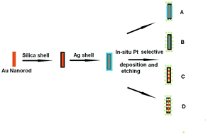
Platinum nanodots have been regiospecifically assembled on a robust core–shell nanorod via an in situ selective deposition and etching protocol. Therefore, different assembled superstructures, including tip coated nanorod/nanodots, tip/edge coated nanorod/nanodots, and core–satellite nanorod/nanodots, have been yielded. In addition, the SERS properties of the assembled superstructures have been investigated, suggesting that the tip coated or tip/edge coated superstructures are superior to the core–satellite superstructures. On the other hand, studies suggest that the catalytic properties of the tip/edge coated and core–satellite superstructure can be better than the tip coated superstructures. Moreover, the tip/edge coated and core–satellite nanorod/nanodots superstructures have been employed as nanoreactors for the real-time SERS monitoring of catalytic reactions.


1. Introduction

Assembling noble metal nanoparticles to form superstructures has been attracting more interest due to their unique optical properties, e.g., a higher refractive index sensitivity, in contrast with the individual nanocrystals or bulk materials. As far as superstructures fabricated bottom-up from noble metal nanocrystals are concerned, the collective oscillation of the free electrons, i.e., the plasmon resonance originating from the “hot-spot” region in the superstructure, results in the concentration of light below the optical diffraction limit. This has led to extensive applications in surface enhanced Raman scattering (SERS), LSPR based chemo-sensors or biosensors, plasmon rulers, optical waveguides, etc.1–7 As an example of a typical Au nanoassembly, core–satellite Au superstructures have been efficiently made via biological or chemical approaches. Schlücker et al. synthesized 3D Au superstructures via a silane reaction between a silica shell isolated Au nanoparticle as the Au core and small Au satellites.8 In addition, the click reaction, that is the azide–alkyne Huisgen 1,3-dipolar cycloaddition, has been utilized for generating plasmonic core–satellite superstructures on the basis of gold nanoparticles.9 Also, a common bifunctional linker molecule, p-aminothiophenol, has been used for producing different superstructures, e.g., Au core–satellite heterostructures and Ag nanoleaves.10,11 Very recently, hyperbranched polymer linkers have been used to fabricate gold nanoassemblies with a core–satellite morphology.12 Rossner et al. reported that a planet-satellite assembly can be produced via a star polymer functionalized with trithiocarbonate groups.13 Apart from the chemical approaches for assembly, DNA, as a representative biomolecule, has been extensively used for assembling Au nanoparticles. Kotov et al. constructed a complex nanoassembly of a Au nanorod and Au nanospheres by the aid of hybridized DNA oligomers; furthermore, three kinds of isomers, i.e., end, side and satellite, could be yielded.14 Bach et al. successfully fabricated highly SERS-active gold superstructures composed of 30 nm cores and 20 nm satellites by using an electrostatic and DNA-directed self-assembly technique.15

Herein, we present a metal shell assisted bottom-up approach for the regiospecific assembly of Pt nanodots on Au@silica@Ag nanorods for high-yielding complicated anisotropic plasmonic superstructures. The bottom-up method is highly tailorable, allowing the Pt density to be controlled on the large-scaled assembled architectures. Generally, the cross section of a nanodot with a size lower than 10 nm is quite small, making it difficult to generate SERS effects; on the other hand, the catalytic efficiency of large nanorods is quite low. Hence, the generated heterogeneous superstructures involving nanodots and nanorods can be used as an ideal bifunctional nanoplatform for the in situ SERS monitoring of catalytic reactions by nanorods/nanodots. This is due to the Au@SiO2@Ag nanorods having sensitive plasmonic activities and Pt nanodots having excellent catalytic activities.

2. Experimental

2.1 Materials and chemicals

HAuCl4·4H2O (99.9%), NaBH4 (99%), ascorbic acid (99.9%), CTAB (99%), AgNO3 (99.9%), SH-PEG (MW 6000), PVP (MW 29[thin space (1/6-em)]000), 3-mercaptopropyl trimethoxysilane (MPTMS), tetraethyl orthosilicate (TEOS), and H2PtCl6·6H2O were purchased from Sigma-Aldrich. The Milli-Q water that was used throughout the experiments was purified using a Milli-Q system.

2.2 Instruments and measurements

UV-Vis-NIR absorption spectra were recorded using a Solidspec-3700 spectrophotometer. The transmission electron microscopy (TEM) images were obtained with a JEOL JEM-2010 instrument operated at 100 kV. Surface enhanced Raman spectra were collected using a Renishaw Invia Reflex Raman spectrometer.

2.3 The synthesis of Au nanorods

The gold nanorods were synthesized by modified seed-mediated approach.16 The preparation of the gold nanorods was performed by three steps. As the initial step, 10 mL 0.5 mM HAuCl4 was mixed with 10 mL 0.2 M CTAB. Subsequently, 600 μL 0.02 M ice-cold NaBH4 was added to the mixed solution. Vigorous stirring of the seed solution was performed for 120 s; the seed solution was then left to settle at room temperature for 1 h. As the second step, the growth solution of the gold nanorods was prepared. 100 mL 0.2 M CTAB was mixed with 2.5 mL 4 mM AgNO3 solution at 27–30 °C. Then, 100 mL 1 mM HAuCl4 was added to the growth solution, followed by the addition of 1.2 mL 0.08 M ascorbic acid. Finally, the gold nanorods were prepared by the addition of 240 μL of the seed solution to the growth solution. The nanorods were concentrated to a 20 mL solution.

2.4 The synthesis of Au@SiO2@Ag nanorods

A thin silica film on the gold nanorods has several functions and advantages: (1) the improvement of the colloidal stability thus avoiding aggregation; (2) the improvement of the stability of the nanorod shapes in the following deposition and etching process; (3) providing a robust framework for forming the complicated nanorod/nanodots with a tailorable superstructure.

The silication of the nanorod was divided into two steps. MPTMS was first reacted onto the Au nanorod to enhance its affinity to the following silica coating on its surface. The 1 mL 0.0049 M as-prepared Au nanorods were then centrifuged once to remove the excess CTAB and mixed with an SH-PEG 100 μL (1 mg mL−1, MW 6000) and diluted to a 10 mL solution and sonicated for 30 min. Then, an ethanolic MPTMS solution (10 wt%) 200 μL was added to the solution for 2 h, under vigorous stirring, and the excess MPTMS was removed by ethanol and water 2 times. The pH value of the 10 mL Au@MPTMS nanorods solution was tuned to approximately 9 via the addition of ammonia aqueous solution. Subsequently, 0.1 mL 10 mM TEOS ethanol solution was added to the Au@MPTMS nanorods solution to complete the silica coating with a thickness ca. 2–3 nm. After the addition of TEOS, the solution was vigorously vibrated for 2 h to promote the hydrolysis and condensation of TEOS on the surface of the Au@MPTMS nanorods, the solution was then left overnight until the reaction was completed. The Au@MPTMS@TEOS nanorods with a 2–3 nm thickness were rinsed by ethanol and water repeatedly 2 times and collected by centrifugation to yield well-dispersed Au@MPTMS@TEOS nanorods for the subsequent preparation of Au@MPTMS@TEOS@Ag core–shell nanorods.

The fresh as-prepared Au@MPTMS@TEOS nanorods were then redispersed in 10 mL 0.1 M CTAB solution, and mixed with 1.5 mL 4 mM AgNO3 solution, 200 μL 0.1 M ascorbic acid, and 400 μL 0.1 M NaOH to efficiently produce the Ag nanoshell on the surface of the Au@SiO2@Ag nanorod. The Au@SiO2@Ag nanorods were centrifuged and redispersed in a 10 mL aqueous solution.

2.5 Assembling the site-specific Pt nanodots onto the nanorod

The site-specific Pt nanodots were assembled onto the surface of Au nanorods via an in situ deposition and etching protocol so that Au@SiO2@AgPt nanorod/nanodots assemblies with mesoporous cavities were efficiently yielded.

10 mL 0.0049 M as-prepared Au@SiO2@Ag core–shell nanorods were centrifuged once to remove excess CTAB surfactant and redispersed in 10 mL Milli-Q water. Subsequently, different amounts of 38.6 mM H2PtCl6, i.e., 50 μL, 200 μL, 600 μL, and 1 mL, were added to the core–shell nanorods solution via a syringe pump (NE 1000) with a rate of 7.2 mL h−1 at a temperature of 55 °C and rapidly stirred for 1 h. Finally, the colloidal solution was left to settle overnight and then centrifuged for characterization.

In order to perform a comparison of the catalytic behavior between the assembled Pt nanodots and discrete Pt nanodots, we synthesized Pt nanodots via a wet-chemical approach. 0.1 g PVP (MW 29[thin space (1/6-em)]000) was added to 8 mL 0.008 M H2PtCl6 solution and diluted to 50 mL. Subsequently, 0.035 g NaBH4 was dissolved in a 30 mL aqueous solution, dripped into the PVP/H2PtCl6 solution and stirred for 2 hours. The color of the solution changed to brown, suggesting that the Pt nanodots were yielded. Finally, the Pt nanodots were centrifuged and redispersed into a 12 mL aqueous solution.

2.6 Catalysis and SERS

The catalytic reduction of 4-nitrophenol by the Au@SiO2@AgPt nanorod/nanodots in an aqueous NaBH4 solution is described in detail. The catalytic reduction process was setup in a standard cell with a 1 cm path length and 3 mL volume. After NaBH4 (0.5 mL of a 0.6 M solution) was added to the quartz cell including 4-nitrophenol (2 mL of a 0.1 mM solution), the color changed from light yellow to yellow-green. Immediately after the addition of 30 μL 0.0049 M nanorod/nanodots and the discrete Pt nanodots solution, the absorption spectra were continually recorded by a solidspec-3700 spectrophotometer at a scanning range of 200–500 nm at room temperature.

1 mL 0.0049 M nanorod/nanodots solution was centrifuged and redispersed in 0.5 mL 10−7 M R6G solution. The sample was then drop-casted on a silicon chip before the Raman measurement. The surface enhanced Raman spectrum was collected with an integrated time of 10 s using 0.85 mW power of 632.8 nm radiation from a laser and a 50× objective lens.

3. Results and discussions

3.1 The assembled superstructure of the Pt nanodots on the Au@SiO2@Ag nanorod

The assembly strategy, shown as a schematic in Fig. 1, has been divided into three steps: (1) the silica coating of the nanorod, (2) the formation of the Au@SiO2@Ag core–shell nanorod, and (3) the formation of the Au@SiO2@AgPt superstructure via in situ regiospecific deposition and etching. According to previous studies on the direct deposition of Pt on Au nanorods, the ratio of Pt/Au and the ratio of ascorbic acid/Pt and Ag+ can finely tune the epitaxial deposition on the endcap, edge and flat surface of the Au nanorod.17,18 In the present case, the Pt nanodots are grown on the Au nanorod by a different strategy, i.e., the facets of the nanorod provide the possibility for the selective deposition of Pt nanodots on the nanorod. Moreover, the Pt nanodots can be deposited regiospecifically on the endcap, edge and flat face of the nanorod via a galvanic reaction between Ag and Pt. Simultaneously, the mesoporous silica shell provides the solid framework for supporting the Pt nanodots distributed around the nanorod, instead of them directly absorbing onto the surface of the nanorod, which is quite different from the previous reported heterostructure.19 Instead of the complicated fabrication procedures of the core–satellite superstructures,20,21 the obvious simplification for constructing a core–satellite assembly can be realized via template sacrification in the present case. The gap between the core and the satellite can be tuned by the thickness of the silica shell.
image file: c5ra04672f-f1.tif
Fig. 1 Schematic of the formation of Au@SiO2@Ag nanorod/Pt nanodots superstructures. (A) Tip coated Au@SiO2@AgPt nanorod/nanodots superstructure; (B) tip/edge coated Au@SiO2@AgPt nanorod/nanodots superstructure; (C) core–satellite Au@SiO2@AgPt nanorod/nanodots with a low Pt density superstructure; (D) core–satellite Au@SiO2@AgPt nanorod/nanodots with a high Pt density superstructure.

As the core, a nanorod provides an ideal platform with different active facets, which can lead to assembly at multiple sites.21–23 Also, gold nanorods are superior to nanospheres for assembly due to their longitudinal plasmonic bands being tunable in the NIR region.24 On the other hand, as satellites, the AgPt nanodots play an important role in the bifunctional heterogeneous plasmonic nanoassembly, i.e., a combination of SERS optical property and catalytic function.25–28

The MPTMS mediation for the silica-coating of the gold nanorods is used to strengthen the affinity of gold for the TEOS silica shell. A TEM image of the silica-coated Au nanorods with the mesoporous thin silica shell is shown in Fig. S1A. According to the magnified TEM of silica-coated Au nanorods in Fig. S2A, the thin silica shell can be clearly observed. Fig. S3 shows that the averaged edge length and width of the Au@SiO2 nanorods are 61.15 nm and 22.87 nm, respectively. As shown in Fig. S4, the longitudinal plasmon band of the gold nanorods is redshifted from 667 nm to 673 nm after the silica coating.

As far as the Au@SiO2@Ag core–shell nanorods are concerned, the longitudinal peak of the nanorods shifts from 667 nm to 642 nm compared to the original gold nanorods (shown in Fig. S4); moreover, the Ag plasmonic band is located at 422 nm, confirming the formation of the effective silver coating. The blueshift can be ascribed to two reasons, i.e., the different dielectric function of Ag in contrast with Au, and the overall decrease of the aspect ratio due to the homogeneous Ag shell. The silver-coated Au@SiO2 nanorods are shown in the TEM image of Fig. S1B, suggesting that Au@SiO2@Ag core–shell nanorods can be effectively yielded. As shown from magnified TEM image of the Ag coated nanorods (Fig. S2B), an obvious Ag shell coating can be observed. The averaged edge length and width of the Au@SiO2@Ag core–shell nanorods are 66.78 nm and 24.93 nm, respectively. Additionally, the averaged shell thickness of the core–shell nanorods is 6.08 nm (shown in Fig. S5).

The in situ assembly of the Pt nanodots onto the Au@SiO2@Ag core can lead to an obvious plasmon coupling, which forms a strong redshift of the plasmon band. As shown in Fig. 2, when the Pt nanodots are assembled at the end of the Au@SiO2@Ag nanorod, the longitudinal LSPR is redshifted from 642 nm to 654 nm; simultaneously, the Ag shell plasmonic shell is slightly redshifted from 422 nm to 428 nm. If the Pt nanodots are assembled around the tip and edge of Au@SiO2@Ag nanorod, the LSPR band of the longitudinal mode and the Ag plasmonic mode are both redshifted by 23 nm and 15 nm, respectively, compared to original core–shell nanorods (shown in Fig. 2). Furthermore, when the density of Pt nanodots is increased to form a core–satellite superstructure, the LSPR of nanorods can be dramatically changed, which is reflected by the large-scale broadening and redshifting. As shown in Fig. 2, the longitudinal plasmonic band of nanorods has been changed by 68 nm in contrast with the core–shell nanostructure. Finally, when the density of the Pt nanodots is increased, so as to form an isolated shell, i.e., nanodots covering the whole nanorod, the LSPR characteristics of the superstructure is totally changed. As seen from Fig. 2, a very broad longitudinal plasmonic band can be observed in the near-infrared region; moreover, the sharp band of the Ag shell has almost disappeared. It should be mentioned that for the assembled superstructures with Pt nanodots, no obvious characteristic absorption for Pt over the entire spectral range can be observed, in contrast with the Ag and Au components.


image file: c5ra04672f-f2.tif
Fig. 2 UV-Vis-NIR spectra of (A) Au@SiO2@Ag nanorods; (B) tip coated Au@SiO2@AgPt nanorod/nanodots superstructures; (C) tip/edge coated Au@SiO2@AgPt nanorod/nanodots superstructures; (D) core–satellite Au@SiO2@AgPt nanorod/nanodots superstructures with a low Pt density; (E) core–satellite Au@SiO2@AgPt nanorod/nanodots superstructures with a high Pt density.

Transmission electron microscopy (TEM) images of the assembled superstructures are shown in Fig. 3. As observed from TEM (Fig. 3A), Pt nanodots with a size of 2–5 nm can be assembled in situ at two tips of the nanorod instead of a random nucleation. When increasing the Pt nanodots surrounding the nanorod, independent Pt nanodots can be assembled along both the endcaps and the edges of the nanorod, which can be observed in Fig. 3B. The high purity of the tip-/edge-coated Au@SiO2@AgPt nanorod/nanodots can be observed from the large field view of the TEM in Fig. S6, where no nanodots can be found on the plane facets.


image file: c5ra04672f-f3.tif
Fig. 3 TEM images of (A) tip coated Au@SiO2@AgPt nanorod/nanodots superstructures; (B) tip/edge coated Au@SiO2@AgPt nanorod/nanodots superstructures; (C) core–satellite Au@SiO2@AgPt nanorod/nanodots superstructures with a low Pt density; (D) core–satellite Au@SiO2@AgPt nanorod/nanodots superstructures with a high Pt density.

Moreover, a whole isolated Pt shell composed of small-sized nanodots can be formed, reflected in Fig. 3C and D. As far as superstructures with a low density of Pt nanodots are concerned, the Pt ions tend to be reduced and assembled in situ at the endcap facet instead of the plane facet and the edge facet. The selective deposition of the platinum nanodots could be initiated from the end, to the edge and then to the plane. The possible reason is that the radius of the curvature of the ends of the nanorod may be affecting the packing of the CTAB surfactants, compared to the flatter edge of the nanorod with more tightly packed CTAB. As far as the tips of nanorods are concerned, the Pt cations preferentially displace the more loosely bound CTAB at the tips in contrast with the flatter edges. As a result, the corresponding three different assembled superstructures with good monodispersity and high reproducibility, i.e., end-, edge- and face-, can be efficiently yielded.

The EDX data reveals that the percentage of the silver component in the Au@SiO2@Ag core–shell nanorods decreases and the amount of the platinum component simultaneously increases with the addition of the platinum salt solution (Fig. S7 and S8), suggesting that the in situ assembly of Pt nanodots on the Au@SiO2 surface can be efficiently produced by a nanoscale galvanic reaction.

Additionally, the loading amount of the Pt nanodots on the Au@SiO2@Ag nanorods can be dependent on the alteration of the morphology of the nanorods, e.g., the Ag shell thickness. We adjusted the amount of added silver salt solution to form the Ag shell with different thicknesses. Subsequently, identical maximized amounts of platinum salt was added to the as-prepared Au@SiO2@Ag nanorods with different shell thicknesses. As a result, as shown in Fig. S9, it can be seen that the number of Pt nanodots on the surface of the nanorods is efficiently increased to accompany the increase of the shell thickness. However, it is difficult to accurately count the number of Pt nanodots to obtain the function between the loading number of the Pt nanodots and the Ag shell thickness due to the overlapping between these nanodots.

3.2 Catalysis and SERS behavior of the nanorod/nanodots superstructures

The present bifunctional nanoassembly with a combination of the SERS property and catalytic activity requires a model reaction to evaluate the properties. Therefore, a model reaction, i.e., para-nitrophenol reduction in the presence of NaBH4, has been used to examine the catalytic properties of these assemblies. In order to evaluate the catalytic properties of these assemblies, all the other reaction parameters, e.g., the amount of assembled nanoparticles, NaBH4, reactant etc., have been controlled with identical conditions to obtain different catalytic rates. The catalytic reaction with the different nanorod/nanodots assemblies was monitored via UV-Vis-NIR apparatus over time. The reaction kinetics with respect to the para-nitrophenol belongs to first-order kinetics, which exhibits a nearly linear correlation for the para-nitrophenol reduction.

As shown in Fig. S10, the absorption band of p-nitrophenol at 313 nm is obviously redshifted to 400 nm when NaBH4 is added to the p-nitrophenol solution due to the generation of p-nitrophenolate anions. As observed from the alteration of the color in the quartz cell, the color of the solution is changed from light yellow to yellow-green. Subsequently, although the reduction of the p-nitrophenol (E0 for p-nitrophenol/p-aminophenol = −0.76 V and H3BO3/BH4 = −1.33 V) is thermodynamically favorable, the absorption band at 400 nm remains for a long time without alteration, which implies that the p-nitrophenolate anions could not be reduced by the aqueous NaBH4 in the present system. However, when the nanorod/nanodots were added to the reaction system, the para-nitrophenolate anions were efficiently reduced via the nanocatalyst, e.g., the Au@SiO2@AgPt nanorod/nanodots superstructures, which led to a gradual decrease in the absorption band at 400 nm with different times depending on the type of the assembled nanoparticles (observed in Fig. S11). As shown in Fig. 4, the reaction with the Pt nanodots assembled on the endcap of the nanorod is slightly quicker than that with core–shell nanorod, due to the fact that the reaction constant of the former (2.27 × 10⁻³ s−1) is higher than that of the latter. As far as the tip/edge coated superstructures being the catalyst are concerned, i.e., Pt nanodots assembled on both the endcap and edge of the nanorod, the reaction is ca. 2 fold faster than the core–shell nanorod. When the Pt nanodots are assembled around the nanorods to form a core–satellite superstructure, the catalytic property of the nanorods is dramatically improved, in that the reaction constant is increased to 5.48 × 10⁻³ s−1, i.e., ca. 3 fold larger than the Au@SiO2@Ag nanorod. Moreover, when the Pt nanodots are assembled on the endcap, edge and flat planes of nanorod to form a core–satellite assembly with a high Pt density, the catalytic efficiency is remarkably further improved compared to the core–satellite assembly with a low Pt density, due to the reaction constant (7.73 × 10⁻² s−1) in the presence of the assembly contrasting with 5.48 × 10⁻³ s−1. In order to compare the catalytic ability of the assembled Pt nanodots to discrete nanodots, we synthesized the Pt nanodots and carried out an analogous catalytic experiment. As shown in Fig. S12, the reaction constant (1.48 × 10⁻³ s−1) is obviously lower than those of assembled Pt nanodots on the surface of nanorod.


image file: c5ra04672f-f4.tif
Fig. 4 Plots of ln[thin space (1/6-em)]A versus time for the catalytic reduction of para-nitrophenol by (A) Au@SiO2@Ag nanorods; (B) tip coated Au@SiO2@AgPt nanorod/nanodots superstructures; (C) tip/edge coated Au@SiO2@AgPt nanorod/nanodots superstructures; (D) core–satellite Au@SiO2@AgPt nanorod/nanodots superstructures with a low Pt density; (E) core–satellite Au@SiO2@AgPt nanorod/nanodots superstructures with a high Pt density.

The SERS properties of the nanorod/nanodots was also examined. Typically, Rhodamine 6G on the assembled nanostructures as a SERS substrate exhibits a strong enhancement of the vibrational modes. The vibrational modes of R6G observed in the present SERS spectra (shown in Fig. 5) are consistent with the previous assignments.29,30 The fingerprint Raman active vibrational modes of R6G, including the bands at 1647 cm−1 and 1360 cm−1, associated with aromatic C–C stretching vibrations, and the bands at 1511 cm−1, 774 cm−1 and 613 cm−1, are dramatically enhanced on the nanorod/nanodots superstructure. Reproducibility of the SERS response is quite crucial for obtaining the SERS enhancement ability of the different superstructures. The variations of the SERS signal intensities at the different locations on the substrate, i.e., spot-to-spot, were considered (see Fig. S13 in ESI). The coefficients of the variation with the Au@SiO2@Ag core–shell nanorods, the tip-coated nanorod/nanodots superstructures, the tip-/edge-nanorod/nanodots, the core–satellite nanorod/nanodots with a low Pt density, and the core–satellite nanorod/nanodots with a high Pt density are 6.54%, 7.28%, 5.19%, 4.84%, 6.23%, respectively, which suggests that signal reproducibility for the SERS substrates is quite good.


image file: c5ra04672f-f5.tif
Fig. 5 SERS spectra of R6G absorbed on the superstructures (A) Au@SiO2@Ag nanorods; (B) tip coated Au@SiO2@AgPt nanorod/nanodots superstructures; (C) tip/edge coated Au@SiO2@AgPt nanorod/nanodots superstructures; (D) core–satellite Au@SiO2@AgPt nanorod/nanodots superstructures with a low Pt density; (E) core–satellite Au@SiO2@AgPt nanorod/nanodots superstructures with a high Pt density.

Moreover, the analytical enhanced factors (AEF) of each assembly under the excitation wavelength (632.8 nm) were evaluated via a comparison of the intensity of the vibration peak at 1647 cm−1. The AEFs at the optimized excitation wavelengths are 1.45 × 10⁷ for Au@SiO2@Ag nanorods, 0.98 × 10⁷ for the Au@SiO2@AgPt tip coated nanorod/nanodots superstructures, 0.58 × 10⁷ for the tip/edge coated nanorod/nanodots superstructures, 0.56 × 10⁷ for the core–satellite nanorod/nanodots superstructures with a low Pt density, 2.6 × 10⁶ for the core–satellite nanorod/nanodots superstructures with a high Pt density, according to the results from Fig. 5. The AEF of the tip/edge coated nanorod/nanodots superstructures and the core–satellite nanorod/nanodots superstructures with a low Pt density is weaker than those of the Au@SiO2@Ag nanorods. In contrast, the AEF of the core–satellite nanorod/nanodots superstructures with a high Pt density is dramatically lower than the others. Compared to the other superstructures, the longitudinal LSPR band of the core–satellite nanorod/nanodots superstructures with a high Pt density is redshifted and broadened, which is away from the 632.8 nm laser line and weakens the overlapping between the incident radiation and the LSPR. Also, accompanying the decrease of the amount of the silver component, the increase in the amount of platinum should lead to a loss of the SERS enhancement. It should be pointed out that for all of the tip/edge coated and core–satellite nanorod/nanodots superstructures with a low Pt density composed of Au, Ag, and Pt, the results suggest that these superstructures could be beneficial for catalysis and SERS due to the strong SERS ability of the Ag components and the excellent catalytic properties of the Pt nanodots with the cavities of the Au@SiO2 nanorod acting as nanoreactors. Therefore, it can be expected that Au@SiO2@AgPt superstructures could be utilized for monitoring some catalytic reactions via a SERS pattern.

3.3 In situ SERS monitoring of the catalytic reduction of para-nitrophenol

In situ SERS kinetic monitoring of model reactions cannot only provide the molecular transformation in heterogeneously catalyzed reactions, but it can also extend the applications of noble metal nanoparticles in label-free monitoring of catalytic reactions. In the present case, integration of a sensitivity to SERS activity with the excellent catalytic properties can contribute to the gain of insight into the reaction mechanism for heterogeneous catalysis.

As shown in the SERS spectra of the 4-nitrophenolate ions in Fig. 6, the band observed at 1334 cm−1 can be ascribed to be symmetric stretching mode of the nitrogroup. The downshift of the nitrogroup stretching mode from 1342 cm−1 in the Raman spectra to 1334 cm−1 in the SERS spectra suggest that the adsorption is performed via the nitrogroup, which is consistent with DFT calculation results and other nitroarenes adsorbed on Ag.31,32 As mentioned above, the quinonoid form of the p-nitrophenolate anions can contribute to the yielding of a large polarizability change in the electron density, leading to the formation of electrostatic interactions between the nitro groups and the CTAB coated Au@SiO2@AgPt superstructures. In addition, another strong stretching band, located at 1309 cm−1, can be assigned as mixed vibrational modes including a ring deformation mode and a nitrogroup stretching mode. The nitrogroup bending mode can be observed at 858 cm−1. The characteristic band at 1594 cm−1 is attributable to the phenyl ring mode of para-nitrophenol. When para-nitrophenol is reduced to para-aminophenol, the phenyl ring mode is upshifted to 1606 cm−1.


image file: c5ra04672f-f6.tif
Fig. 6 In situ SERS spectra from the reduction reaction of para-nitrophenol, collected at different times by using core–satellite Au@SiO2@AgPt nanorod/nanodots superstructures with a low Pt density.

The in situ kinetic monitoring of the catalytic reduction in Fig. 6 suggests that the intensity ratio of 1309 cm−1 and 1334 cm−1 reflects the yield of the para-aminophenol. In the initial stage of the reaction, the intensity of the peak located at 1334 cm−1 is stronger than 1309 cm−1, indicating that the percentage of para-aminophenol is quite low. With the progress in reduction by aid of the Au@SiO2@AgPt superstructures, the intensity of the band at 1334 cm−1 rapidly decreases compared to the band at 1309 cm−1, implying that a large quantity of para-nitrophenol was transformed to para-aminophenol. Finally, the disappearance of the vibrational band at 1334 cm−1 suggests that all the para-nitrophenol was changed to para-aminophenol.

4. Conclusions

Herein, we present a strategy for assembling Pt nanodots on Au@SiO2@Ag nanorods to form robust plasmonic superstructures. The site-specific assembly of molecular-scale Pt nanodots on a single gold nanorod surface can be finely controlled. The detailed catalytic and SERS optical comparisons amongst the different superstructures, including tip coated, tip/edge coated, core–satellite with a low Pt density and a core–satellite with high Pt density, have been performed. As a result, both the tip/edge coated and the core–satellite superstructures with a low Pt density possess good catalytic and SERS optical properties. Moreover, the application for the real-time monitoring of the catalytic reduction of para-nitrophenol has been carried out. Compared to some other assembly approaches, the present assembly pattern can be well controlled through selective deposition and etching and a corresponding robust superstructure can be realized via a silica framework.

Acknowledgements

This work is supported by the MOST-TEKES China-Finland International Cooperation Project (Grant no. 2014DFG42290), and the National Natural Science Foundation of China (Grant no. 21077106).

References

  1. Y. Y. Zhu, H. Kuang, L. G. Xu, W. Ma, C. F. Peng, Y. Hua, L. B. Wang and C. L. Xu, J. Mater. Chem., 2012, 22, 2387 RSC.
  2. D. D. Li, J. Wang, G. C. Zheng, J. H. Liu and W. H. Xu, Nanotechnology., 2013, 24, 235502 CrossRef CAS PubMed.
  3. Z. N. Zhu, H. F. Meng, W. J. Liu, X. F. Liu, J. X. Gong, X. H. Qiu, L. Jiang, D. Wang and Z. Y. Tang, Angew. Chem., Int. Ed., 2011, 50, 1593 CrossRef CAS PubMed.
  4. M. Potara, A. M. Gabudean and S. Astilean, J. Mater. Chem., 2011, 21, 3625 RSC.
  5. H. Lee, J. H. Lee, S. M. Jin, Y. D. Suh and J. M. Nam, Nano Lett., 2013, 13, 6113 CrossRef CAS PubMed.
  6. N. Liu, M. Hentschel, T. Weiss, A. P. Alivisatos and H. Giessen, Science, 2011, 332, 1407 CrossRef CAS PubMed.
  7. P. Alonso-Gonzalez, M. Schnell, P. Sarriugarter, H. Sobhani, C. Wu, N. Arju, A. Khanikaev, F. Golmar, P. Albella, L. Arzubiaga, F. Casanova, L. E. Nordlander, G. Shvets and R. Hillenbrand, Nano Lett., 2011, 11, 3922 CrossRef CAS PubMed.
  8. W. Xie, B. Walkenfort and S. Schlücker, J. Am. Chem. Soc., 2013, 135, 1657 CrossRef CAS PubMed.
  9. N. Gandra and S. Singamaneni, Chem. Commun., 2012, 48, 11540 RSC.
  10. N. Gandra, A. Abbas, L. Tian and S. Singamaneni, Nano Lett., 2012, 12, 2645 CrossRef CAS PubMed.
  11. L. Li and Q. B. Wang, ACS Nano, 2013, 7, 3053 CrossRef CAS PubMed.
  12. P. Dey, S. L. Zhu, K. J. Thurecht, P. M. Fredericks and I. Blakey, J. Mater. Chem. B, 2014, 2, 2827 RSC.
  13. C. Rossner and P. Vana, Angew. Chem., Int. Ed., 2014, 53, 12639 CAS.
  14. L. G. Xu, H. Kuang, C. L. Xu, W. Ma, L. L. Wang and N. A. Kotov, J. Am. Chem. Soc., 2012, 134, 1699 CrossRef CAS PubMed.
  15. Y. H. Zheng, T. B. Thai, P. Reineck, L. Qiu, Y. M. Guo and U. Bach, Adv. Funct. Mater., 2013, 23, 1519 CrossRef CAS PubMed.
  16. B. Nikoobakht and M. A. El-Sayed, Chem. Mater., 2003, 15, 1957 CrossRef CAS.
  17. M. Grzelczak, J. Pérez-Juste, F. Javier García de Abajo and L. M. Liz-Marzán, J. Phys. Chem. C, 2007, 111, 6183 CAS.
  18. L. L. Feng, X. C. Wu, L. R. Ren, Y. J. Xiang, W. W. He, K. Zhang, W. Y. Zhou and S. S. Xie, Chem.–Eur. J., 2008, 14, 9764 CrossRef CAS PubMed.
  19. Z. Q. Weng, H. B. Wang, J. Vongsvivut, R. Q. Li, A. M. Glushenkov, J. He, Y. Chen, C. J. Barrow and W. R. Yang, Anal. Chim. Acta, 2013, 803, 128 CrossRef CAS PubMed.
  20. D. S. Sebba, J. J. Mock, D. R. Smith, T. H. Labean and A. A. Lazarides, Nano Lett., 2008, 8, 1803 CrossRef CAS PubMed.
  21. S. Pierrat, I. Zins, A. Breivogel and C. Sönnichsen, Nano Lett., 2007, 7, 259 CrossRef CAS PubMed.
  22. A. Sánchez-Iglesias, M. Grelczak, J. Pérez-Juste and L. M. Liz-Marzán, Angew. Chem., Int. Ed., 2010, 49, 9985 CrossRef PubMed.
  23. A. McLintock, N. Hunt and A. W. Wark, Chem. Commun., 2011, 47, 3757 RSC.
  24. X. C. Ye, L. H. Jin, H. Caglayan, J. Chen, G. Z. Xing, C. Zheng, V. Doan-Nguyen, Y. J. Kang, N. Engheta, C. R. Kagan and C. B. Murray, ACS Nano, 2012, 6, 2804 CrossRef CAS PubMed.
  25. F. Ye, H. Liu, W. W. Hu, J. Y. Zhong, Y. Y. Chen, H. B. Cao and J. Yang, Dalton Trans., 2012, 41, 2898 RSC.
  26. H. Liu, F. Ye, Q. F. Yao, H. B. Cao, J. P. Xie, J. Y. Lee and J. Yang, Sci. Rep., 2014, 4, 3969 Search PubMed.
  27. M. Y. Duan, R. Liang, N. Tian, Y. J. Li and E. S. Yeung, Electrochim. Acta, 2013, 87, 432 CrossRef CAS PubMed.
  28. H. J. Zhang and N. Toshima, J. Colloid Interface Sci., 2013, 394, 166 CrossRef CAS PubMed.
  29. J. A. Dieringer, K. L. Wustholz, D. J. Masiello, J. P. Camden, S. L. Kleinman, G. C. Schatz and R. P. Van Duyne, J. Am. Chem. Soc., 2009, 131, 849 CrossRef CAS PubMed.
  30. M. F. Peng, J. Gao, P. P. Zhang, Y. Li, X. H. Sun and S. T. Lee, Chem. Mater., 2011, 23, 3296 CrossRef CAS.
  31. T. Tanaka, A. Nakajima, A. Watanabe, T. Ohno and Y. Ozaki, Vib. Spectrosc., 2004, 34, 157 CrossRef CAS PubMed.
  32. V. Chis, M. M. Venter, N. Leopold and O. Cozar, Vib. Spectrosc., 2008, 48, 210 CrossRef CAS PubMed.

Footnotes

Electronic supplementary information (ESI) available. See DOI: 10.1039/c5ra04672f
These authors contributed equally to this work.

This journal is © The Royal Society of Chemistry 2015
Click here to see how this site uses Cookies. View our privacy policy here.