Sandhya Rai,
Harjinder Singh and
U. Deva Priyakumar*
Center for Computational Natural Science and Bioinformatics, International Institute of Information Technology, Hyderabad, India. E-mail: deva@iiit.ac.in
First published on 19th May 2015
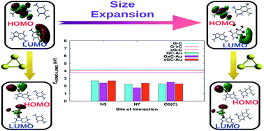
DNA molecules tagged to metal nanoparticles, especially gold nanoparticles (AuNPs), have been shown to have potential applications in the design and fabrication of novel electronic nano-devices, but the binding mechanism between gold nanoparticles and DNA bases and its implications are not completely understood. In this work, a comprehensive study to examine the effect of structural perturbations caused to DNA base pairs in terms of size expansion and adsorption on a gold cluster (Au3) has been carried out using density functional theory. The geometric and electronic features of these complexes provide evidence for the distortion of certain base pairs depending on the binding site of the cluster. This is further substantiated via normal mode, natural bond orbital (NBO) and atoms in molecules (AIM) analyses. The natural population analysis (NPA) and NBO analysis indicate that complexation greatly affects the charge distribution on the base pairs due to charge transfer between the base pair and gold cluster. This charge redistribution may offer the possibility of higher order interactions. Upon complexation, a marked decrease in the HOMO–LUMO gap is observed, which is more profound in cases where size-expanded bases are involved due to the extended π-conjugation of the fused benzene rings. This study demonstrates the possibility of combining structural modifications to DNA base pairs and subsequent binding to gold nanoparticles to modulate and achieve molecular systems with desired optoelectronic properties.
It is well known that bulk gold is inert and biomolecules show very low adsorption on its surface. To determine what it is in AuNPs that is responsible for the stability of these complexes, investigation of the nature of these interactions is essential for understanding the physisorption/chemisorption regime, which modulates the transport, catalytic and sensing mechanisms of these complexes. A plethora of literature is directed towards understanding the alteration in the photochemical properties of nucleic acids on being tagged with gold clusters.21–24 Most of them have focused on understanding the influence of metal complexation on the hydrogen bonding patterns of the canonical base pairs. Kryachko et al. have reported that the interaction between DNA bases and gold clusters occurs via the N and O atoms in the bases and one gold atom.21 They have also shown that this interaction could be further fortified by ‘non-conventional’ NH⋯Au bonds. In another work carried out by the same group on DNA–Au complexes, the authors compared the geometrical parameters, vibrational frequencies and nuclear magnetic resonance (NMR) signatures of the NH⋯Au bonding with the prerequisites of conventional weak hydrogen bonds to substantiate their finding of the presence of ‘non-conventional’ hydrogen bonds in these complexes.25 Recently, an experimental work carried out by Cao et al. on nucleobase–gold complexes using anion photoelectron spectroscopy supported by density functional theory (DFT) calculations also confirmed the existence of NH⋯Au hydrogen bonds.26 Our recent studies on nucleobase–gold interactions also confirm the presence of NH⋯Au bonds via NBO studies.27
In spite of the wide applications of AuNP-tagged DNA systems, their inherently low thermal stability does not allow us to achieve the long standing goal of designing a genetic system with a greater capacity of storing and transmitting information than that of natural DNA.28 Designing synthetic analogues of natural DNA bases, such as size-expanded DNA bases, may possibly realize more thermally stable molecular wires. This suggests a way to go beyond the limits of natural DNA in nanotechnological applications and might eventually lead to entirely new genetic systems.22–24,29–33 Kool et al. synthesized size-expanded oligomers of DNA which are known to possess high thermal stability due to better stacking interactions when compared to native DNA base pairs.28,31 Both theoretical and experimental studies suggest that, owing to larger base size, all the pairs of this helical system are fluorescent, which suggests practical applications in the detection of natural DNA and RNA using imaging techniques.28,31,34–36 Recent studies show that modified bases possess significantly divergent photophysical and photochemical properties, although the basic structural and bonding aspects remain the same.37 This is driven by the presence of an aromatic ring that causes an alteration of the electronic distribution over the frontier molecular orbitals, thereby causing the optical response diverge from that of natural bases.38 Some theoretical reports address the interaction of these size-expanded bases and base pairs with small metal clusters. Brancolini et al. have studied the interaction of size-expanded bases with copper and silver metal atoms and suggest that these complexes can be the most promising candidates for nanowires with enhanced electron transfer and also for the modification of the DNA double-helix for signal detection.23 The binding of gold nanoclusters with size-expanded bases has also been studied, indicating a modification of electronic structure that leads to some interesting properties, such as high conductivity and charge transfer.39,40 The interaction between size-expanded guanine and small gold clusters has been investigated, the results of which suggest that the introduction of an aromatic ring in guanine reduces the HOMO–LUMO gap and delocalizes the spatial distribution of electrons, making it ideal for charge transfer.41,42
Previous studies have reported the interaction of DNA base pairs with metal atoms and clusters; however, the structural and electronic perturbations induced due to size-expansion in addition to metal binding has not been examined in a systematic manner. Size-expansion may lead to exciting new physico-chemical properties, like reduced HOMO–LUMO gaps, and increased dipole moments and polarizability values, which may be useful in designing improvised nano-electronic devices. Tagging to gold clusters is known to reduce the cytotoxicity of a drug/biomolecule, which can be theoretically accounted for in terms of its electrophilicity index.43 In this work, we investigate the effect of adsorption of natural and size-expanded base pairs on an Au3 gold cluster on the electronic properties of the system, using DFT. Our study focuses on how the use of size-expanded bases (Fig. 1) and gold nanoclusters alters the electronic properties and hydrogen bonding patterns of the natural base pairs, justifying the suitability of the newly designed complexes for molecular conduction applications.
![]() | ||
Fig. 1 Optimized geometries of the size-expanded bases synthesized by Kool et al.28 |
A benchmark study of Wu et al. performed on transition elements suggests that PBE0, which is the generalized-gradient-approximation exchange–correlation functional of Perdew, Burke, and Ernzerhof,48–50 reproduces the experimental value of the dissociation energy,51 which is also supported in another work by Zhao and Truhlar.52,53 In yet another work, Knal et al. have shown PBE0 to be the best method for describing the ground state thermodynamic stabilities of intermolecular charge transfer complexes.54 Therefore, all the calculations reported in this paper were done using the PBE0 functional. Stuttgart–Dresden 19-electron effective core potentials (ECPs), designated as SDD, were used for gold.55–58 These energy-consistent ECPs work within the relativistic Dirac–Fock theory and significantly remove the spin contamination. A split valence double-ζ augmented with a d-type polarization function for all non-hydrogen atoms and a p-type polarization function for hydrogen atoms, including an s–p and a p–d diffusion function, 6-311++G(2d,2p), was used as a basis set for the bases.59–62 The initial geometries of the complexes were generated by placing the gold cluster at the electronegative sites of the base and were subjected to further optimization. Hence, the interaction was studied for the N3 and N7 sites of purines/x-purines and the O2 site of pyrimidines/x-pyrimidines. All possible orientations of the gold cluster (Au3) with respect to the base pair at above mentioned titratable sites were considered, which resulted in more than one stationary point. Vibrational frequency analysis was carried out to check if all the optimized structures were at minima. Real frequencies were obtained for all the optimized structures, indicating that all the stationary points reported here are minima on the respective potential energy surfaces. The interaction energies (Eint) reported are zero-point vibrational energy (ZPVE) corrected. Details of the Eint calculations are available in the ESI.†
Analysis of charge distribution was carried out based on (i) electrostatic potential (ESP) surfaces (mapped with the total electron density) and (ii) natural population analysis (NPA). NBO analysis63 was performed on these complexes to examine all possible stabilizing interactions. All the calculations were performed using the Gaussian 09 suite of programs.64 AIM analysis was performed with the AIMAll package65 to calculate the properties of the bond critical points (BCPs).
System | Site of interaction | Eint | Ebase pairdef | Eclusterdef | H–L (isolated) | H–L (complexed) |
---|---|---|---|---|---|---|
AT | N3 | −29.68 | 1.28 | 0.24 | 5.16 | 2.62 |
N7 | −31.35 | 1.22 | 0.35 | 5.16 | 2.68 | |
O2(T) | −15.45 | 1.36 | 0.07 | 5.16 | 2.27 | |
A–xT | N3 | −28.91 | 1.26 | 0.52 | 4.76 | 2.55 |
N7 | −31.39 | 1.27 | 0.27 | 4.76 | 2.70 | |
O2(T) | −21.42 | 1.91 | 0.08 | 4.76 | 2.49 | |
xA–T | N3 | −34.22 | 0.97 | 0.08 | 4.51 | 2.24 |
N7 | −32.35 | 0.89 | 0.06 | 4.51 | 2.43 | |
O2(T) | −16.80 | 1.37 | 0.07 | 4.51 | 2.27 | |
GC | N3 | −30.29 | 1.85 | 0.21 | 4.24 | 2.70 |
N7 | −32.86 | 0.90 | 0.19 | 4.24 | 2.23 | |
O2(C) | −16.78 | 1.86 | 0.08 | 4.24 | 2.29 | |
G–xC | N3 | −30.16 | 1.86 | 0.23 | 3.76 | 2.42 |
N7 | −32.89 | 0.92 | 0.20 | 3.76 | 1.90 | |
O2(C) | −22.11 | 2.20 | 0.10 | 3.76 | 2.51 | |
xG–C | N3 | −32.31 | 1.43 | 0.25 | 4.08 | 2.70 |
N7 | −35.48 | 0.93 | 0.22 | 4.08 | 2.39 | |
O2(C) | −16.68 | 1.88 | 0.07 | 4.08 | 2.30 |
The deformation energies (details of calculation available in the ESI†) of the base pair and the gold cluster occurring due to complexation were also calculated to give a quantitative insight into the instability arising in each of the monomers due to the complex formation. The values for the cluster suggest that very little deformation occurs on interaction with the base pair. This indicates that adsorption of the base pair on gold surface would not cause major alteration in the lattice structure of the AuNP. The deformation energy corresponding to the base pair lies in the range of 0.89–1.91 kcal mol−1. A careful observation of Table 1 suggests that the lower the deformation energy of the base-pair, the higher the corresponding interaction energy, which suggests that the stability of the base pair also plays an important role in enhancing the affinity towards the gold cluster. For example, GC has three possible sites interacting with Au3 that result in different deformation energy values, and amongst them N7 shows the minimum deformation energy value of 0.90 kcal mol−1. This particular complex shows the maximum interaction energy value with Au3.
The anchoring bond lengths are referred to as the bond length between the gold atom (acceptor) directly interacting with the active site of the base (donor). The anchor and nonconventional N–H⋯Au hydrogen bonds are considered to be the two major factors that govern the hybridization between the nucleobases and gold clusters, as reported in some earlier works.21,25,27 This causes the electron density of the nucleobases to change, particularly at the sites which are involved in the intermolecular H-bonds with the WC complementary ones, which was also predicted in our earlier work on base–gold interactions.27
In all cases, the interaction with the N7 site exhibits the shortest bond length. This suggests that the N7 site has the highest affinity for gold, an observation in line with previous results.25 This may be due to the easily available lone pair of electrons on the nitrogen. The nitrogen here is sp2 hybridised and hence has a higher electron donating capacity than any other site considered. The N3 site of purines lying on the sugar edge have longer anchor bond lengths (by 0.02 Å) when compared to the N7 site, even though they are also sp2 hybridised. This may be explained on the basis of the steric hindrance provided by the methyl group present on the adjacent N9 position. It is also observed that the interaction with the gold cluster is non-planar wherever the carbonyl oxygen is involved in the interaction, as discussed above.
The introduction of a spacer ring is also responsible for altering the anchoring bonds and reducing the bond lengths by around 0.01 Å. In the case of the N3 site, the spacer ring is also responsible for reducing the steric hindrance caused by the methyl group. For the O2 site, the effect is even more prominent. The planarity of the complex is regained on the insertion of a spacer ring into the corresponding base (A–xT (O2),G–xC (O2)). This structural perturbation is additionally stabilized by the formation of a ‘non-conventional’ C/N–H⋯Au bond.
The results of natural bond orbital (NBO) analysis and the shapes of the frontier molecular orbitals (Fig. 4 and 5) indicate that the anchoring bond is obtained by charge transfer from the N or O lone pair to the antibonding orbital of gold (Table S5 of the ESI†). Also, the ‘non-conventional’ N–H⋯Au bonds are characterized by charge transfer from the LP of gold to the BD* orbital of the NH groups. The detailed perturbation theory energy analysis is reported in the ESI (Table S5†). In addition to this, the nature of the bonding present in the formation of the anchoring bond is studied using AIM parameters. Table 2 indicates a positive ∇2ρ(r) in all cases, supporting the existence of ionic interactions in the formation of the anchoring bonds. In contrast, Table 2 also indicates negative values for H(r) in all cases, which supports the covalent character of these bonds. From this it can be concluded that the anchoring bonds are partially ionic and partially covalent in nature. For complexes with a non-planar geometry and lower interaction energy, the value of H(r) is also less, suggesting H(r) to be an indicator of complex stability.
System | Site of interaction | ∇2ρ(r) | G(r) | V(r) | H(r) |
---|---|---|---|---|---|
AT | N3 | 3.72 × 10⁻¹ | 1.23 × 10⁻¹ | −1.53 × 10⁻¹ | −3.01 × 10⁻² |
N7 | 3.88 × 10⁻¹ | 1.30 × 10⁻¹ | −1.63 × 10⁻¹ | −3.32 × 10⁻² | |
O2(T) | 3.27 × 10⁻¹ | 8.91 × 10⁻² | −9.65 × 10⁻² | −7.42 × 10⁻³ | |
A–xT | N3 | 3.66 × 10⁻¹ | 1.21 × 10⁻¹ | −1.50 × 10⁻¹ | −2.93 × 10⁻² |
N7 | 4.09 × 10⁻¹ | 1.31 × 10⁻¹ | −1.59 × 10⁻¹ | −2.85 × 10⁻² | |
O2(T) | 3.70 × 10⁻¹ | 1.04 × 10⁻¹ | −1.14 × 10⁻¹ | −1.10 × 10⁻² | |
xA–T | N3 | 3.64 × 10⁻¹ | 1.21 × 10⁻¹ | −1.51 × 10⁻¹ | −3.02 × 10⁻² |
N7 | 3.90 × 10⁻¹ | 1.30 × 10⁻¹ | −1.63 × 10⁻¹ | −3.29 × 10⁻² | |
O2(T) | 3.25 × 10⁻¹ | 8.86 × 10⁻² | −9.60 × 10⁻² | −7.38 × 10⁻³ | |
GC | N3 | 3.58 × 10⁻¹ | 1.19 × 10⁻¹ | −1.49 × 10⁻¹ | −2.98 × 10⁻² |
N7 | 4.05 × 10⁻¹ | 1.28 × 10⁻¹ | −1.55 × 10⁻¹ | −2.69 × 10⁻² | |
O2(C) | 3.31 × 10⁻¹ | 9.11 × 10⁻² | −9.95 × 10⁻² | −8.40 × 10⁻³ | |
G–xC | N3 | 3.58 × 10⁻¹ | 1.19 × 10⁻¹ | −1.49 × 10⁻¹ | −2.98 × 10⁻² |
N7 | 3.85 × 10⁻¹ | 1.28 × 10⁻¹ | −1.59 × 10⁻¹ | −3.15 × 10⁻² | |
O2(C) | 3.80 × 10⁻¹ | 1.07 × 10⁻¹ | −1.19 × 10⁻¹ | −1.22 × 10⁻² | |
xG–C | N3 | 3.55 × 10⁻¹ | 1.19 × 10⁻¹ | −1.50 × 10⁻¹ | −3.07 × 10⁻² |
N7 | 4.09 × 10⁻¹ | 1.31 × 10⁻¹ | −1.59 × 10⁻¹ | −2.84 × 10⁻² | |
O2(C) | 3.29 × 10⁻¹ | 9.06 × 10⁻² | −9.90 × 10⁻² | −8.35 × 10⁻³ |
System | Site of interaction | Atoms | Δr* | Δ∠* | Δν* |
---|---|---|---|---|---|
a Δr* = rcomplex − risolated, Δ∠* = ∠complex − ∠isolated, Δν* = νcomplex − νisolated. | |||||
AT | N3 | A(N1)–T(N3) | 0.04 | −0.9 | 199.5 |
A(N6)–T(O4) | −0.04 | 1.8 | −63.5 | ||
A(C2)–T(O2) | 0.03 | −2.4 | 22.7 | ||
N7 | A(N1)–T(N3) | −0.03 | −0.8 | 123.8 | |
A(N6)–T(O4) | 0.05 | 2.0 | −56.0 | ||
A(C2)–T(O2) | 0.03 | 0.9 | 0.7 | ||
O2(T) | A(N1)–T(N3) | −0.11 | 0.4 | −233.1 | |
A(N6)–T(O4) | 0.07 | −1.5 | 38.2 | ||
A(C2)–T(O2) | 0.10 | 0.0 | 3.9 | ||
A–xT | N3 | A(N1)–T(N3) | 0.04 | −0.5 | 180.4 |
A(N6)–T(O4) | −0.03 | 1.7 | −68.8 | ||
A(C2)–T(O2) | 0.00 | −2.2 | 21.4 | ||
N7 | A(N1)–T(N3) | 0.03 | −0.6 | 114.7 | |
A(N6)–T(O4) | 0.00 | 2.1 | −53.2 | ||
A(C2)–T(O2) | 0.01 | 1.3 | −11.8 | ||
O2(T) | A(N1)–T(N3) | −0.04 | 0.4 | −192.0 | |
A(N6)–T(O4) | 0.00 | −1.4 | 25.0 | ||
A(C2)–T(O2) | −0.12 | −0.2 | 5.2 | ||
xA–T | N3 | A(N1)–T(N3) | 0.02 | −0.9 | 137.7 |
A(N6)–T(O4) | 0.02 | 2.0 | 0.8 | ||
A(C2)–T(O2) | 0.05 | −2.1 | 1.7 | ||
N7 | A(N1)–T(N3) | 0.02 | −0.8 | 67.5 | |
A(N6)–T(O4) | −0.02 | 0.2 | −26.4 | ||
A(C2)–T(O2) | 0.05 | −0.5 | −12.1 | ||
O2(T) | A(N1)–T(N3) | −0.04 | 0.2 | −242.6 | |
A(N6)–T(O4) | 0.01 | −1.3 | 37.2 | ||
A(C2)–T(O2) | −0.10 | −0.1 | 3.2 |
The hydrogen bonding patterns in the G–C counterparts are also similarly affected, owing to tagging with the gold cluster (Table 4). When the cluster interacts from the N3 site of G–C, the G(N1)–C(N3) and G(N2)–C(O2) bond distances are reduced, whereas the G(O6)–C(N4) bond distance is increased (Table 4). The complexes with xG–C also follow the same pattern. In the case of the complex with G–xC, there is a slight lengthening in the G(N1)–C(N3) bond by only 0.004 Å. Vibrational frequency analysis suggests a weakening of the hydrogen bond strength in the G–C base pair, where as in G–xC the bonds are expected to be stronger, which is also supported by the NBO data (Table S5 of the ESI†). In the case of xG–C, the G(N1)–C(N3) bond is weakened, whereas the other two bonds become more linear. The interaction with the N7 site causes a lengthening of the G(O6)–C(N4) bond and a shortening of the other two bonds in all three cases. The vibrational frequency analysis suggests that the hydrogen bonds are weakened in the case of G–C, and strengthened in the case of G–xC. The G(O6)–C(N4) bond is weakened in the case of xG–C and the other two bonds are strengthened. The interaction with the O2 site causes the shortening of the G(O6)–C(N4) bond and the lengthening of the other two bonds in all three complexes. The angles are reduced in their linearity in all three hydrogen bonds. This analysis suggests that tagging the gold at the N3 or N7 site of guanine would facilitate the base-pair opening from the major groove, whereas the same would be favored from the minor groove if the gold is tagged to the O2 site of cytosine. A comparison of the hydrogen bonding data obtained via NBO calculations for the natural and modified (size-expanded and tagged with gold) base pairs also suggests the weakening of these bonds, resulting in their opening from the major groove if the cluster is tagged at the purines/x-purines (Table S5 of the ESI†). The opening is facilitated from the minor groove if the cluster is tagged to the pyrimidines/x-pyrimidines. A comparison of the BCP parameters for the hydrogen bonds present in the (un)complexed base pairs also indicates that the opening of the base pair is site-specific, i.e. it can be controlled by varying the site with which the gold cluster interacts. The details of these calculations are provided in Table S6 of the ESI.† This site-specific behavior of the alteration in hydrogen bonding patterns is a useful tool for tailoring the unwinding of DNA strands.
System | Site of interaction | Atoms | Δr* | Δ∠* | Δν* |
---|---|---|---|---|---|
a Δr* = rcomplex − risolated, Δ∠* = ∠complex − ∠isolated, Δν* = νcomplex − νisolated. | |||||
GC | N3 | G(O6)–C(N4) | 0.03 | −2.1 | 113.2 |
G(N1)–C(N3) | −0.02 | −0.7 | −109.1 | ||
G(N2)–C(O2) | −0.07 | −0.8 | −106.8 | ||
N7 | G(O6)–C(N4) | 0.03 | −2.1 | 131.7 | |
G(N1)–C(N3) | −0.03 | −0.9 | −110.9 | ||
G(N2)–C(O2) | −0.04 | −1.1 | −51.6 | ||
O2(C) | G(O6)–C(N4) | −0.04 | −1.2 | −83.4 | |
G(N1)–C(N3) | 0.05 | −2.1 | 92.1 | ||
G(N2)–C(O2) | 0.13 | −3.2 | 123.2 | ||
G–xC | N3 | G(O6)–C(N4) | 0.03 | 0.8 | 131.5 |
G(N1)–C(N3) | 0.00 | 0.2 | −96.5 | ||
G(N2)–C(O2) | −0.08 | 1.1 | −67.1 | ||
N7 | G(O6)–C(N4) | 0.04 | 1.3 | 119.5 | |
G(N1)–C(N3) | 0.00 | 0.5 | −106.6 | ||
G(N2)–C(O2) | −0.05 | 0.8 | −116.0 | ||
O2(C) | G(O6)–C(N4) | −0.03 | −1.4 | −63.9 | |
G(N1)–C(N3) | 0.08 | −1.2 | 86.5 | ||
G(N2)–C(O2) | 0.10 | −2.6 | 133.8 | ||
xG–C | N3 | G(O6)–C(N4) | 0.02 | −1.5 | 41.7 |
G(N1)–C(N3) | −0.02 | 0.5 | −89.3 | ||
G(N2)–C(O2) | −0.08 | 2.8 | −31.3 | ||
N7 | G(O6)–C(N4) | 0.02 | −0.7 | 57.7 | |
G(N1)–C(N3) | −0.01 | 0.6 | −113.3 | ||
G(N2)–C(O2) | −0.04 | 1.7 | −106.3 | ||
O2(C) | G(O6)–C(N4) | −0.04 | 0.1 | −116.7 | |
G(N1)–C(N3) | 0.06 | −1.2 | 85.9 | ||
G(N2)–C(O2) | 0.12 | −2.2 | 107.7 |
![]() | ||
Fig. 4 The distribution of the HOMO−1, HOMO, LUMO and LUMO+1 levels of the complexed and uncomplexed AT base pairs, calculated at the UHF level of theory using the 6-31+G(2d,2p)∪SDD basis set. |
![]() | ||
Fig. 5 The distribution of the HOMO−1, HOMO, LUMO and LUMO+1 levels of the complexed and uncomplexed GC base pairs calculated at the UHF level of theory using the 6-31+G(2d,2p)∪SDD basis set. |
The dipole moments (μ) and polarizabilities (α) were computed in order to examine the effect of complexation on the electronic properties of the base pairs. The values are available in Table S4 of the ESI.† The dipole moment is higher for GC than for AT. It is observed that μ varies on ring expansion (Fig. 6). Pairing a size-expanded pyrimidine with a purine causes the dipole moment to increase, whereas the opposite happens when size-expanded purines are paired with pyrimidines. Tagging with the gold cluster causes the dipole moment to increase in all cases, except when G–C and its variants are made to interact with the cluster from the O2 site of cytosine. Fig. 7 clearly shows an increase in the value of polarizability (α) on ring expansion in all cases. This value is further raised on tagging the base pair with the gold cluster. It is found that size-expanded purines tagged with the cluster at the N7 site show the maximum value of polarizability in both xA–T and xG–C base pairs. The increase in polarizability causes the dispersion forces to increase, resulting in an increase in the melting and boiling points of the complex.68 Hence, we can expect that these structural perturbations lead to the formation of more thermally stable complexes that find a more relevant place in the design of nanoelectronic devices.
It has been reported that the global electrophilic power of a molecule can be quantified in terms of a global electrophilicity index (ω).69 The value of ω gives a measure of the propensity of the molecule to act as a nucleophile. It is known from previous studies that DNA acts as a nucleophile and tends to attack certain water-soluble epoxides that are carcinogenic.70 This results in the complexation of DNA with these molecules, causing damage to the DNA. The increase in the electrophilicity index values suggests that the tendency to react with epoxides would possibly decrease. The values of ω for the complexed systems are reported. It is clear from Fig. 8 that the electrophilicity index values increase on size-expansion, which is further enhanced by complexation. The enhancement in this value is dependent on the site with which the gold interacts. However, in both cases (AT and GC) it is observed that the ω value is at its maximum when the interaction is taking place with the O2 site. This information is useful in producing drugs that have a high degree of selectivity and specificity.
The structural perturbations introduced induce a base pair distortion which is found to be binding site-specific. This feature can be exploited in designing highly selective and specific nano-devices. Following the gold binding, a redistribution of electronic charge around the Hoogsteen and sugar edges takes place, suggesting that these systems favor higher order interactions. This distinctive property may provide a promising scaffold for the synthesis of 3D metalized objects that find application in nano-electronics.
The electronic coupling between the metal atoms and nucleic acid base pairs results in the reduction of the HOMO–LUMO gaps, suggesting that these systems might behave as good conductors. It is also found that on complexation, a considerable amount of electronic charge is transferred from the base pair to the gold cluster. Thus, the gold clusters oxidize the base/x-base. The increase in α values indicates greater thermal stability. Increasing ω values emphasizes the importance of these systems in designing drugs related to curing cancer. Hence, it can be concluded that an alteration in geometric and electronic properties on tagging the base pairs with gold clusters opens up an avenue for developing novel nano-bioelectronic devices/sensors with tailored molecular properties.
Footnote |
† Electronic supplementary information (ESI) available: Geometric features, frequency analysis, discussion of dipole moments, polarizability values, electrophilicity index, NBO and AIM details. See DOI: 10.1039/c5ra04668h |
This journal is © The Royal Society of Chemistry 2015 |