Hydrothermal syntheses, structural characterizations, and magnetic properties of five MOFs assembled from C2-symmetric ligand of 1,3-di(2′,4′-dicarboxylphenyl)benzene with various coordination modes

Liming Fanab, Weiliu Fana, Bin Lib, Xian Zhao*a and Xiutang Zhang*ab
aState Key Laboratory of Crystal Materials, Shandong University, Jinan 250100, China. E-mail: zhaoxian@sdu.edu.cn
bAdvanced Material Institute of Research, College of Chemistry and Chemical Engineering, Qilu Normal University, Jinan, 250013, China. E-mail: xiutangzhang@163.com

Received 15th March 2015 , Accepted 14th April 2015

First published on 15th April 2015


Abstract

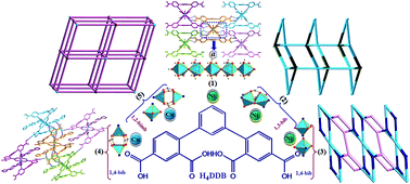
Five new complexes, namely, [Ni(H2DDB)(H2O)22-H2O)]n (1), [Ni1.5(DDB)(1,4-bib)1.5(H2O)]n (2), {[Ni2(DDB)(1,3-bib)22-H2O)]·2H2O}n (3), [Cu2(H2DDB)2(1,4-bib)2]·H2O (4), and {[Cu1.5(HDDB)(1,2-bimb)]·H2O}n (5), have been synthesized using the solvothermal reaction of 1,3-di(2′,4′-dicarboxylphenyl)benzene (H4DDB) with nickel(II) or copper(II) salts in the presence of ancillary ligands of bis(imidazole) linkers (1,4-bib = 1,4-bis(1H-imidazol-4-yl)benzene, 1,3-bib = 1,3-bis(1H-imidazol-4-yl)benzene, and 1,2-bimb = 1,2-bis(imidazol-1-ylmethyl)benzene). Their structures have been determined by single-crystal X-ray diffraction analyses and further characterized by elemental analyses, IR spectroscopy, powder X-ray diffraction (PXRD), and thermogravimetric (TG) analyses. In complex 1, NiII ions are bridged by associated water molecules to form an interesting [Ni(H2DDB)(H2O)22-H2O)]n chain structure, which is further extended to a 3D supramolecular structure via the hydrogen bonds. In complex 2, a novel 3D (3,3,6)-connected (63)4(65·88·102) net is generated from the synergistic effect of 1D [Ni(1,4-bib)]n zigzag chains and 2D [Ni3(DDB)2]n bilayers. For complex 3, with the employment of 1,3-bib bis(imidazole) linker instead of 1,4-bib, an unprecedented binuclear {Ni2(COO)(μ2-H2O)} SBUs based 3D (3,6)-connected (3·6·7)(32·43·54·63·7·82) net was obtained. Furthermore, when NiII ions were replaced by CuII ions, a paddle wheel {Cu2(COO)4} SBUs based [Cu2(H2DDB)(1,4-bib)2] (4) complex was formed. For complex 5, 2D [Cu3(HDDB)2]n sheets are further linked by 1,2-bimb pillars to expand a paddle wheel {Cu2(COO)4} SBUs based 3D architecture with 4-connected (65 8)-cds topology. Moreover, the magnetic properties of 1, 3, and 5 have been investigated.

Introduction

During the past few decades, extensive experimental and theoretical efforts towards metal–organic frameworks (MOFs) have attracted a great deal of interest for their regulated and interesting structural topologies as well as their potential applications in the fields of photoluminescence, magnetism, catalysis, gas storage, conductivity, ion exchange, ferroelectricity, optoelectronic effect, non-linear optics, and spin-transition behavior.1–3 Such materials are usually constructed from metal ions as connected centers and multifunctional organic ligands as linkers.4 In principle, the targeting assemblies with desired structural features and physicochemical properties greatly depend on the nature of the organic ligands and metal ions, among which the appropriate choice of well-designed organic building blocks and metal ions or clusters is one of the most effective ways.5,6

However, despite the breathtaking achievements in this aspect, the prediction and further accurate control over the framework array of a given crystalline product still remain a considerable challenge at this stage. This limitation mainly arises from the fact that the subtle assembly progress may be influenced by many intrinsic and external parameters such as the different coordination preferences of the metal ion, templating agents, metal–ligand ratio, pH value, counter anion, and number of coordination sites provided by organic ligands.7–9

Among the numerous organic ligands, polycarboxylic acids and the N-donor ligands are favored for their strong coordinating ability, which can stabilize the packing architecture, including that of honeycomb, grid, T-shaped, ladder, diamondoid, and octahedral structures.10,11 The ancillary ligands containing N-donors such as bipyridine have been widely used with polycarboxylates to construct the desired structures.12 For example, using a bipyridine linker as an N-donor ligand is beneficial to the syntheses of extended MOFs and can generate high dimensional structures owing to its simple bridging mode and strong coordination ability; however, the utilization of bis(imidazole) linkers (1,4-bib, 1,3-bib, 1,2-bimb) ligands as co-ligands to react with polycarboxylates has been rarely reported in detail.13 In addition, the cis- or trans-configuration of bis(imidazole) linkers often causes structural diversity when they coordinate to metal centers.14

A recent study on coordination assemblies using 3,5-bis(3-carboxyphenyl)pyridine (H2bcpb) and different N-donor ancillary ligands states a reliable strategy for obtaining new topological prototypes of coordination nets.14e Moreover, a minor change in the polycarboxylic acid building blocks may be applied to realize good structural control of the resulting metal–organic polymers. Thus, these considerations inspired us to explore new coordination frameworks with the designed 1,3-di(2′,4′-dicarboxylphenyl)benzene (H4DDB) ligand and different metal salts under solvothermal conditions in the presence of bis(imidazole) linkers (shown in Scheme 1). In this study, we report the syntheses and characterization of five novel coordination complexes, which exhibit systematic structural variation from 0D paddle wheel {Cu2(COO)4} SBUs based complex (4), 1D [Ni(H2DDB)(H2O)22-H2O)]n chain (1), 3D 4-connected cds net (5), 3D (3,6)-connected (3·6·7)(32·43·54·63·7·82) net (3), to 3D (3,3,6)-connected (63)4(65·88·102) net (2). These results revealed that the H4DDB ligand is a good candidate to construct inorganic building block based coordination complexes, in which metal ions as well as the bis(imidazole) ancillary linkers have a great influence on the final structures.


image file: c5ra04559b-s1.tif
Scheme 1 The structure of H4DDB and bis(imidazole) ancillary ligands.

Experimental section

Reagents and physical measurements

All the chemical reagents were purchased from Jinan Henghua Sci. & Tec. Co. Ltd. and used without further purification. Elemental analyses were carried out on a CE instruments EA 1110 elemental analyzer. TGA was measured from 25 to 800 °C on a SDT Q600 instrument at a heating rate of 5 °C min under an N2 atmosphere (100 mL min−1). X-ray powder diffractions were measured on a Panalytical X-Pert pro diffractometer with Cu-Kα radiation. The variable-temperature magnetic susceptibility measurements were performed on Quantum Design SQUID MPMS XL-7 instruments in the temperature range of 2–300 K under a field of 1000 Oe. The PXRD patterns of 1–5 were obtained, and are shown in Fig. S1. IR spectra were obtained on a NEXUS 670 FTIR spectrometer in the range of 600–4000 cm−1 and are given in Fig. S2.
Synthesis of [Ni(H2DDB)(H2O)22-H2O)]n (1). A mixture of H4DDB (0.10 mmol, 0.041 g), NiCl2·6H2O (0.20 mmol, 0.048 g), 6 mL H2O, and 3 mL CH3CN was sealed in a 25 mL Teflon-lined stainless steel vessel and heated to 130 °C for 5 days, followed by slow cooling to room temperature at a descent rate of 10 °C h−1. Green block crystals of 1 were obtained in 76% yield (based on H4DDB). Anal. (%) calcd for C22H18NiO11: C, 51.10; H, 3.51. Found: C, 51.17; H, 3.67. IR (KBr pellet, cm−1): 3225 (s), 2110 (w), 1686 (s), 1607 (m), 1547 (s), 1425 (m), 1367 (s), 1220 (s), 1164 (w), 1124 (s), 979 (w), 896 (w), 792 (m), 767 (w), 669 (s), 623 (vs).
Synthesis of [Ni1.5(DDB)(1,4-bib)1.5(H2O)]n (2). The synthetic method was similar to that used for the preparation of complex 1 except that 1,4-bib (0.30 mmol, 0.063 g) was added as an ancillary ligand. Green block crystals of 2 were obtained in 61% yield (based on H4DDB). Anal. (%) calcd for C80H56N12Ni3O18: C, 58.32; H, 3.32; N, 10.20. Found: C, 58.47; H, 3.32; N, 10.41. IR (KBr pellet, cm−1): 3363 (m), 3134 (s), 2113 (m), 1591 (vs), 1511 (vs), 1445 (m), 1378 (s), 1318 (m), 1281 (m), 1240 (m), 1065 (s), 1005 (w), 776 (m), 731 (m), 694 (m), 658 (m).
Synthesis of {[Ni2(DDB)(1,3-bib)22-H2O)]·2H2O}n (3). The synthetic method was similar to that used for the preparation of complex 1 except that 1,3-bib (0.30 mmol, 0.063 g) was added as an ancillary ligand. Green block crystals of 3 were obtained in 82% yield (based on H4DDB). Anal. (%) calcd for C46H34N8Ni2O10: C, 56.60; H, 3.51; N, 11.48. Found: C, 56.72; H, 3.53; N, 11.50. IR (KBr pellet, cm−1): 3457 (m), 31[thin space (1/6-em)]657 (m), 3138 (m), 1627 (m), 1603 (m), 1549 (s), 1524 (s), 1496 (m), 1448 (m), 1399 (m), 1367 (m), 1309 (s), 1065 (s), 826 (m), 776 (m), 734 (m), 653 (w).
Synthesis of [Cu2(H2DDB)2(1,4-bib)2]·H2O (4). The synthetic method was similar to that used for the preparation of complex 2 except that CuSO4·5H2O (0.20 mmol, 0.050 g) replaced NiCl2·6H2O (0.20 mmol, 0.048 g). Blue block crystals of 4 were obtained in 31% yield (based on H4DDB). Anal. (%) calcd for C68H44Cu2N8O16: C, 60.22; H, 3.27; N, 8.26. Found: C, 60.61; H, 3.30; N, 8.42. IR (KBr pellet, cm−1): 3186 (s), 2164 (m), 1686 (vs), 1607 (s), 1562 (m), 1546 (s), 1439 (m), 1422 (s), 1366 (vs), 1215 (s), 1163 (m), 1123 (s), 896 (m), 766 (s), 680 (m), 648 (w).
Synthesis of {[Cu1.5(HDDB)(1,2-bimb)]·H2O}n (5). The synthetic method was similar to that used for the preparation of complex 4 except that 1,2-bib (0.30 mmol, 0.063 g) replaced 1,3-bib (0.30 mmol, 0.063 g). Purple blue crystals of 5 were obtained in 57% yield (based on H4DDB). Anal. (%) calcd for C72H54Cu3N8O18: C, 57.27; H, 3.60; N, 7.42. Found: C, 57.47; H, 3.73; N, 7.38. IR (KBr pellet, cm−1): 3124 (s), 2659 (m), 2116 (s), 1620 (s), 1525 (s), 1439 (m), 1403 (s), 1293 (m), 1240 (s), 1093 (s), 837 (s), 776 (m), 724 (s), 668 (m), 622 (vs).

X-ray crystallography

Intensity data collection was carried out on a Siemens SMART diffractometer equipped with a CCD detector using Mo-Kα monochromatized radiation (λ = 0.71073 Å) at 296(2) K. The absorption correction was based on multiple and symmetry-equivalent reflections in the data set using the SADABS program based on the method of Blessing. The structures were solved by direct methods and refined by full-matrix least-squares using the SHELXTL package.15 All non-hydrogen atoms were refined anisotropically. Hydrogen atoms except those for water molecules were generated geometrically with fixed isotropic thermal parameters and included in the structure factor calculations. The approximate positions of the water H atoms, obtained from a difference Fourier map, were restrained to the ideal configuration of the water molecule and fixed in the final stages of refinement. Some carbon atoms and two nitrogen atoms of 1,3-bib in 3 and three lattice water molecules were refined with split positions and an occupancy ratio of 45.6[thin space (1/6-em)]:[thin space (1/6-em)]54.4 for C(13)–C(21) and N(6), 91.9[thin space (1/6-em)]:[thin space (1/6-em)]8.1 for C(4)–C(9), 88[thin space (1/6-em)]:[thin space (1/6-em)]12 for O(1W), and 37[thin space (1/6-em)]:[thin space (1/6-em)]13 for both O(2W) and O(3W). For complex 5, five carbon atoms of 1,2-bimb and the lattice water molecule were refined with split positions and an occupancy ratio of 39[thin space (1/6-em)]:[thin space (1/6-em)]61 for C(28)–C(32) and 70.4[thin space (1/6-em)]:[thin space (1/6-em)]29.6 for O(1W) (Table 1).
Table 1 Crystal data for complexes 1–5a
Complex 1 2 3 4 5
a R1 = ∑‖Fo| − |Fc‖/∑|Fo|, wR2 = [∑w(Fo2Fc2)2/∑w(Fo2)2]1/2.
Empirical formula C22H18NiO11 C80H56N12Ni3O18 C46H36N8Ni2O11 C68H48Cu2N8O18 C72H54Cu3N8O18
Formula weight 517.07 1649.50 994.25 1392.22 1509.85
Crystal system Orthorhombic Monoclinic Monoclinic Orthorhombic Monoclinic
Space group Pnma P21/n C2/c Pbca P21/n
a (Å) 7.7017(3) 12.2234(4) 42.0250(14) 19.4760(7) 17.8722(10)
b (Å) 22.7746(7) 10.2921(4) 11.1915(4) 10.5905(4) 10.9304(6)
c (Å) 11.5195(4) 28.1648(10) 23.6784(8) 29.7263(11) 19.1523(11)
α (°) 90 90 90 90 90
β (°) 90 98.6459(11) 120.5704(9) 90 115.5103(16)
γ (°) 90 90 90 90 90
V3) 2020.56(12) 3503.0(2) 9588.6(6) 6131.4(4) 3376.6(3)
Z 4 2 8 4 2
Dcalcd (Mg m−3) 1.700 1.564 1.377 1.508 1.485
μ (mm−1) 1.028 0.884 0.851 0.777 1.015
T (K) 296(2) 296(2) 296(2) 296(2) 296(2)
Rint 0.0408 0.0499 0.0542 0.0980 0.1415
θ range (°) 3.21–25.00 3.11–25.00 2.96–25.00 3.00–25.00 2.95–25.50
F(000) 1064 1696 4096 2856 1546
Data/restraints/parameters 1823/45/172 6155/3/517 8387/329/786 5382/0/485 6273/97/504
R indices (all data) 0.0290, 0.0506 0.0365, 0.0863 0.0602, 0.1274 0.0886, 0.1560 0.1161, 0.1078
Gof 1.051 1.000 1.064 1.057 1.046


Result and discussion

Synthesis

Five title complexes were synthesized under hydrothermal conditions and their formation were strongly influenced by the reaction conditions such as metal ions and ancillary linkers. The diverse coordination modes of H4DDB proved that different bis(imidazole) linkers (1,4-bib, 1,3-bib, 1,2-bimb) also play an important role in adjusting the coordination mode Scheme 2 of polycarboxylic acid and crystal packing structure.
image file: c5ra04559b-s2.tif
Scheme 2 The coordination modes of H4DDB in complexes 1–5.
Structural description of [Ni(H2DDB)(H2O)22-H2O)]n (1). X-ray single-crystal determination reveals that complex 1 possessed a three-dimensional supramolecular structure, built from 1D [Ni(H2O)22-H2O)]n chains with the help of O–H⋯O hydrogen bonds. Complex 1 crystallizes in the orthorhombic system, space group Pnma. The asymmetric unit consists of half of NiII ion, half of H2DDB2− ligand, one and a half water molecules. In the building unit of 1, the nickel center adopts a distorted octahedral geometry by coordinating to four water molecules and two oxygen atoms of two monodentate carboxyl groups from one H2DDB2−ligand (Fig. 1a). The bond lengths and angles of Ni–O are similar to those in other nickel–carboxylate coordination polymers.16a–c It is worth mentioning that Ni–μ2-OH2 bond length is slightly longer than the other five Ni–O bonds.
image file: c5ra04559b-f1.tif
Fig. 1 (a) Crystal structure of complex 1 (symmetry codes: A: x, 3/2 − y, z; B: 1/2 + x, 3/2 − y, 1/2 − z). (b) The 1D coordinated water molecule bridged [Ni(H2O)22-H2O)]n chain. (c) Schematic view of O–H⋯O hydrogen bond based 3D supramolecular structure of 1 along the a direction.

In 1, H2DDB2− is partly deprotonated and adopts a coordination mode of I, as shown in Scheme 1. The two dihedral angles between the central phenyl ring and the two side phenyl rings are equal (49.3(4)°). In addition, the dihedral angle between the two side phenyl rings is 31.9(1)°. Two 2-position carboxyl groups are protonated and coordinated to one NiII cation. Furthermore, the NiII ions are bridged by μ2-coordinated water molecules to form a 1D [Ni(H2O)22-H2O)]n chain with the nearest Ni⋯Ni distance being 3.862 Å (Fig. 1b). Moreover, the neighbouring chains are interconnected with each other through O–H⋯O hydrogen bonds (Table S2) to generate a 3D supramolecular structure (Fig. 1c).

Structural description of [Ni1.5(DDB)(1,4-bib)1.5(H2O)]n (2). X-ray crystallography reveals that 2 has a 3D (63)4(65·88·102) framework and crystallizes in the monoclinic system, space group P21/n. As shown in Fig. 2a, the asymmetric unit consists of one and a half NiII ions, one completely deprotonated DDB4− ligand, one and a half 1,4-bib ligands, and one coordinated water molecule. Ni(1) is located in a slightly distorted {NiN2O4} octahedral coordination environment, completed by four carboxyl groups at the 4-position from four distinct DDB4− ligands, and two N atoms from two 1,4-bib ligands. Ni(2) is hexacoordinated by four O atoms from two 2-position carboxyl groups of one DDB4− ligand and one coordinated water molecule, and two N atoms from two different 1,4-bib ligands, showing a distorted octahedral geometry. The Ni–O/N bond lengths are in the normal range of 2.0383(18)–2.1669(14) Å.16d,e
image file: c5ra04559b-f2.tif
Fig. 2 (a) Crystal structure of complex 2 (symmetry codes: A: 2 − x, 1 − y, 1 − z; B: 3/2 − x, 1/2 + y, 1/2 − z; C: 1/2 + x, 1/2 − y, 1/2 + z; D: 1 + x, y, z; G: 2 − x, −y, −z). (b) The 2D [Ni3(DDB)2]n bilayer view along the a direction. (c) Schematic view of the 3D frameworks of 2 along the b direction. (d) The 3D novel (3,3,6)-connected (63)4(65·88·102) net of 2 (green nodes: Ni(1) ions, rose nodes: Ni(2) ions, dark blue nodes: DDB4− ligands).

H4DDB is completely deprotonated and adopts a (κ10)-(κ11)-(κ10)-(κ10)-μ3 coordination mode (Mode II) to link three NiII ions. Ni(2) ions are chelated by the monodentate 2-position carboxyl groups of DDB4− ligands, which further connect Ni(1) ions via the terminal 4-position monodentate carboxyl groups to generate a 2D [Ni3(DDB)2]n bilayer (Fig. 2b). Moreover, the NiII ions are linked by 1,4-bib linkers to construct a 1D [Ni(1,4-bib)]n zigzag chain with Ni⋯Ni distances being 13.596 Å and 13.530 Å, in which the 1,4-bib linkers adopts a trans-configuration (Fig. S4). Finally, 1D [Ni(1,4-bib)]n zigzag chains and 2D [Ni3(DDB)2]n bilayers are expanded to a 3D framework by sharing NiII ions (Fig. 2c).

To better understand the final structure of complex 2, the topology analysis was introduced to simplify the networks.17 The final structure of 2 can be defined as an unprecedented (3,3,6)-connected net with the Schläfli symbol of (63)4(65·88·102) by attributing the Ni(1) ions to 6-connected nodes, DDB4− ligands and the Ni(2) ions to 3-connected nodes (Fig. 2d).

Structural description of {[Ni2(DDB)(1,3-bib)22-H2O)]·2H2O}n (3). Sequentially, when we used 1,3-bib instead of 1,4-bib as the bridging co-ligand, a {Ni2(COO)(μ2-H2O)} SBUs based 3D (3,6)-connected (3·6·7)(32·43·54·63·7·82) net (3) was obtained. Complex 3 crystallizes in the monoclinic system C2/c and the asymmetric unit contains two NiII ions, one DDB4− ligand, two 1,3-bib ligands, one μ2-coordinated water molecule, and two lattice water molecules (Fig. 3a). Both the Ni(1) and Ni(2) ions are located in distorted {NiN2O4} octahedral coordination environments, surrounded by two oxygen atoms from two carboxylate groups at the 2-position of one DDB4− ligand, one carboxylate oxygen atom at the 4-position to form another DDB4− ligand, one μ2-H2O molecule, and two nitrogen atoms from two 1,3-bib ligands. Ni–N and Ni–O bond lengths are in the normal range of 2.057(3)–2.072(3) Å and 2.050(2)–2.179(2) Å, respectively.
image file: c5ra04559b-f3.tif
Fig. 3 (a) The crystal structure of complex 3 (symmetry codes: A: 2 − x, 1 − y, −z; E: x, 1 − y, −1/2 + z; F: 1/2 + x, 3/2 + y, 1/2 + z). (b) The space-filling of 2D [Ni2(DDB)(μ2-H2O)]n networks view along the c direction. (c) A schematic view of the 3D frameworks of 3 along the b direction. (d) The unprecedented binuclear {Ni2(COO)(μ2-H2O)} SBUs based 3D (3,6)-connected (3·6·7)(32·43·54·63·7·82) net of 3 (green spheres: binuclear {Ni2(COO)(μ2-H2O)} clusters based SBUs; dark green spheres: DDB4− ligands).

H4DDB is completely deprotonated and exhibits a (κ10)-(κ11)-(κ20)-(κ10)-μ4 coordination mode (Mode III), different from that found in complex 1 and 2. The Ni(1) and Ni(2) ions are connected by one μ2-η1:η1-syn-anti 2-position carboxyl groups and one μ2-H2O molecule to form an unprecedented binuclear {Ni2(COO)(μ2-H2O)} SBUs, which are further linked by the 4-position carboxyl groups to generate a 2D sheet with an opening area of about 11.192 × 26.839 Å2 (Fig. 3b). From another point of view, it is worth mentioning that four NiII ions are linked by four 1,3-bib to form an interesting [Ni4(1,3-bib)4] loop with the Ni⋯Ni distances of 9.532 and 11.430 Å (Fig. S5). In addition, the [Ni4(1,3-bib)4] loops are hinged by adjacent 2D sheets to result in a 3D framework (Fig. 3c).

From the standpoint of topology, the final structure of 3 can be defined as a (3,6)-connected net with the Schläfli symbol of (3·6·7)(32·43·54·63·7·82) by attributing the DDB4− ligands to 3-connected nodes and the binuclear {Ni2(COO)(μ2-H2O)} SBUs to 6-connected nodes (Fig. 3d).

Structural description of [Cu2(H2DDB)2(1,4-bib)2]·H2O (4). Although the reaction conditions were similar to those used to prepare complex 2, the final packing diagram of 4 exhibited an entirely different 0D paddle wheel {Cu2(COO)4} SBUs based complex, which may be attributed to the different coordination preferences between the CuII and NiII cations. Structural analysis reveals that complex 4 crystallizes in the orthorhombic system, space group Pnca.

As shown in Fig. 4a, there was one crystallographically independent CuII ion, one H2DDB2− ligand, and one 1,4-bib ligand in the asymmetric unit. Each CuII centre was pentacoordinated by four 2-position carboxylate groups from two H2DDB2− ligands and one N atom from the 1,4-bib ligand, exhibiting a distorted square pyramidal coordination geometry. In the paddle wheel {Cu2(COO)4} SBUs, the Cu⋯Cu distance was 2.778(8) Å. The dihedral angles between the two side phenyl rings and central phenyl ring in H2DDB2− are 43.9(4) and 42.2(2)°, respectively. In addition, the dihedral angle between the two side phenyl rings is 48.6(7)°. {Cu2(COO)4} SBUs are interconnected with each other via C–H⋯O hydrogen bonds [C(27)–H(27)⋯O(4)#1 = 3.436 Å, C(24)–H(24)⋯O(4)#1 = 3.632 Å, symmetry code: #1 x, y − 1, z] to form 1D chains along the c axis (Fig. 4b), which are further linked by O–H⋯O/N hydrogen bonds [O(2)–H(2)⋯O(8)#2 = 2.638 Å, O(7)–H(7)A⋯N(4)#3 = 2.638 Å, symmetry codes: #2 −x + 3/2, −y + 2, −z + 1/2; #3 x, −y + 1/2, z − 1/2.] to generate a 3D supramolecular structure (Fig. 4c). The detailed hydrogen bonds are listed in Table S3.


image file: c5ra04559b-f4.tif
Fig. 4 (a) Crystal structure of complex 4 (symmetry code: A: 1 − x, 2 − y, 1 − z). (b) Schematic view of the hydrogen bond based 1D chain and (c) 3D supramolecular structure of 4.
Structural description of {[Cu1.5(HDDB)(1,2-bimb)]·H2O}n (5). Structural analysis reveals that complex 5 crystallizes in the monoclinic system P21/n. There are one and a half crystallographically independent CuII ions, one HDDB3− ligand, one 1,2-bimb ligand, and one lattice water molecule in the asymmetric unit (Fig. 5a). Cu(1) is pentacoordinated by four 2-position carboxylate groups from two HDDB3− ligands and one N atom from the 1,2-bib ligand, showing a distorted square pyramidal coordination environment. While the Cu(2) ions are surrounded by four O atoms from two distinct HDDB3− ligands and two N atoms from two 1,2-bimb ligands, showing a distorted {CuO4N2} octahedral coordination geometry.
image file: c5ra04559b-f5.tif
Fig. 5 (a) Crystal structure of complex 5 (symmetry codes: A: 1 − x, 1 − y, 2 − z; B: 1/2 − x, −1/2 + y, 3/2 − z; C: 1 − x, 1 − y, 1 − z). (b) The [Cu3(1,2-bimb)2] (the above) and 1D [Cu3(HDDB)2]n chain (the below). (c) Schematic view of the 3D frameworks of 5 along the b direction. (d) The 3D 4-connected (65 8)-cds net of 5 (green nodes: paddle wheel {Cu2(COO)4} SBUs, dark red nodes: Cu(2) ions).

The ligand of H4DDB is partly deprotonated and acts as a μ3 node to coordinate with three CuII ions, in which the 2-position and 4-position carboxylate groups adopt synsyn μ211 and μ110 coordination modes, respectively (Mode V). Two Cu(1) ions are connected by four μ211 carboxyl groups to form a binuclear paddle wheel {Cu2(COO)4} SBUs with the nearest Cu(1)⋯Cu(1) distance being 2.692(8) Å are further bridged by two 4-position carboxylate groups from two other neighbouring SBUs to form a 1D [Cu3(HDDB)2]n chain (Fig. 5b). Moreover, 1,2-bimb ligands expand those 1D [Cu3(HDDB)2]n chains to generate a 3D framework by alternately connecting the {Cu2(COO)4} SBUs and Cu(2) ions (Fig. 5c).

From a topological perspective, {Cu2(COO)4} SBUs and Cu(2) ions act as 3-connected and 4-connected nodes, respectively, giving rise to a 3D 4-connected (65·8)-cds net (Fig. 5d).

Structural comparison and discussion

As shown in Scheme 1 and Table 2, H4DDB exhibits versatile coordination modes including ((κ10)-(κ10)-μ1 (Mode I, in 1), (κ10)-(κ11)-(κ10)-(κ10)-μ3 (Mode II, in 2), (κ10)-(κ11)-(κ20)-(κ10)-μ4 (Mode III, in 3), (κ11)-(κ11)-μ2 (Mode IV, in 4), and (κ11)-(κ11)-(κ11)-μ3 (Mode V, in 5)). The H4DDB ligands act as μ1-to μ4-linkers to connect the transition metal centers, giving 0D paddle wheel {Cu2(COO)4} SBUs to form 3D frameworks, which further interact with the ancillary ligands or hydrogen bonds, leaving 3D high-connected frameworks. This proved the fact that the subtle assembly progress is influenced by many intrinsic and external parameters, such as the different coordination preferences of the metal ion as well as the number of coordination sites provided by the organic ligands. Moreover, this also illustrated that the carboxyl groups fixed in different phenyl positions have different coordination ability to the metal ions. It is also worth noting that further linkage via the auxiliary ligands with different configurations can result in novel interestingly structures.
Table 2 The detailed comparison of complexes 1–5
Complex Coordination modes Ancillary ligands/role Dihedral angles (°) of HxDDB Final structure and topology
1 Mode I N/A 49.3(4)/31.9(1)/49.3(4) 1D water bridged Ni–H2O chain
2 Mode II 1,4-bib/bridging 32.9(4)/59.8(7)/60.5(7) 3D (3,3,6)-connected (63)4(65·88·102) net
3 Mode III 1,3-bib/bridging 41.0(3)/53.8(3)/64.0(8) 3D (3,6)-connected (3·6·7)(32·43·54·63·7·82) net
4 Mode IV 1,4-bib/bridging 43.9(4)/48.6(7)/43.2(2) 0D supramolecular structure
5 Mode V 1,2-bimb/bridging 51.4(9)/42.7(8)/46.6(1) 3D 4-connected (65·8)-cds net


Thermal analyses

The thermogravimetric analysis (TGA) was performed on samples 1–5 under an N2 atmosphere, and the TGA curves shown in Fig. S6. Complex 1 loses its coordinated water molecules gradually (obsd 6.8%, calcd 6.9%) before 140 °C, and then starts to lose its ligands. There are two main weight losses in the thermal decomposition process of 2, the first weight loss of 2.4% in the range of 75–120 °C was consistent with the release of coordinated water molecules (2.2%); then, an abrupt weight loss up to 330 °C corresponds to the loss of the organic ligands. For 3, the weight loss of 5.36% from 70 to 190 °C was attributed to the loss of coordinated and lattice water molecules (calcd 5.45%). The network then began to collapse with the release of the organic ligands. For 4, the weight loss was found from room temperature to 140 °C, corresponding to the dehydration process. Upon further heating, the second weight loss appeared, which was attributed to the decomposition of the H2DDB2− and 1,4-bib ligands. For 5, the first weight loss was measured to be 2.46%, which appears from room temperature to 105 °C, corresponding to the dehydration process (calcd 2.38%). Upon further heating, the second weight loss took place due to the decomposition of the organic ligands.

Magnetic properties

The variable-temperature magnetic susceptibility measurements of 1, 3, and 5 were investigated. As shown in Fig. 6, the χMT value of 1 is 1.39 cm3 K mol−1 at room temperature, larger than the expected value for one isolated NiII ion (S = 1) (1.05 cm3 K mol−1) and considerably lower than two isolated ones (2.10 cm3 K mol−1), which is consistent with that of the reported [Ni(μ2-H2O)]n chain. With decreasing temperature, the χMT value decreases continuously to 0.04 cm3 K mol−1 at about 2 K. Moreover, the temperature dependence of χM followed the Curie–Weiss law χM = C/(Tθ) with C = 1.87 cm3 K mol−1 and θ = −107.75 K (Fig. S7). The curve of χMT at 2 K to 300 K and the θ value indicates that complex 1 displays antiferromagnetic properties.18a–c For complex 3 (Fig. 7), the χMT value at room temperature was 2.91 cm3 K mol−1, larger than that for two magnetically isolated NiII ions (2.1 cm3 K mol−1), which can be attributed to the contribution of the susceptibility from orbital angular momentum at higher temperature. With decreasing temperature, the χMT value decreases continuously to 0.83 cm3 K mol−1 at about 2 K. The temperature dependence of χM followed the Curie–Weiss law χM = C/(Tθ) with C = 3.18 cm3 K mol−1 and θ = −29.78 K (Fig. S8). In addition, the negative value of θ also indicates that complex 3 also displays antiferromagnetic properies.18d,e For 5, the χMT value at room temperature is 1.32 cm3 K mol−1, and the χMT value steadily decreases with decreasing temperature, reaching a minimum value of 0.86 cm3 at 59 K (Fig. 8). Upon further cooling, the χMT value increases up to a maximum of 1.54 cm3 K mol−1 at about 2 K. The increase of χMT value between 60 and 300 K is due to the antiferromagnetic behaviour of the neighbouring CuII ions. In addition, the decrease in the value of χMT at low temperature reveals a strong ferromagnetic coupling between the adjacent units.19
image file: c5ra04559b-f6.tif
Fig. 6 The temperature dependence of the magnetic susceptibility of 1 under a static field of 1000 Oe.

image file: c5ra04559b-f7.tif
Fig. 7 The temperature dependence of the magnetic susceptibility of 3 under a static field of 1000 Oe.

image file: c5ra04559b-f8.tif
Fig. 8 The temperature dependence of the magnetic susceptibility of 5 under a static field of 1000 Oe.

Conclusions

In summary, five new complexes based on the 1,3-di(2′,4′-dicarboxylphenyl)benzene (H4DDB) ligand and bis(imidazole) linkers have been successfully synthesized under solvothermal conditions. Compounds 1–5 displayed appealing structural features from 0D paddle wheel {Cu2(COO)4} SBUs to 3D frameworks such as the novel and unprecedented 3D (3,3,6)-connected (63)4(65·88·102) host-framework of 2 and the 3D (3,6)-connected (3·6·7)(32·43·54·63·7·82) network of 3. A structural comparison of these networks reveals that H4DDB is an effective ligand with rich coordination modes, which is useful to better understand the synthon selectivity in multifunctional crystal structures. In addition, the employment of bis(imidazole) bridging ligand during the assembly of the metal–polycarboxylate system often leads to structural changes and affords new frameworks. The variable-temperature magnetic susceptibility measurements showed that complexes 1 and 3 display antiferromagnetic properties, whereas complex 5 shows ferromagnetic properties.

Conflict of interest

The authors declare no competing financial interest.

Acknowledgements

The work was supported by financial support from the Natural Science Foundation of China (Grant no. 21101097, 21451001), key discipline and innovation team of Qilu Normal University.

References

  1. (a) G. Férey and C. Serre, Chem. Soc. Rev., 2009, 38, 1380 RSC; (b) Z. H. Wang, D. F. Wang, T. Zhang, R. B. Huang and L. S. Zheng, CrystEngComm, 2014, 16, 5028 RSC; (c) M. Zhang, W. Lu, J. R. Li, M. Bosch, Y. P. Chen, T. F. Liu, Y. Liu and H. C. Zhou, Inorg. Chem. Front., 2014, 1, 159 RSC; (d) W. Shi, S. Song and H. Zhang, Chem. Soc. Rev., 2013, 42, 5714 RSC; (e) K. Wang, S. Zeng, H. Wang, J. Dou and J. Jiang, Inorg. Chem. Front., 2014, 1, 167 RSC; (f) W. L. Leong and J. J. Vittal, Chem. Rev., 2011, 111, 688 CrossRef CAS PubMed.
  2. (a) T. R. Cook, Y. R. Zheng and P. J. Stang, Chem. Rev., 2013, 113, 734 CrossRef CAS PubMed; (b) B. L. Chen, N. W. Ockwig, A. R. Millward, D. S. Contreras and O. M. Yaghi, Angew. Chem., Int. Ed., 2005, 44, 4745 CrossRef CAS PubMed; (c) M. Kim, J. F. Cahill, H. Fei, K. A. Prather and S. M. Cohen, J. Am. Chem. Soc., 2012, 134, 18082 CrossRef CAS PubMed; (d) S. L. Huang, A. Q. Jia and G. X. Jin, Chem. Commun., 2013, 49, 2403 RSC; (e) D. S. Li, Y. P. Wu, J. Zhao, J. Zhang and J. Y. Lu, Coord. Chem. Rev., 2014, 261, 1 CrossRef CAS PubMed.
  3. (a) E. D. Bloch, W. L. Queen, R. Krishna, J. M. Zadrozny, C. M. Brown and J. R. Long, Science, 2012, 335, 1606 CrossRef CAS PubMed; (b) S. Chen, R. Shang, K. L. Hu, Z. M. Wang and S. Gao, Inorg. Chem. Front., 2014, 1, 83 RSC; (c) X. T. Zhang, D. Sun, B. Li, L. M. Fan, B. Li and P. H. Wei, Cryst. Growth Des., 2012, 12, 3845 CrossRef CAS; (d) X. J. Kong, Y. Wu, L. S. Long, L. S. Zheng and Z. Zheng, J. Am. Chem. Soc., 2009, 131, 6918 CrossRef CAS PubMed; (e) W. Liu, X. Bao, L. L. Mao, J. Tucek, R. Zboril, J. L. Liu, F. S. Guo, Z. P. Ni and M. L. Tong, Chem. Commun., 2014, 50, 4059 RSC; (f) S. L. Huang, Y. J. Lin, T. S. A. Hor and G. X. Jin, J. Am. Chem. Soc., 2013, 135, 8125 CrossRef CAS PubMed.
  4. (a) T. Wu, Y. J. Lin and G. X. Jin, Dalton Trans., 2014, 2356 RSC; (b) X. T. Zhang, L. M. Fan, X. Zhao, D. Sun, D. C. Li and J. M. Dou, CrystEngComm, 2012, 14, 2053 RSC; (c) J. J. Liu, Y. J. Lin and G. X. Jin, Organometallics, 2014, 33, 1283 CrossRef CAS; (d) J. B. Lin, W. Xue, B. Y. Wang, J. Tao, W. X. Zhang, J. P. Zhang and X. M. Chen, Inorg. Chem., 2012, 51, 9423 CrossRef CAS PubMed; (e) X. Zhang, L. Fan, W. Zhang, Y. Ding, W. Fan and X. Zhao, Dalton Trans., 2013, 16562 RSC; (f) H. Li, Y. F. Han, Y. J. Lin, Z. W. Guo and G. X. Jin, J. Am. Chem. Soc., 2014, 136, 2982 CrossRef CAS PubMed.
  5. (a) R. Pedrido, M. Vázquez López, L. Sorace, A. M. González-Noya, M. Cwiklinska, V. Suárez-Gómez, G. Zaragozad and M. R. Bermejoa, Chem. Commun., 2010, 46, 4797 RSC; (b) C. Zhan, C. Zou, G. Q. Kong and C. D. Wu, Cryst. Growth Des., 2013, 13, 1429 CrossRef CAS; (c) X. Chang, Y. Zhao, M. Han, L. Ma and L. Wang, CrystEngComm, 2014, 16, 6417 RSC; (d) D. S. Li, X. J. Ke, J. Zhao, M. Du, K. Zou, Q. F. He and C. Li, CrystEngComm, 2011, 13, 3355 RSC; (e) M. Meng, D. C. Zhong and T. B. Lu, CrystEngComm, 2011, 13, 6794 RSC.
  6. (a) U. J. Williams, B. D. Mahoney, A. J. Lewis, P. T. DeGregorio, P. J. Carroll and E. J. Schelter, Inorg. Chem., 2013, 52, 4142 CrossRef CAS PubMed; (b) T. Liu, S. Wang, J. Lu, J. Dou, M. Niu, D. Li and J. Bai, CrystEngComm, 2013, 15, 5476 RSC; (c) Y. W. Li, D. C. Li, J. Xu, H. G. Hao, S. N. Wang, J. M. Dou, T. L. Hu and X. H. Bu, Dalton Trans., 2014, 15708 RSC; (d) H. Deng, S. Grunder, K. E. Cordova, H. Furukawa, M. Hmadeh, F. Gandara, A. C. Whalley, Z. Liu, S. Asahina, H. Kazumori, M. O'Keefee, O. Terasaki, J. F. Stoddart and O. M. Yaghi, Science, 2012, 336, 1018 CrossRef CAS PubMed.
  7. (a) R. E. Osta, M. Frigoli, J. Marrot, N. Guillou, H. Chevreau, R. I. Walton and F. Millange, Chem. Commun., 2012, 48, 10639 RSC; (b) J. Zhao, D. S. Li, X. J. Ke, B. Liu, K. Zou and H. M. Hu, Dalton Trans., 2012, 2560 RSC; (c) W. Liu, L. Ye, X. Liu, L. Yuan, J. Jiang and C. Yan, CrystEngComm, 2008, 10, 1395 RSC; (d) M. Yan, F. Jiang, Q. Chen, Y. Zhou, R. Feng, K. Xiong and C. Hong, CrystEngComm, 2011, 13, 3971 RSC; (e) X. S. Wang, S. Ma, K. Rauch, J. M. Simmons, D. Yuan, X. Wang, T. Yildirim, W. C. Cole, J. J. Lopez, A. de Meijere and H. C. Zhou, Chem. Mater., 2008, 20, 3145 CrossRef CAS; (f) M. D. Zhang, Z. Q. Shi, M. D. Chen and H. G. Zheng, Dalton Trans., 2015, 5818 RSC.
  8. (a) J. L. Liu, Y. C. Chen, Y. Z. Zheng, W. Q. Lin, L. Ungur, W. Wernsdorfer, L. F. Chibotaru and M. L. Tong, Chem. Sci., 2013, 4, 3310 RSC; (b) X. H. Chang, Y. Zhao, M. L. Han, L. F. Ma and L. Y. Wang, CrystEngComm, 2014, 16, 6417 RSC; (c) L. P. Xue, C. X. Chang, S. H. Li, L. F. Ma and L. Y. Wang, Dalton Trans., 2014, 7219 RSC; (d) M. L. Han, X. C. Chang, X. Feng, L. F. Ma and L. Y. Wang, CrystEngComm, 2014, 16, 1687 RSC; (e) L. L. Liu, C. X. Yu, J. Sun, P. P. Meng, F. J. Ma, J. M. Du and L. F. Ma, Dalton Trans., 2014, 2915 RSC; (f) T. Wang, C. Zhang, Z. Ju and H. Zheng, Dalton Trans., 2015, 6926 RSC.
  9. (a) L. Liu, X. Lv, L. Zhang, L. Guo, J. Wu, H. Hou and Y. Fan, CrystEngComm, 2014, 16, 8736 RSC; (b) G. Yu, N. Y. Li, X. Y. Ji, J. F. Wang, D. Liu and X. Y. Tang, CrystEngComm, 2014, 16, 6621 RSC; (c) X. C. Yi, M. X. Huang, Y. Qi and E. Q. Gao, Dalton Trans., 2014, 3691 RSC; (d) J. Yang, X. Wang, F. Dai, L. Zhang, R. Wang and D. Sun, Inorg. Chem., 2014, 53, 10649 CrossRef CAS PubMed; (e) H. Jia, Y. Li, Z. Xiong, C. Wang and G. Li, Dalton Trans., 2014, 3704 RSC; (f) J. Cui, Y. Li, Z. Guo and H. Zheng, Chem. Commun., 2013, 49, 555 RSC.
  10. (a) L. Fan, W. Fan, W. Song, G. Liu, X. Zhang and X. Zhao, CrystEngComm, 2014, 16, 9191 RSC; (b) X. Zhang, L. Fan, Z. Sun, W. Zhang, W. Fan, L. Sun and X. Zhao, CrystEngComm, 2013, 15, 4910 RSC; (c) X. Q. Song, Y. K. Lei, X. R. Wang, M. M. Zhao, Y. Q. Peng and G. Q. Cheng, J. Solid State Chem., 2014, 210, 178 CrossRef PubMed; (d) X. Song, Z. Xiao, M. Zhao and W. Qian, Z. Anorg. Allg. Chem., 2014, 640, 1769 CrossRef CAS PubMed.
  11. (a) Y. Q. Chen, S. J. Liu, Y. W. Li, G. R. Li, K. H. He, Z. Chang and X. H. Bu, CrystEngComm, 2013, 15, 1613 RSC; (b) T. Cao, Y. Peng, T. Liu, S. Wang, J. Dou, Y. Li, C. Zhou, D. Li and J. Bai, CrystEngComm, 2014, 16, 10658 RSC; (c) L. Zhang, J. Guo, Q. Meng, R. Wang and D. Sun, CrystEngComm, 2013, 15, 9578 RSC; (d) Z. H. Yan, X. W. Zhang, H. Pang, Y. Zhang, D. Sun and L. Wang, RSC Adv., 2014, 4, 53608 RSC; (e) F. Y. Yi, S. Dang, W. Yan and Z. M. Sun, CrystEngComm, 2013, 15, 8320 RSC; (f) L. Fan, W. Fan, B. Li, X. Liu, X. Zhao and X. Zhang, Dalton Trans., 2015, 2380 RSC.
  12. (a) X. H. Chang, J. H. Qin, M. L. Han, L. F. Ma and L. Y. Wang, CrystEngComm, 2014, 16, 8720 Search PubMed; (b) L. Fan, X. Zhang, W. Zhang, Y. Ding, W. Fan, L. Sun, Y. Pang and X. Zhao, Dalton Trans., 2014, 6701 RSC; (c) X. Zhang, L. Fan, W. Zhang, W. Fan, L. Sun and X. Zhao, CrystEngComm, 2014, 16, 3203 RSC; (d) L. Fan, Y. Gao, G. Liu, W. Fan, W. Song, L. Sun, X. Zhao and X. Zhang, CrystEngComm, 2014, 16, 7649 RSC; (e) W. Yang, C. Yang, Q. Ma, C. Li, H. Wang and J. Jiang, CrystEngComm, 2014, 16, 4554 RSC.
  13. (a) Z. Wu, W. Sun, Y. Chai, W. Zhao, H. Wu, T. Shi and X. Yang, CrystEngComm, 2014, 16, 406 RSC; (b) F. Guo, B. Zhu, M. Liu, X. Zhang, J. Zhang and J. Zhao, CrystEngComm, 2013, 15, 6191 RSC; (c) L. Chen, L. Zhang, S. L. Li, Y. Q. Qiu, K. Z. Shao, X. L. Wang and Z. M. Su, CrystEngComm, 2013, 15, 8214 RSC; (d) X. Zhang, L. Fan, W. Song, W. Fan, L. Sun and X. Zhao, RSC Adv., 2014, 4, 30274 RSC; (e) L. Fan, W. Fan, B. Li, X. Liu, X. Zhao and X. Zhang, RSC Adv., 2015, 5, 14897 RSC.
  14. (a) L. Fan, W. Fan, W. Song, L. Sun, X. Zhao and X. Zhang, Dalton Trans., 2014, 15979 RSC; (b) L. Fan, X. Zhang, Z. Sun, W. Zhang, Y. Ding, W. Fan, L. Sun, X. Zhao and H. Lei, Cryst. Growth Des., 2013, 13, 2462 CrossRef CAS; (c) X. T. Zhang, L. M. Fan, Z. Sun, W. Zhang, D. C. Li, J. M. Dou and L. Han, Cryst. Growth Des., 2013, 13, 792 CrossRef CAS; (d) T. Wu, Y. J. Lin and G. X. Jin, Dalton Trans., 2014, 82 Search PubMed; (e) L. Fan, X. Zhang, W. Zhang, Y. Ding, L. Sun, W. Fan and X. Zhao, CrystEngComm, 2014, 16, 2144 RSC.
  15. (a) G. M. Sheldrick, SHELXTL, version 5.1, Bruker Analytical X-ray Instruments Inc., Madison, WI, 1998 Search PubMed; (b) G. M. Sheldrick, SHELX-97, PC Version, University of Gottingen, Gottingen, Germany, 1997 Search PubMed.
  16. (a) H. S. Jena, S. Goswami, S. Snada, S. Parshamoni, S. Biswas and S. Konar, Dalton Trans., 2014, 16996 RSC; (b) S. D. Liu, B. C. Kuo, Y. W. Liu, J. Y. Lee, K. W. Wong and H. M. Lee, CrystEngComm, 2014, 16, 8874 RSC; (c) F. Su, L. Lu, S. Feng and M. Zhu, Dalton Trans., 2014, 7990 CAS; (d) J. W. Lin, P. Thanasekaran, J. S. Chang, J. Y. Wu, L. L. Lai and K. L. Lu, CrystEngComm, 2013, 15, 9798 RSC; (e) F. L. Hu, Y. Mi, Y. Q. Guo, L. G. Zhu, S. L. Yang, H. Wei and J. P. Lang, CrystEngComm, 2013, 15, 9553 RSC.
  17. (a) V. A. Blatov, A. P. Shevchenko and V. N. Serezhkin, J. Appl. Crystallogr., 2000, 33, 1193 CrossRef CAS; (b) The network topology was evaluated by the program “TOPOS-4.0”, see: http://www.topos.ssu.samara.ru; (c) V. A. Blatov, M. O'Keeffe and D. M. Proserpio, CrystEngComm, 2010, 12, 44 RSC.
  18. (a) S. K. Lee, K. W. Tan and S. W. Ng, RSC Adv., 2014, 4, 60280 RSC; (b) Y. H. Cui, J. Wu, A. M. Kirillov, J. Z. Gu and W. Dou, RSC Adv., 2015, 5, 10400 RSC; (c) J. Z. Qiao, M. S. Zhan and T. P. Hu, RSC Adv., 2014, 4, 62285 CAS; (d) K. Ohno, A. Nagasawa and T. Fujihara, Dalton Trans., 2015, 368 RSC; (e) Y. Y. Yang, Z. J. Lin, T. T. Liu, J. Liang and R. Cao, CrystEngComm, 2015, 17, 1381 RSC.
  19. (a) B. Shen, P. F. Shi, Y. L. Hou, F. F. Wan, D. L. Gao and B. Zhao, Dalton Trans., 2013, 3455 RSC; (b) Y. Pang, D. Tian, X. F. Zhu, Y. H. Luo, X. Zheng and H. Zhang, CrystEngComm, 2011, 13, 5142 RSC; (c) L. F. Ma, J. W. Zhao, M. L. Han, L. Y. Wang and M. Du, Dalton Trans., 2012, 2078 RSC.

Footnote

Electronic supplementary information (ESI) available: Additional figures, powder XRD patterns, TG curves, and X-ray crystallographic data. CCDC 1046680–1046684 for 1–5. For ESI and crystallographic data in CIF or other electronic format see DOI: 10.1039/c5ra04559b

This journal is © The Royal Society of Chemistry 2015
Click here to see how this site uses Cookies. View our privacy policy here.