DOI:
10.1039/C5RA04276C
(Paper)
RSC Adv., 2015,
5, 35033-35041
Size-and phase-dependent structure of copper(II) oxide nanoparticles†
Received
11th March 2015
, Accepted 1st April 2015
First published on 2nd April 2015
Abstract
Copper(II) oxide (CuO) nanoparticles (NPs) have found numerous applications in electronics, optics, catalysis, energy storage, health, and water purification. Controlled synthesis of CuO NPs requires information on their nanoscale structure, which is expected to vary depending on the size, shape, phase, and most importantly, on the surface morphology. In this work, we report a detailed analysis of the structure of solid and melted CuO nanoparticles as functions of size and temperature at global and local scales, using molecular dynamics simulations. Comparisons of simulated X-ray diffraction profiles, mean bond lengths, average coordination numbers, and melting points with available experimental data support the modeling results. Melting points of CuO NPs vary linearly with the reciprocal of the diameters of NPs. The long-range order seen in solid nanoparticles with diameters greater than 6 nm gradually vanishes as size decreases, indicating the loss of translational symmetry of the lattice structure and the emergence of amorphous-like structure even below the melting point. Melted nanoparticles show liquid-like characteristics with only a short-range order. Mean bond lengths and average Cu–O coordination numbers of both solid and melted NPs indicate weakening of the structural stabilization for smaller NPs that leads to an increased deformation in the local atomic arrangement because of the lack of long-range interactions. For the cases studied, most of the structural features are independent of temperature, with the notable exception of the number of oxygen atoms coordinated to Cu. This latter quantity is indeed indicative of melting phase transition and can be used to compute the melting point accurately. Atoms on the surface of solid NPs show amorphous-like behavior even at temperatures well below the melting point of the NPs due to the limited coordination environment. This study represents a useful step towards the establishment of a structure–property relationship for CuO nanoparticles.
Introduction
Many of the fascinating features of nanoparticles (NPs) are due to their large surface volume ratio, structure and morphology that dictate surface-mediated reactions, nucleation, and growth mechanisms.1–4 Copper(II) oxide NPs have found a multitude of applications5–10 in the fields of catalysis,11,12 semiconducting and superconducting materials, lubricants, electronics, optics, sensors,13 spintronics, and health.14 Examples of cutting-edge utilizations of CuO NPs include arsenic removal from water,15 fabrication of lithium ion battery,16 rocket propellant combustion, and antimicrobial.17 Despite the remarkable technological importance of this material, the literature on atomic scale investigations of CuO NPs is very limited as compared with other metal oxide nanoparticles, such as TiO2,18–20 Al2O3,21 SiO2,22,23 CeO2, and ZnO.
Recently, Rodrigo24 has observed different XANES (X-ray absorption near edge structure) spectra between solid and bulk phases of CuO, and he attributed this feature to the different coordination geometry of Cu atoms during the synthesis process. Zhang et al. have highlighted the importance of surface characteristics, consisting of under-coordinated25 atoms of dissimilar ionic radii and dangling bonds, in catalysis and surface mediated reactions for CuO NPs.13
The nature of the atomic interactions at the nanoscale,26,27 however, depends on the environment and thermophysical conditions.28 This information is hard to obtain experimentally, not only because of the length and timescales involved, but mostly because the synthesis of monodisperse nanoparticles is difficult to realize and hence the structure, thermodynamics, and dynamics measures become complicated by polydispersity, impurities, and defects.
Atomistic simulations represent a powerful tool that can provide insights on the molecular mechanisms that drive and control the interactions at the nanoscale. Furthermore, atomic scale data of NPs via molecular simulations are also important for understanding and interpreting the experimental information obtained from X-ray diffraction (XRD), neutron scattering, EXFAS (extended X-ray fine absorption structure), XANES, WAXS (wide-angle X-ray scattering), XAS (X-ray absorption spectroscopy).1,18,19,29 For these reasons, a considerable effort has been devoted to understand the structures of solid, amorphous, and liquid nanoparticles, including a few metal oxides,18,19,30 using molecular simulation techniques.18,19,22,23,31,32 For CuO, however, there is still a limited understanding of its growth mechanisms as well as its physicochemical properties as a function of size, shape, and temperature.
In this paper, we report a comprehensive analysis of the atomic scale properties of solid and melted CuO NPs and their dependence on size and temperature using molecular dynamics simulation techniques. We examine structural properties using simulated XRD profiles, mean bond length, and coordination numbers.
Methodology
Force field
Although, a number of force fields (FFs) are available for pure metals or for specific metal oxides (e.g., TiO2, Al2O3), the number of FFs for CuO is limited. On one side the use of fixed charge ionic potentials has shortcomings, as indicated by Zhou et al.33 and Stolbov and Rahman,34 on the other hand, the application of first principles methods to simulate nanoparticles is prohibitively expensive in terms of computational resources due to the number of atoms involved. A new version of the ReaxFF variable charge force field,35,36 optimized for CuO and applicable to a wide range of temperatures,36–38 was employed in this study. This force field was evaluated previously for bulk CuO by computing heat of formation and comparing it with DFT prediction which were −43.2 kcal mol−1 and −37.3 kcal mol−1, respectively.36,39,40 Our choice was also motivated by the ability of ReaxFF to perform dynamic charge equilibrations dependent on local geometries at each time step.
ReaxFF force field computes the energy of a system considering the following partial contributions:
|
Esystem = Ebond + Elp + Eover + Eunder + Eval + Epen + E3conj + Etors + E4conj + EH-bond + EvdWaals + ECoulomb
| (1) |
where from first to the last symbolic terms represent energies due to bond, lone pair, over-coordination, under-coordination, valence angle (3-body), penalty, 3-body conjugated, torsion angle (4-body), 4-body conjugation, hydrogen bond interactions, van der Waals interactions, and Coulomb interactions, respectively. Details of each energy contributions including complete functional forms can be found in the work by van Duin
et al.41,42 In contrast to fixed charge force fields, ReaxFF uses variable charges computed on the fly using electron equilibrium method
43 and considering charge transfer and polarization. Also, instead of fixed connectivity between atoms, ReaxFF computes bond order from instantaneous interatomic distances, which allows bond breaking and formation. The pairwise van der Waals interactions and electrostatic interactions are modeled using distance corrected (to limit excessive repulsive interactions between bonded atoms and atoms shearing a valence angle) Morse-potential and Coulomb potential, respectively. ReaxFF also applies a Taper correction (7th order polynomial function) to avoid energy discontinuities when charged species move in and out of the non-bonded cutoff radius. The parameters for CuO relevant to the energy terms in
eqn (1) can be found in the ESI of the literature by van Duin.
36
Simulation details
Molecular dynamics simulations were carried out for four spherical CuO nanoparticles with diameters of 2 nm, 3 nm, 4 nm, and 6 nm consisting of 416, 1404, 3362, and 11
326 atoms, respectively. The initial configurations were obtained via a spherical cut of a superlattice generated from monoclinic CuO lattice parameters, using Accelrys Materials Studio software. To maintain charge balance, excess ions were removed from the surface. All the nanoparticles considered were spherical, as reported from different synthesis routes,44 as well as by the size range chosen for our work.
Molecular dynamics simulations were carried out in vacuum using the LAMMPS package.45 The simulations were run at constant temperature employing the Berendsen thermostat46,47 without periodic boundary conditions and with an integration step of 0.01 fs. All the systems were equilibrated for up to 250 ps depending on the system size, temperature and the analysis performed.
X-ray diffraction (XRD) profiles at a wavelength of 1.5418 Å were computed using Debye scattering formula:48–50
|
 | (2) |
where
I(
Q) is the scattering intensity,
rij = |
ri −
rj| is the distance between atoms
i and
j and
Q is the atomic scattering vector which is a momentum transfer vector defined by
|
 | (3) |
where
θ is the diffraction half-angle and
λ is the radiation wavelength.
Results and discussion
The effect of structural properties on melting point is an important information and as first step we investigated the size dependence of the melting points. Empirical models51–53 for different metal oxides can be found in the literature, but none of them is specific for CuO. To determine the melting points of NPs of various sizes, we carried out simulations at intervals of 100 K. We monitored the potential energy profile, isochoric heat capacity, and radial distribution function to locate the melting point. Fig. 1 shows the computed potential energy profile, isochoric heat capacities, and radial distribution functions at melting point (1100 ± 50 K) for a 4 nm CuO NP.
 |
| Fig. 1 Computed (a) potential energy profile, (b) isochoric heat capacity profile, and (c) radial distributions as functions of temperature for a 4 nm CuO NP. | |
Fig. 2 shows the comparison between our MD simulations (filled circles) and the values obtained from the empirical model proposed by Zhang et al.53 The latter bases its prediction on the bulk properties of the materials and has been used for other metal oxides.54,55 We extended the empirical model to include the physical properties of CuO; further details can be found in the ESI.†
 |
| Fig. 2 Melting temperatures of spherical CuO NPs as a function of the inverse of diameters computed with MD (solid circles) and predicted by the model of Zhang et al. Solid and dashed lines show the linear fits of the results. The bulk melting temperature as determined with MD is 1657 ± 132 K. Dotted line shows the melting temperature for the bulk (1600 K) as determined experimentally.57 | |
Molecular dynamics results show the expected decrease in melting temperatures as the diameter of the NP increases, a phenomenon commonly observed for other nanomaterials, including metals and metal oxides.51–53,56 An important point is the ability of MD simulations to reproduce the experimental value of the bulk melting point: despite the limited number of simulated NPs, the extrapolation of the bulk melting point (1657 ± 132 K) is in agreement with the experimental value of 1600 K.57
While both the MD simulations and the empirical model predict the same trend, the difference between them increases rapidly as the diameters of NPs are reduced. Without an experimental measure of the melting point of the nanoparticles is hard to conclude the relative quality of these two methods. However, the MD method shows consistency and applicability to a broad range of sizes. It can predict the bulk melting point using information on the computed melting temperatures of nanoparticles. In addition, the empirical model is applicable only to nanoparticles as small as 4 nm in diameter.53,58,59
Based on these results, we simulated solid NPs at 300 K and melted NPs at 1700 K. To examine their structures, we computed their XRD profiles and compared with the experimental XRD patterns of bulk CuO. Fig. 3 shows the simulated XRD profiles and the experimental pattern (ICSD: 67850).60,61 XRD data clearly differentiates between the two states (solid and melted), except for very small NPs (2 nm in diameter). Peaks in the XRD plots of solid NPs (Fig. 3a) indicate the presence of a CuO monoclinic phase. The match between the XRD profiles of the solid NPs and the bulk increases with the diameters of NPs, transitioning from the amorphous-like spectrum of the smallest (2 nm) NP to the biggest NP (6 nm). NP melts are characterized by a disordered structure (so called “X-ray amorphous”18) that results in flattened characteristic peaks as reported in Fig. 3b.
 |
| Fig. 3 Simulated X-ray diffraction (XRD) profiles for (a) solid (300 K) and (b) melted (1700 K) CuO nanoparticles of different diameters. The positions of the peaks of the experimental60,61 XRD profile of bulk CuO are shown as dashed vertical lines. | |
While XRD diffraction profiles describe the global structure, the average Cu–O and Cu–Cu first nearest neighbor distances can provide information on the short range atom arrangement. These values are computed as the first moment of the distribution of the Cu–O and Cu–Cu minimum distances.
For solid NPs, the Cu–O bond length slightly decreases with the size of NPs (Fig. 4a). Linear fit of the mean bond lengths as a function of the reciprocal diameters predicts bulk Cu–O bond length of 0.194 nm that is in great agreement with the experimental value of 0.1954 ± 0.0005 nm,62,63 obtained from XRD at low temperature and the value of 0.1944 ± 0.0010 nm (ref. 63) measured with EXAFS for the bulk crystal. This value is higher than the theoretical value of 0.181 nm (ref. 64) obtained from density functional theory calculation using B3LYP/LANL2DZ and experimental value of 0.1704 nm (ref. 65) obtained for a single CuO molecule at 0 K. This discrepancy is unsurprising as single molecules are expected to have a different bond length than NPs or bulk systems. Similar size-dependent bond lengths of solid NPs were reported earlier66 for metal and metal oxide NPs.
 |
| Fig. 4 Mean Cu–O and Cu–Cu first neighbor distances of (a) solid (300 K) and (b) melted (1700 K) CuO NPs as a function of the inverse diameter. Dashed lines show the linear fits. | |
Differently from solid NPs, the mean bond lengths of melted NPs (Fig. 4b) are slightly shorter and marginally vary with the system size between 0.191 ± 0.004 nm (2 nm NP) and 0.193 ± 0.004 (6 nm NP). A similar shortening of metal–oxygen bond length has been previously reported for amorphous TiO2 nanoparticles compared to bulk anatase,18 once again confirming the amorphous nature of CuO NP melts.
The results for the Cu–Cu nearest neighbor distances indicate opposite phase trends. Solid phase Cu–Cu nearest neighbor distances are more sensitive to the size of the system than to those of the melted phase, even though the predicted bulk values are the same (0.275 nm). In fact, the Cu–Cu nearest neighbor distances for melted NPs is all within their standard errors. In contrast, for solid NPs Cu–Cu nearest neighbor distances vary between 0.270 ± 0.003 nm (for 2 nm NP) and 0.274 ± 0.003 nm (for 6 nm NP).
To augment the information on local structure given by the average first neighbor distances, we analyzed the local environment of the atoms by computing the average number of oxygen coordinated to Cu in the melt and solid states. The coordination number (CN) was obtained by computing the ensemble average of the RDF integral up to the minimum after the first significant peak. The computed coordination numbers as a function of the inverse of NP diameter (Fig. 5) show that the CNs are smaller in the melt than the solid state, as expected due to the change in entropy associated with melting. For both solid and melted NPs, however, the CN has strong size dependence, increasing with decreasing NP size. Extrapolated CNs of solid NPs are in reasonable agreement with the standard bulk value of 4 for the Cu–O coordination.63,67 Cu–O coordination of a solid nanoparticle with a diameter of 2 nm is mostly pentahedral (CN = 5) instead of tetrahedral. This increased CN is a consequence of the reduced stabilization of surface atoms from long range interactions compensated by an increase of short-range interactions. This increased “effective surface pressure”18 is reflected in the deformation of monoclinic crystallographic planes observed in smaller CuO NPs. The same effect is visible in the melted NPs, even though in this case the extrapolated bulk CN value is 3.7. Similar reduction in CN of oxide melts (Y2O3, Ho2O3, La2O3, ZrO2, and Al2O3) has been very recently reported using both experiments (combining aerodynamic levitation, neutron and X-ray diffraction) and MD simulations.68–70
 |
| Fig. 5 Coordination numbers as a function of the inverse of diameter of nanoparticles. Dashed lines show the linear fit of the data. | |
The understanding of the phase transition mechanism in CuO NPs requires a different approach, as it involves exploring a first order phase transition (see Fig. 1). However, information can be inferred by looking at the thermal dependence of structural data. Therefore, we studied a CuO NP with a diameter of 4 nm in two temperature intervals: 300–600 K for the solid state and 1500–1800 K for the melted phase (Fig. S2 in the ESI†). We chose a size of 4 nm, since it is the smallest size for which we can still observe a clearly distinct behavior between solid and melted phases.
Fig. 6 shows the mean Cu–O bond lengths at different temperatures in both solid and melted NPs. Unsurprisingly, the bonding distance depends on the phase with a slight contraction observed in the melted NP. Similar contraction of bond distances has been recently reported for the melted Cu and for other pure metal nanoparticles using both experiments (XRD and/or EXAFS) and molecular dynamics techniques.71,72 Moreover, a similar behavior was previously found for the melts of other pure metals72–74 and alloys.75,76 Both in the solid and melted phases Cu–O bond distances are independent of temperature, in agreement with previous results for CuO NPs and bulk, obtained respectively, from XRD and EXAFS63 measurements. The temperature dependence of the CN of oxygen atoms around Cu atoms (Fig. 7) is striking.
 |
| Fig. 6 Mean Cu–O bond lengths as a function of temperature in the solid and melted phases. | |
 |
| Fig. 7 Temperature dependence of the numbers of oxygen atoms coordinated around Cu in a 4 nm (diameter) CuO NP. | |
In both phases the CN decreases with increasing temperature, and while the effect is almost negligible in the solid phase, it is very pronounced for the melted NPs. This result is in agreement with the CNs' trends shown above and a consequence of the balance between enthalpic and entropic contributions. Interestingly, the temperature dependence is linear within the limit of the error and therefore CN can be used to estimate the melting temperature. For example for the 4 nm NP, extrapolation of the CN predicts a melting temperature of 1160 ± 50 K in great agreement with the 1100 ± 50 K value obtained from Fig. 1.
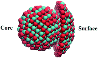
Due to the high surface to volume ratio typical of NPs, many properties are potentially affected by surface behavior, which is normally negligible in bulk. Therefore, we decided to investigate to which extent outer and inner layers of the CuO NPs differ. Since for small NPs it is hard to distinguish between surface and core atoms, we studied NPs with a diameter of 4 nm. Our previous results show that this is the smallest size where “bulk” behavior begins to appear and therefore we expect this NP to show the strongest surface effects. We defined the surface as the NP's outermost layer having a thickness of 0.36 nm (approximately twice Cu–O bond length), as a compromise between statistical error (from the small number of atoms) and error due to averaging of the surface and core properties.
A comparison of the core and surface XRD profiles for solid NP (see Fig. 8) suggests that surface structure does not have the monoclinic features present in the core structure as shown in Fig. 9.
 |
| Fig. 8 Simulated X-ray diffraction (XRD) profiles of the surface (blue) and core (black) of a CuO NP of 4 nm in diameter at 300 K. The positions of the peaks of the experimental XRD profile of bulk CuO are shown as dashed vertical lines. | |
 |
| Fig. 9 Structures of core and surface of a CuO NP. Red and blue represent oxygen and copper(II) ions, respectively. | |
The XRD profile shows an amorphous-like organization for the surface of a solid 4 nm NP even at room temperature, far below the melting point. This effect is probably caused by the truncation of crystal plane at the surface due to the high curvature of the NP. Similar distorted surface was previously observed experimentally for nearly spherical CuO nanoparticles using transmission electron microscopy (TEM).13 Based on these results, we expected the surface and core of a melted NP to produce similar XRD profile.
While this is generally true, as shown in Fig. 10, there are slight differences. In both cases here is no indication of long-range order in either the core or the surface plot, which confirms the amorphous structure. However, the XRD plot of the core shows a characteristic18 broad and weak hump at 35° whereas the surface profile is flattened, although slightly noisy. This difference suggests amorphous structures of different stoichiometry for the surface and core.
 |
| Fig. 10 Simulated X-ray diffraction (XRD) profiles of the surface (blue) and core (black) of a CuO NP of 4 nm in diameter at 1700 K. The positions of the peaks of the experimental XRD profile of bulk CuO are shown as dashed vertical lines. | |
Conclusions
In this paper, we reported a detailed analysis of the nanoscale structure of CuO nanoparticles using molecular dynamics simulations, and compared the results with available experimental data. We investigated the properties of different phases as function of size and temperatures. As for other inorganic NPs, we found that the melting temperature is inversely proportional to the particle diameter, with a linear fit that predicted the experimental bulk melting temperature very well (1657 K versus 1600 K), and it estimated NP melting point better than generic empirical models. The comparison between predicted and experimental XRD profiles showed a remarkable agreement indicating monoclinic crystal structure for solid particles, as small as for particles of 4 nm in diameter. Global structural information was integrated with data on local geometries, such as first neighbors mean distance and Cu average coordination number. The results suggest distinct nanoscale structural features for the solid and melted NPs. Differently, particles with diameters of 2 nm do not show any clear core–surface or phase dependent distinction. These simulations results were confirmed by experimental observations of amorphous-like structures for NPs of 2 nm or less in diameter. Like other transition metal oxides, the Cu–O bond length in NPs is smaller than that of the bulk, with negligible temperature dependence, again in accordance with experimental data.
For nanoparticles with diameters of 4 nm, we observed key differences in properties depending on spatial position (surface vs. core) and thermodynamic phase (melt vs. solid). The local geometry of CuO NPs at different temperatures is modified not by virtue of bond change, which is only minimally dependent on NP size and phase, but by the atomic arrangement (quantified as coordination number, or CN). Since the phase dependence of the CNs is significant, CN can be used as an indicator of the phase transformation. Atoms on the surface of a solid NP form an amorphous-like layer (or distorted outer shell) at temperatures well below the melting point. This phenomenon explains the existence of a rough spherical surface of CuO nanoparticles observed in TEM images. Notably, even the surface of melted NPs displays differences, from the core.
This study is a useful step towards the establishment of a structure–property relationship for CuO nanoparticles that is important for the understanding of nucleation, melting/freezing transition, sintering, aggregation, nanocomposite, and the growth of nanoparticles on other nanostructures (e.g., multi-walled carbon nanotube, nanoelectrode devices). These results will provide guidance for understanding the phase diagrams of mixed metal oxides and surface mediated reaction mechanisms in catalysis.
Acknowledgements
This research has been funded by NSF through DMR1105361. This research was supported in part through computational resources and services provided by Advanced Research Computing at the University of Michigan, Ann Arbor.
References
- L. X. Chen, T. Rajh, Z. Wang and M. C. Thurnauer, XAFS Studies of Surface Structures of TiO2 Nanoparticles and Photocatalytic Reduction of Metal Ions, J. Phys. Chem., 1997, 101, 10688–10697 CrossRef.
- N. Wang, Y. H. Tang, Y. F. Zhang, C. S. Lee, I. Bello and S. T. Lee, Si nanowires grown from silicon oxide, Chem. Phys. Lett., 1999, 299(2), 237–242 CrossRef.
- N. Wang, Y. Caia and R. Q. Zhang, Growth of nanowires, Mater. Sci. Eng., R, 2008, 60(1–6), 1–51 Search PubMed.
- G. C. Schatz, Using theory and computation to model nanoscale properties, Proc. Natl. Acad. Sci. U. S. A., 2007, 104(17), 6885–6892 CrossRef PubMed.
- J. T. Lue, Physical Properties of Nanomaterials, Encycl. Nanosci. Nanotechnol., 2004, X, 1–46 Search PubMed.
- A. Umar, M. Vaseem and Y. Hahn, Growth, Properties, and Applications of Copper Oxide and Nickel Oxide/Hydroxide Nanostructures, 2010 Search PubMed.
- T. Maruyama, Copper oxide thin films prepared by chemical vapor deposition from copper dipivaloylmethanate, Sol. Energy Mater. Sol. Cells, 1998, 56(1), 85–92 CrossRef.
- Y. P. Sukhorukov, N. N. Loshkareva, A. A. Samokhvalov, S. V. Naumov, A. S. Moskvin and A. S. Ovchinnikov, Magnetic phase transitions in optical spectrum of magnetic semiconductor CuO, J. Magn. Magn. Mater., 1998, 183(3), 356–358 CrossRef.
- M. Frietsch, F. Zudock, J. Goschnick and M. Bruns, CuO catalytic membrane as selectivity trimmer for metal oxide gas sensors, Sens. Actuators, B, 2000, 65(1–3), 379–381 CrossRef.
- C. L. Carnes and K. J. Klabunde, The catalytic methanol synthesis over nanoparticle metal oxide catalysts, J. Mol. Catal. A: Chem., 2003, 194(1–2), 227–236 CrossRef.
- S. Lee, Methanol Synthesis Technology, CRC Press, 1990 Search PubMed.
- B. S. Anandakumar, M. B. M. Reddy, C. N. Tharamani, M. A. Pasha and G. T. Chandrappa, Combustion-derived CuO nanoparticles: An effective and environmentally benign catalyst in the synthesis of aromatic nitriles from aromatic aldehydes, Chin. J. Catal., 2013, 34(4), 704–710 CrossRef.
- J. Zhang, J. Liu, Q. Peng and X. Wang, Nearly Monodisperse Cu2O and CuO Nanospheres: Preparation and Applications for Sensitive Gas Sensors, Chem. Mater., 2006, 18, 867–871 CrossRef CAS.
- D. Gao, J. Zhang, J. Zhu, J. Qi, Z. Zhang, W. Sui, H. Shi and D. Xue, Vacancy-Mediated Magnetism in Pure Copper Oxide Nanoparticles, Nanoscale Res. Lett., 2010, 5(4), 769–772 CrossRef CAS PubMed.
- K. J. Reddy, K. J. McDonald and H. King, A novel arsenic removal process for water using cupric oxide nanoparticles, J. Colloid Interface Sci., 2013, 397, 96–102 CrossRef CAS PubMed.
- P. Poizot, S. Laruelle, S. Grugeon, L. Dupont and J. M. Tarascon, Nano-sized transition-metal oxides as negative-electrode materials for lithium-ion batteries, Nature, 2000, 407(6803), 496–499 CrossRef CAS PubMed.
- A. Azam, A. S. Ahmed, M. Oves, M. S. Khan and A. Memic, Size-dependent antimicrobial properties of CuO nanoparticles against Gram-positive and -negative bacterial strains, Int. J. Nanomed., 2012, 7, 3527–3535 CrossRef PubMed.
- H. Zhang, B. Chen, J. Banfield and G. Waychunas, Atomic structure of nanometer-sized amorphous TiO2, Phys. Rev. B: Condens. Matter Mater. Phys., 2008, 78(21), 214106 CrossRef.
- P. K. Naicker, P. T. Cummings, H. Zhang and J. F. Banfield, Characterization of titanium dioxide nanoparticles using molecular dynamics simulations, J. Phys. Chem. B, 2005, 109(32), 15243–15249 CrossRef PubMed.
- B. Buesser, A. J. Gröhn and S. E. Pratsinis, Sintering Rate and Mechanism of TiO2 Nanoparticles by Molecular Dynamics, J. Phys. Chem. C, 2011, 115(22), 11030–11035 Search PubMed.
- Y. Li, R. K. Kalia, A. Nakano and P. Vashishta, Size effect on the oxidation of aluminum nanoparticle: Multimillion-atom reactive molecular dynamics simulations, J. Appl. Phys., 2013, 114(13), 134312 CrossRef PubMed.
- I. V. Schweigert, K. E. J. Lehtinen, M. J. Carrier and M. R. Zachariah, Structure and properties of silica nanoclusters at high temperatures, Phys. Rev. B: Condens. Matter Mater. Phys., 2002, 65(23), 235410 CrossRef.
- M. R. Zachariah and M. J. Carrier, Properties of Silicon Nanoparticles: A Molecular Dynamics Study, J. Phys. Chem., 1996, 100(36), 14856–14864 CrossRef CAS.
- U. I. Rodrigo, Geometrical and Electronic Structure of Metal Oxide Nanoparticles on Silica and Alumina, Ph.D. Thesis, Lousiana State University, 2012.
- C. Q. Sun, Size dependence of nanostructures: Impact of bond order deficiency, Prog. Solid State Chem., 2007, 35, 1–159 CrossRef CAS PubMed.
- A. Barnard, C. M. Li, R. Zhou and Y. Zhao, Modelling of the nanoscale, Nanoscale, 2012, 4(4), 1042 RSC.
- D. Spagnoli and J. D. Gale, Atomistic theory and simulation of the morphology and structure of ionic nanoparticles, Nanoscale, 2012, 4(4), 1051–1067 RSC.
- Y. Waseda and J. M. Toguri, The Structure and Properties of Oxide Melts: Application of Basic Science to Metallurgical Processing, World Scientific, Singapore, 1998 Search PubMed.
- A. I. Frenkel, A. Yevick, C. Cooper and R. Vasic, Modeling the Structure and Composition of Nanoparticles by Extended X-Ray Absorption Fine-Structure Spectroscopy, Annu. Rev. Anal. Chem., 2011, 4, 23–39 CrossRef PubMed.
- H. Zhang and J. F. Banfield, Understanding Polymorphic Phase Transformation Behavior during Growth of Nanocrystalline Aggregates : Insights from TiO2, J. Phys. Chem. B, 2000, 104, 3481–3487 CrossRef.
- H. Zhang and J. F. Banfield, Phase transformation of nanocrystalline anatase-to-rutile via combined interface and surface nucleation, J. Mater. Res., 2000, 15(2), 437–448 CrossRef CAS.
- G. Jian, N. W. Piekiel and M. R. Zachariah, Time-Resolved Mass Spectrometry of Nano-Al and Nano-Al/CuO Thermite under Rapid Heating: A Mechanistic Study, J. Phys. Chem. C, 2012, 116, 26881–26887 Search PubMed.
- X. W. Zhou, H. N. G. Wadley, J. Filhol and M. N. Neurock, Modified Charge Transfer-Embedded Atom Method Potential for Metal/Metal Oxide Systems, Phys. Rev. B: Condens. Matter Mater. Phys., 2004, 69, 035402 CrossRef.
- S. Stolbov and T. S. Rahman, Relationship between Electronic and Geometric Structures of the O/Cu(001) System, J. Chem. Phys., 2002, 117(18), 8523–8530 CrossRef PubMed.
- T. Liang, T. Shan, Y. Cheng, B. D. Devine, M. Noordhoek, Y. Li, Z. Lu and S. R. Phillpot, et al., Classical atomistic simulations of surfaces and heterogeneous interfaces with the charge-optimized many body (COMB) potentials, Mater. Sci. Eng., R, 2013, 74(9), 255–279 CrossRef PubMed.
- A. C. T. van Duin, V. S. Bryantsev, M. S. Diallo, W. A. Goddard, O. Rahaman, D. J. Doren, D. Raymand and K. Hermansson, Development and Validation of a ReaxFF Reactive Force Field for Cu Cation/Water Interactions and Copper Metal/Metal Oxide/Metal Hydroxide Condensed Phases, J. Phys. Chem., 2010, 114(35), 9507–9514 CrossRef PubMed.
- X. Duan, O. Warschkow, A. Soon, B. Delley and C. Stampfl, Density functional study of oxygen on Cu(100) and Cu(110) surfaces, Phys. Rev. B: Condens. Matter Mater. Phys., 2010, 81(7), 075430 CrossRef.
- B. Jeona, S. K. R. S. Sankaranarayanan, A. C. T. van Duin and S. Ramanathan, Influence of surface orientation and defects on early-stage oxidation and ultrathin oxide growth on pure copper, Philos. Mag., 2011, 91(32), 4073–4088 CrossRef.
- B. Jeon and S. K. R. S. Sankaranarayanan, Influence of Surface Orientation and Defects on Early-Stage Oxidation and Ultrathin Oxide Growth on Pure Copper, Philos. Mag., 2011, 91(32), 4073–4088 CrossRef CAS.
- B. Jeon, S. K. R. S. Sankaranarayanan, A. C. T. Van Duin and S. Ramanathan, Reactive Molecular Dynamics Study of Chloride Ion Interaction with Copper Oxide Surfaces in Aqueous Media, ACS Appl. Mater. Interfaces, 2012, 4(3), 1225–1232 Search PubMed.
- A. C. T. van Duin, S. Dasgupta, F. Lorant and W. A. Goddard, ReaxFF: A Reactive Force Field for Hydrocarbons, J. Phys. Chem. A, 2001, 105(41), 9396–9409 CrossRef.
- A. C. T. van Duin, A. Strachan, S. Stewman, Q. Zhang, X. Xu and W. A. Goddard, Reactive Force Field for Silicon and Silicon Oxide Systems, J. Phys. Chem. A, 2003, 107, 3803–3811 CrossRef.
- W. J. Mortier, S. K. Ghosh and S. Shankar, Electronegativity Equalization Method for the Calculation of Atomic Charges in Molecules, J. Am. Chem. Soc., 1986, 108, 4315–4320 CrossRef CAS.
- C.-Y. Chiang, K. Aroh and S. H. Ehrman, Copper oxide nanoparticle made by flame spray pyrolysis for photoelectrochemical water splitting – Part I. CuO nanoparticle preparation, Int. J. Hydrogen Energy, 2012, 37(6), 4871–4879 CrossRef CAS PubMed.
- S. Plimpton, Fast Parallel Algorithms for Short-Range Molecular Dynamics, J. Comput. Phys., 1995, 117, 1–19 CrossRef CAS.
- H. J. C. Berendsen, J. P. M. Postma, W. F. van Gunsteren, A. DiNola and J. R. Haak, Molecular dynamics with coupling to an external bath, J. Chem. Phys., 1984, 81(8), 3684 CrossRef CAS PubMed.
- R. J. Sadus, Molecular simulation of fluids: theory, algorithms, and object-orientation, Elsevier, Amsterdam, 1999 Search PubMed.
- P. Debye, Zerstreuung von Rontgenstrahlen, Ann. Phys., 1915, 46, 809–823 CrossRef CAS PubMed.
- C. L. Farrow and S. J. L. Billinge, Relationship between the atomic pair distribution function and small-angle scattering: implications for modeling of nanoparticles research papers, Acta Crystallogr., Sect. A: Found. Crystallogr., 2009, 65, 232–239 CrossRef CAS PubMed.
- M. Wojdyr, Debyer, http://debyer.readthedocs.org/en/latest/, 2011 Search PubMed.
- V. N. Koparde and P. T. Cummings, Sintering of titanium dioxide nanoparticles: a comparison between molecular dynamics and phenomenological modeling, J. Nanopart. Res., 2008, 10(7), 1169–1182 CrossRef CAS.
- Y. Xing and D. E. Rosner, Prediction of spherule size in gas phase nanoparticle synthesis, J. Nanopart. Res., 1999, 1, 277–291 CrossRef CAS.
- Z. Zhang, X. X. Lu and Q. Jiang, Finite size effect on melting enthalpy and melting entropy of nanocrystals, Phys. B, 1999, 270, 249–254 CrossRef CAS.
- P. A. Bhatt, A. Pratap and P. K. Jha, Size and Dimension Dependent Diffusion Coefficients of SnO2 Nanoparticles, AIP Conf. Proc., 2013, 1536, 237–238 CrossRef CAS PubMed.
- S. Mishra, S. K. Gupta, P. K. Jha and A. Pratap, Study of dimension dependent diffusion coefficient of titanium dioxide nanoparticles, Mater. Chem. Phys., 2010, 123(2–3), 791–794 CrossRef CAS PubMed.
- P. A. Bhatt, A. Pratap and P. K. Jha, Study of size-dependent glass transition and Kauzmann temperatures of tin dioxide nanoparticles, J. Therm. Anal. Calorim., 2012, 110, 535–538 CrossRef CAS PubMed.
- Copper (II) oxide [Material Safety Data Sheet], Iowa State University, 2 August 2000, retrieved from http://avogadro.chem.iastate.edu/MSDS/CuO.htm Search PubMed.
- X. Zheng, C. Xu, K. Nishikubo, K. Nishiyama, W. Higemoto, W. Moon, E. Tanaka and E. Otabe, Finite-size effect on Néel temperature in antiferromagnetic nanoparticles, Phys. Rev. B: Condens. Matter Mater. Phys., 2005, 72(1), 014464 CrossRef.
- X. Lang and Q. Jiang, Size dependence of phase transition temperatures of ferromagnetic, ferroelectric and superconductive nanocrystals, Front. Phys. China., 2007, 2(3), 289–311 CrossRef PubMed.
- G. Bergerhoff and I. D. Brown, Crystallographic Databases, in Crystallogr. Databases. International Union of Crystallography, 1987 Search PubMed.
- A. Belsky, M. Hellenbrandt, V. L. Karen and P. Luksch, New developments in the Inorganic Crystal Structure Database (ICSD): accessibility in support of materials research and design research papers, Acta Crystallogr., Sect. B: Struct. Sci., 2002, 58, 364–369 CrossRef PubMed.
- S. Asbrink and L.-J. Norrby, A Refinement of the Crystal Structure of Copper(II) Oxide with a Discussion of Some Exceptional E.s.d.'s, Acta Crystallogr., Sect. B: Struct. Crystallogr. Cryst. Chem., 1970, 26(8), 8–15 CrossRef CAS.
- L. Troger, T. Yokoyama, D. Arvanitis, T. Lederer, M. Tischer and K. Baberschke, Determination of bond lengths, atomic mean-square relative displacements, and local thermal expansion by means of soft-X-ray photoabsorption, Phys. Rev. B: Condens. Matter Mater. Phys., 1994, 49(2), 888–904 CrossRef.
- G. Bae, B. Dellinger and R. W. Hall, Density functional calculation of the structure and electronic properties of CunOn (n = 1–8) clusters, J. Phys. Chem., 2011, 115(11), 2087–2095 CrossRef CAS PubMed.
- M. L. Polak, M. K. Gilles, J. Ho and W. C. Lineberger, Photoelectron Spectroscopy of CuO−, J. Phys. Chem., 1991, 95, 3460–3463 CrossRef CAS.
- R. Vajtai, Science and Engineering of Nanomaterials, in Springer Handb. Nanomater, ed. R. Vajtai, Springer, New York, 2013 Search PubMed.
- M. S. P. Francisco, V. R. Mastelaro, A. O. Florentino and D. Bazin, Structural study of copper oxide supported on a ceria-modified titania catalyst system, Top. Catal., 2002, 18, 105–111 CrossRef CAS.
- L. B. Skinner, C. J. Benmore, J. K. R. Weber, J. Du, J. Neuefeind, S. K. Tumber and J. B. Parise, Low Cation Coordination in Oxide Melts, Phys. Rev. Lett., 2014, 112(15), 157801 CrossRef CAS.
- L. B. Skinner, C. J. Benmore, J. K. R. Weber, S. Tumber, L. Lazareva, J. Neuefeind, L. Santodonato and J. Du, et al., Structure of Molten CaSiO3: Neutron Diffraction Isotope Substitution with Aerodynamic Levitation and Molecular Dynamics Study, J. Phys. Chem. B, 2012, 116, 13439–13447 CrossRef CAS PubMed.
- L. B. Skinner, A. C. Barnes, P. S. Salmon, L. Hennet, H. E. Fischer, C. J. Benmore, S. Kohara and J. K. R. Weber, et al., Joint diffraction and modeling approach to the structure of liquid alumina, Phys. Rev. B: Condens. Matter Mater. Phys., 2013, 87, 024201 CrossRef.
- H. Lou, X. Wang, Q. Cao, D. Zhang, J. Zhang, T. Hu and H. Mao, Negative expansions of interatomic distances in metallic melts, Proc. Natl. Acad. Sci. U. S. A., 2013, 110(25), 10068–10072 CrossRef CAS PubMed.
- D. Holland-Moritz, T. Schenk, R. Bellissent, V. Simonet, K. Funakoshi, J. M. Merino, T. Buslaps and S. Reutzel, Short-range order in undercooled Co melts, J. Non-Cryst. Solids, 2002, 312–314, 47–51 CrossRef CAS.
- T. Schenk, V. Simonet, R. Bellissent and D. M. Herlach, Icosahedral Short-Range Order in Deeply Undercooled Metallic Melts, Phys. Rev. Lett., 2002, 89(7), 075507 CrossRef CAS.
- T. Itami, S. Munejiri, T. Masaki, H. Aoki, Y. Ishii, T. Kamiyama, Y. Senda and F. Shimojo, et al., Structure of liquid Sn over a wide temperature range from neutron scattering experiments and first-principles molecular dynamics simulation: A comparison to liquid Pb, Phys. Rev. B: Condens. Matter Mater. Phys., 2003, 67(6), 064201 CrossRef.
- K. Georgarakis, D. V. Louzguine-luzgin and J. Antonowicz, Variations in atomic structural features of a supercooled Pd–Ni–Cu–P glass forming liquid during in situ vitrification, Acta Mater., 2011, 59(2), 708–716 CrossRef CAS PubMed.
- N. A. Mauro, W. Fu, J. C. Bendert, Y. Q. Cheng, E. Ma and K. F. Kelton, Local atomic structure in equilibrium and supercooled liquid Zr(75.5)Pd(24.5), J. Chem. Phys., 2012, 137(4), 044501 CrossRef CAS PubMed.
Footnote |
† Electronic supplementary information (ESI) available: Potential energy profiles versus time to demonstrate convergence; details about the empirical model to determine melting point; and temperature dependence of X-ray diffraction (XRD) profiles are given. See DOI: 10.1039/c5ra04276c |
|
This journal is © The Royal Society of Chemistry 2015 |
Click here to see how this site uses Cookies. View our privacy policy here.