Cu doped Fe3O4 magnetic adsorbent for arsenic: synthesis, property, and sorption application

Ting Wanga, Weichun Yangab, Tingting Songa, Chaofang Lia, Liyuan Zhanga, Haiying Wang*ab and Liyuan Chai*ab
aDepartment of Environmental Engineering, School of Metallurgy and Environment, Central South University, Changsha, 410083, P. R. China. E-mail: haiyw25@163.com; haiyw25@yahoo.com; lychai@csu.edu.cn; Fax: +86 0731 88710171
bChinese National Engineering Research Center for Control & Treatment of Heavy Metal Pollution, Changsha, 410083, P. R. China

Received 5th March 2015 , Accepted 29th May 2015

First published on 29th May 2015


Abstract

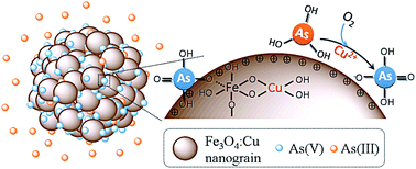
Cu doped Fe3O4 (Fe3O4:Cu) particles were synthesized and applied for arsenic adsorption. As the copper ions increase, the adsorption capacity of Fe3O4:Cu towards As(V) and As(III) increases from 7.32 to 42.90 mg g−1 and from 8.12 to 37.97 mg g−1, respectively. The incorporation of copper decreased the particle size, increased the surface area, porosity and zeta potential, leading to the increase of the adsorption sites and affinity toward negative As(V) species. More importantly, the doped copper ions catalyzed the efficient oxidation of As(III) to As(V) by O2 followed by As(V) adsorption. The Fe3O4:Cu particles also exhibited good performance toward low level arsenic removal, excellent separation, and satisfactory regeneration property. The results indicate Fe3O4:Cu particles possess great potential for both As(III) and As(V) adsorption.


1. Introduction

Arsenic contamination has greatly threatened water safety due to its high toxicity and carcinogenicity.1,2 It is urgent and significant to develop simple and effective approaches for arsenic removal. Among various arsenic treatment technologies, adsorption has been recognized as the best available method, because of its effectiveness, low cost, low energy requirements and simple operation.3–7 As(V) and As(III) are the most observed arsenic species in natural water.8 In comparison, As(V), generally existing as negative species (5 < pH < 9), can be readily removed by adsorbents. As(III), as a more mobile and toxic species than As(V),9 is generally difficult to be adsorbed due to the nonionic H3AsO3 at pH values (5 < pH < 9).10,11 Thus, the transformation of As(III) to As(V) is necessary for arsenic removal.12–15 Another challenge for adsorption is the time consuming and cost ineffective separation procedures.16–17 Magnetic adsorbents, such as Fe3O4, avoid the traditional separation due to their quick and effective magnetic separation.18 However, bare Fe3O4 adsorbent still exhibited limited arsenic absorbance.

Doping is an effective approach to modify particle structure, such as increased amount of surface defects,19,20 increased hydroxyl amount,21,22 tunable surface charge,23 and high electronic conduction properties,24 which could enhance adsorption performance and broaden applications.25 To date, rare earth metals are investigated as doping ions, such as Ce–Fe3O4,26–28 Zr–Fe3O4,29 and La–Fe LDH,30 which are good adsorbents for As(V). However, the rare earth metals doped adsorbents are relatively expensive and still fail to transform of As(III).

Adding oxidant to convert As(III) to As(V) is normal and efficient route. Among various oxidants, such as Cl2, NaClO, O3, ClO2, KMnO4, and H2O2,31 O2 is a green, low cost, and rather feasible oxidant. However, the arsenic oxidation by atmospheric O2 is kinetically slow, and generally needs the addition of catalyst. The photocatalytic oxidation of As(III) by O2 followed by As(V) adsorption using TiO2/UV system has been extensively investigated, which still needs the UV radiation instruments.31,32 Consequently, it is highly desirable to synthesize adsorbents with efficient catalytic property to transform of As(III) to As(V).

Doping metal ions into metal oxide to improve the catalytic activity has attracted ever-lasting attentions since the first report in 1985.33,34 Most recently, Cu doping has attracted great attentions, such as Cu-doped TiO2,35 Cu doped zinc sulfide,36 and multivalent Cu-doped ZnO,37 which are generally applied in photocatalytic oxidation field. The previous investigation demonstrated that copper ions were able to act as electron mediation centers to accelerate the oxidation reaction based on Cu(II)/Cu(I) couple.38–41 It is rational to dope copper ions in magnetic adsorbents, which are expected to simultaneously possess good adsorption performance, catalytic oxidation and readily magnetic separation property.

The main objectives of this research were (i) the preparation of Cu doped Fe3O4 adsorbent, (Fe3O4:Cu), (ii) revealing the structure of Fe3O4:Cu, (iii) the dependence of arsenic adsorption performance on Fe3O4:Cu structure, (iv) the adsorption mechanism for As(V) and As(III) on Fe3O4:Cu.

2. Experiment section

2.1. Materials

Ferric chloride hexahydrate (FeCl3·6H2O), copper chloride dehydrate (CuCl2·2H2O), anhydrous sodium acetate (CH3COONa, NaAc), and ethylene glycol (EG) were obtained from the Sinopharm Group Chemical Reagent Co., Ltd. Na3AsO4·12H2O and NaAsO2 were used as the sources of As(V) and As(III), respectively. All reagents were used without further treatment. Ultrapure water with a resistivity of 18.2 MΩ cm−1, produced with a Milli-Q apparatus (Millipore), was used throughout all of the experiments.

2.2. Preparation of porous Fe3O4:Cu

Solvothermal synthesis of porous Fe3O4:Cu was carried out as follows. 1.35 g of FeCl3·6H2O (5 mM) and different amount of CuCl2·2H2O of 0, 0.05, 0.25, 0.5, 1, and 2.5 mM were fully dispersed in 36 mL of EG via ultrasonification. Then 3.6 g of NaAc was added into the above solution. After vigorous stirring for 30 min, the solution was transferred to a 50 mL Teflon-lined autoclave, which was placed in an oven at 200 °C for 9 h, followed by naturally cooling to room temperature. The precipitate was collected and ultrasonic washed by water and ethanol for three times, respectively, through magnetic separation. Finally, the yielded product was vacuum-dried at 60 °C for 12 h. The magnetic Cu doped Fe3O4 particles with initial dosage Cu2+ amount varied from 0 mM to 2.5 mM were denoted as Fe3O4:Cu-0, Fe3O4:Cu-0.05, Fe3O4:Cu-0.25, Fe3O4:Cu-0.5, Fe3O4:Cu-1.0, and Fe3O4:Cu-2.5, respectively.

2.3. Characterization

The morphology of the nanoparticles was characterized by scanning electron microscopy (SEM, JSM-6360) and transmission electron microscopy equipped with energy-dispersive spectroscopy (EDS) (TEM, TECNAI G2). The X-ray diffraction (XRD) patterns of the Fe3O4:Cu were obtained using Rigaku D/Max-RB diffractometer with Cu-Kα radiation (λ = 0.15406 nm, 35 kV, 40 mA). The X-ray photoelectron spectroscopy (XPS) measurements were conducted on ESCALAB 250Xi spectrometer with Mg/Al X-ray source with 400 W power. All binding energies were calibrated according to the C 1s neutral carbon peak at 284.8 eV. Magnetic properties of the product were investigated using a vibrating sample magnetometer (VSM, EV7, ADE) with an applied field between −7000 and 7000 Oe at room temperature. Specific surface areas of the yielded products were measured by adsorption–desorption of ultrapure N2 on a Quantachrome Instruments system (Adsorb SI) via Brunauer–Emmett–Teller (BET) method. Pore size distribution was determined by N2 desorption isotherm using Barrett–Joyner–Halenda (BJH) method.

XAFS experiments were conducted at 1W2B beamline of the Beijing Synchrotron Radiation Facility. Samples were ground into fine powers and then smeared on Scotch tapes. As K-edge XANES/EXAFS spectra were collected at room temperature in transmission/fluorescence mode. The storage ring was working at 2.5 GeV with a maximum electron current of about 250 mA. Data were collected using a Si (111) double-crystal monochromator and normalized/analyzed using the program ATHENA. Each sample was calibrated along with pre-edge and post-edge normalization using the most intense peak of the first derivative at each elemental foil edge.

The zeta potential of the Fe3O4:Cu particles was analyzed with the Malvern zetasizer instrument (type Nano-ZS, Malvern Instruments Ltd., Britain). Adsorbent with concentration of 0.01 g L−1 was fully dispersed in 20 mL of deionized water via ultrasonification. The solution pH was adjusted with 0.1 M NaOH or HCl solution to a desired value and the supernatant was used to conduct ζ potential measurements.

2.4. Adsorption experiment

Solutions containing different concentrations of As(V) or As(III) were prepared and adjusted to pH 5 ± 0.2 using HCl. Then, 10 mg of the adsorbent sample was added to 20 mL arsenic aqueous solution under stirring. After a specified time, the solid and liquid were magnetically separated and the initial and residual concentrations of arsenic were measured by Inductively Coupled Plasma-Optical Emission Spectroscopy (ICP-OES) (Optima 5300DV). The adsorption isotherm was obtained by varying the initial arsenic concentrations and stirring for 4 h at 25 °C (concentration range: 1–85 mg L−1 for As(III) and 1–45 mg L−1 for As(V), respectively).

The adsorption kinetics was investigated with the initial As(V) concentration of 20 mg L−1 and As(III) concentration of 10 mg L−1 at pH = 5.0 ± 0.2. The solution was allowed to react with the adsorbent for a fixed period (between 10 and 240 min).

The equilibrium adsorption capacity (qe) (mg g−1) for arsenic was calculated according to the following equation:

 
image file: c5ra03951g-t1.tif(1)
where c0 and ce (mg L−1) are the initial and equilibrium arsenic aqueous concentrations, respectively; V is the volume (mL) of arsenic aqueous solution; m is the mass (mg) of adsorbents used in the experiment.

The adsorption of As(III) under N2 protection and O2 sufficient condition was conducted via purging with gas into the suspension at a rate of 50 mL min−1 controlled by gas flow meter.

To investigate the acid effect on adsorption, the initial pH value of arsenic was varied from 3 to 10 with initial As(V) and As(III) concentration of 45 and 85 mg g−1. To reveal the ionic strength effect on adsorption, NaCl with different concentration was added adjusted. To effects of co-existing anions on arsenic removal, NO3, PO43−, SO42−, and CO32− were added to arsenic solution. The initial As(V) and As(III) concentration were 0.6 and 1.13 mM, respectively, and pH values were adjusted as 5.

To test the low-level arsenic removal feasibility of adsorbents, initial arsenic solution with As(V) concentration in the range of 50–1200 μg L−1 and As(III) in the range of 50–1000 μg L−1 were prepared. Atomic fluorescence spectrometer (AFS-8X, Beijing Titan Instruments Co., Ltd.) was exploited to detect the residual arsenic concentration. Other adsorption experiment procedures were the same as above.

2.5. Adsorption modeling

Both Langmuir and Freundlich isotherm models were used to fit the equilibrium adsorption data:
 
image file: c5ra03951g-t2.tif(2)
 
image file: c5ra03951g-t3.tif(3)
where qm and KL are Langmuir constants and represent the maximum adsorption capacity of adsorbents (mg g−1) and the energy of adsorption, respectively. KF and n are Freundlich constants related to adsorption capacity and adsorption intensity, respectively.

The pseudo-second-order rate kinetic model was used to fit the adsorption kinetic experimental data, which is represented as:

 
image file: c5ra03951g-t4.tif(4)
where qe and qt are the amount of arsenic adsorbed at equilibrium and at time t, respectively; k2 is the rate constant of the pseudo-second-order model of adsorption (g mg−1 min−1).

2.6. Regeneration property

The regeneration of the absorbent was conducted using 0.1 M NaOH solution as eluent with adsorbents dose of 1 g L−1 at 25 °C. Briefly, the absorbent was firstly ultrasonificated in NaOH solution for 30 min and then shaken for 2 hours, followed by magnetic separation and washing by water for three times. Then the adsorbent was applied into recycle adsorption study. The recycle adsorption experimental procedure and detection method are in accordance with the first adsorption experiment.

All the experimental data were the average of triplicate determinations with relative errors under 5%.

3. Results and discussion

3.1. Morphology and structure characterization

Fig. 1 shows the microscopic images of synthesized Fe3O4:Cu particles. As the initial amount of Cu2+ increased from 0 to 2.5 mM, the sphere size decreased from ∼560 nm to ∼120 nm. As reflected by the TEM images in the inset of Fig. 1, the particle structure transformed from solid spheres to porous spheres with decreased size accordingly. The EDS results in Fig. S1 and Table S1 confirmed the atom ratio of Cu on Fe3O4:Cu particles increased from 0% to 31.31% with Cu amount increase from 0 to 2.5 mM. Taking Fe3O4:Cu-2.5 particle as an example, detailed structure information was obtained. EDS mapping images of Fe3O4:Cu-2.5 in Fig. 1G–I revealed that Cu, Fe, and O elements distributed uniformly on the Fe3O4:Cu-2.5 particles. HRTEM images of Fe3O4:Cu-2.5 in Fig. S2 revealed a hierarchical and porous sphere consisting of the nanograins assembly. Microscopic images indicated that porous Fe3O4:Cu particles with Cu, Fe, O uniformly distributed shows particle size and structure dependent on initial Cu2+ dosage.
image file: c5ra03951g-f1.tif
Fig. 1 SEM of Fe3O4:Cu-0 (A), Fe3O4:Cu-0.05 (B), Fe3O4:Cu-0.25 (C), Fe3O4:Cu-0.5 (D), Fe3O4:Cu-1.0 (E), Fe3O4:Cu-2.5 (F) particles, EDS-mapping images Cu (G), Fe (H) and O (I) of Fe3O4:Cu-2.5 particles. The inset shows the corresponding TEM image.

The XRD spectra of all Fe3O4:Cu particle in Fig. 2 show well-defined diffraction lines suggesting the well-crystallized structure. As expected, the typical crystal planes (111), (220), (311), (400), (511), and (440) of Fe3O4 were observed for all samples Fe3O4:Cu exhibit XRD pattern of face centered Fe3O4 (JCPDS no. 86-1344, a = b = c = 8.3925 Å).18 With Cu2+ initial dosage from 0 to 2.5 mM, the magnified peak for (311) crystal plane shifted slightly from 35.46° to 35.61°, corresponding to the decreased length of the a-axis from 8.39 to 8.35 Å, as listed in Table S2. The decreased a-axis might be caused by the doping of small radium copper ions (0.57 Å, which is smaller than 0.63 Å of iron ions), where some Fe–O was substituted by Cu–O in the Fe3O4 crystal.42,43 On the other hand, with the increase of initial Cu2+ dosage, the intensity of diffraction peaks at 43.31°, 50.44°, and 74.10° correspond to the crystal planes (111), (200), and (220) of Cu increase, indicating the augment in the amount of metal Cu (JCPDS no. 85-1326, a = b = c = 3.615 Å).44–46 The metal Cu might be produced due to the reduction of Cu2+ by ethylene glycol.47 No peaks attributed to CuO or Cu2O were detected.48,49 Moreover, the HRTEM images of Fe3O4:Cu-2.5 showed the lattice distance of 0.22 nm and 0.26 nm, in accordance with the typical lattice plane for Cu (111)50,51 (Fig. S2B) and Fe3O4 (311)52 (Fig. S2C), respectively.


image file: c5ra03951g-f2.tif
Fig. 2 XRD spectra (A) and the magnified peak (311) (B) of Fe3O4:Cu-0 (1), Fe3O4:Cu-0.05 (2), Fe3O4:Cu-0.25 (3), Fe3O4:Cu-1.0 (4), and Fe3O4:Cu-2.5 (5) particles. Rhombic and stellate icons represent the crystal place of Fe3O4 and Cu, respectively.

As revealed by XPS spectra (Fig. S3), compared with Fe3O4, the Fe 2p3/2 and Fe 2p1/2 peak of Fe3O4:Cu-2.5 shifted to the higher energy region by 0.3 and 0.2 eV, respectively, indicating the higher oxidation state compared to that of the Fe3O4, which might be caused by the doping of copper ions.53 For the Fe3O4:Cu-2.5 sample, the broad photoelectron peak at above 934.1 eV (Cu 2p3/2) along with the presence of the characteristic shakeup satellite peaks suggested the presence of Cu(II),48 while the peaks located at 932.4 and 952.3 eV correspond to Cu 2p3/2 and Cu 2p1/2 of metallic copper.54

The combined microscopic, XRD and XPS results indicated the concurrent presence of doped Cu2+ and the Cu nanocrystal on Fe3O4:Cu particles. The mixture of Cu0 and Cu2+ doped Fe3O4 nanograin constituted Fe3O4:Cu particles with Fe, Cu, and O uniformly distributed. The Cu loading decreased the particle size, which should be ascribed to the nucleation effect of the doped Cu2+.33,55 On the other hand, Cu nano-crystals was observed in the formation and assembly of Fe3O4 nanograins, which might hinder the growth of nanograins, leading to the size decrease of Fe3O4:Cu particles, as discussed in the ESI (Fig. S4).

The specific surface area of the Fe3O4:Cu particles (Fig. S5) increased from 41.11 to 93.94 m2 g−1 and the pore volume of Fe3O4:Cu particles increased from 5.24 to 12.81 cm3 g−1, with Cu dosage varied from 0.05 to 2.5 mM. Thus, the addition of Cu ions dosage decreased the particle size, increased the particle porosity and surface area, thus providing lots of active sites for adsorption. The zeta potential of Fe3O4:Cu sample as function of pH was given in Fig. 3. From the intersection at the zeta potential = 0 line, pH point of zero charged (pH PZC) was estimated. The pH PZC of Fe3O4:Cu increased from 6.97 to 8.86 as increasing initial dosage of copper ions from 0 to 2.5 mM. The function of Cu incorporation in enhancing pH PZC should be that the doping of Cu2+ cause more defects in Fe3O4 crystals, which was favorable for the adsorption of hydroxyl on particles and then increased the affinity to proton, eventually leading to the increase in pH PZC.56 The increased pH PZC indicate that the adsorbent has a high tendency to be positive and thus has the potential of high affinity to negative arsenic species.57,58


image file: c5ra03951g-f3.tif
Fig. 3 Zeta potential of Fe3O4:Cu particles as a function of pH. The dashed line represents zero zeta potential.

Fig. S6 shows the magnetization curves of Fe3O4:Cu particles measured at room temperature. The small hysteresis loops reflected by the magnification of low field region given in the inset of Fig. S6 indicates the superparamagnetic nature of the Fe3O4:Cu particles. As the amount of copper ions varied from 0 to 2.5 mM, the magnetic saturation (Ms) values of the Fe3O4:Cu particles are in the ranges of 66.86–108.4 emu g−1, which is higher than reported magnetic nanoparticles.22,42,59,60 Further increasing copper ions dosage, the particles exhibited weak magnetic response, and thus was not investigated in our system. The magnetism decrease should be caused by the decreased size and the Cu2+ doping induced a disordered structure.22 The high magnetic property greatly facilitate its facile magnetic separation.

3.2. Adsorption performance for arsenic species

Fig. 4A and C showed the adsorption isotherms of As(V) and As(III) on Fe3O4:Cu particles. As can be seen clearly, as the initial dosage of copper ions increase from 0 to 2.5 mM, the adsorption capacity for As(V) and As(III) increases from 7.32 to 42.90 mg g−1 and from 8.12 to 32.97 mg g−1, respectively. The adsorbent capacity of Fe3O4:Cu-2.5 particle is much higher than many reported related adsorbents, such as Fe3O4 nanoparticles,18 flowerlike α-Fe2O3,61 commercial α-Fe2O3,61 commercial CuO nanoparticles,62 Mg-doping α-Fe2O3,42 and CeO2–ZrO2,15 as listed in Table S3. The specific binding sites (ss, nm−2) of arsenic species on Fe3O4:Cu samples were also shown in Fig. S7, which is defined as the number of arsenic adsorbed per specific area and quantitatively evaluates the binding density.
 
image file: c5ra03951g-t5.tif(5)
where qm is the adsorption capacity (mg g−1) and S is the surface area of adsorbent (m2 g−1). As calculated by eqn (5), ss for As(V) gradually increased from 2.54 for Cu–Fe3O4-0 to 5.35 for Cu–Fe3O4-2.5 and ss for As(III) increased from 2.82 for Cu–Fe3O4-0 to 4.85 for Cu–Fe3O4-2.5. As the increase of Cu2+ initial dosage, the variation tendency of ss for As(V) and As(III) is in accordance with that of zeta potential. Adsorbent with higher zeta potential possessed positive charged surface and thus high affinity toward negative As(V) species. However, the modified surface charged state could not explain increased affinity for As(III). The speciation of adsorbed As(III) was then characterized in detail to explain the phenomenon.

image file: c5ra03951g-f4.tif
Fig. 4 Adsorption isotherms of As(V) (A and B) and As(III) (C and D) on Fe3O4:Cu-0 (1), Fe3O4:Cu-0.05 (2), Fe3O4:Cu-0.25 (3), Fe3O4:Cu-0.5 (4), Fe3O4:Cu-1 (5), and Fe3O4:Cu-2.5 (6) particles at 298 K.

The experimental data were fitted by two empirical equations, Langmuir and Freundlich models. It can be found that the Langmuir models can well describe the adsorption behavior of As(V) on Fe3O4:Cu (Fig. 4B), while Freundlich models well describe the adsorption behavior of As(III) on Fe3O4:Cu (Fig. 4D). The two different adsorption isotherm models may be attributed to the different surface charge of arsenic species, namely negative charged As(V) and noncharged As(III).18,63 The calculated isotherm parameters of Fe3O4:Cu for As(V) and As(III) are summarized in Table S4. With the increase of Cu amount, the calculated Langmuir maximum adsorption capacities for As(V) increased from 7.90 to 43.37 mg g−1. As the Cu amount increased, a decrease in 1/n value of As(III) adsorption was observed, indicating that the adsorption process becomes easier and more efficient.64

The adsorption kinetics of As(V) or As(III) are shown in Fig. S8. The equilibrium data are evidently described better by the pseudo-second order model for both As(V) and As(III), suggesting that the adsorption rate are affected by the concentration of both As species and adsorption sites on particles. The sorption was rapid in the first 30 min, then a relatively slower rate, and an equilibrium after 120 min for both As(V) and As(III), respectively. The rapid adsorption may be attributed to its high surface area of Fe3O4:Cu.

The effect of acid, ionic strength, and co-existing ions on arsenic removal were investigated. As shown in Fig. S9, as pH value increased from 3 to 10, both the adsorption capacity of Fe3O4:Cu-2.5 particles for As(V) and As(III) decreased from 61.76 to 13.38 mg g−1 and from 51.50 to 12.72 mg g−1, respectively, indicating that weak acid condition is beneficial for arsenic adsorption while basic condition was adverse due to the competition of OH. The low pH was favorable for the protonation of sorbent surface and thus increased the electrostatic attraction force between the sorbent surface and negative arsenic anions, leading to high adsorption capacity. The effect of acidity on As(III) adsorption was similar to that of As(V), which would be explained after analyzing speciation of adsorbed As(III).

As shown in Fig. S10, it was clear that a change in ionic strength from 0.1 to 10 mM NaCl had little effect on the removal of arsenic by the Fe3O4:Cu-2.5 adsorbent. Fig. S11 showed the effects of co-existing anions on arsenic removal. The adsorbent followed the interference order phosphate > carbonate > sulfate > nitrate. The high interfering effect of phosphate on the arsenic removal can be explained by the chemical similarity between them.

In actual drinking water treatment, arsenic concentration is normally below 1 mg L−1 and the final arsenic concentration in the effluent must be below the standard of 10 μg L−1.12 As shown in Fig. S12, Fe3O4:Cu-2.5 adsorbent is able to decrease the initial As(V) concentration from 800 μg L−1 and that of As(III) from 400 μg L−1 to lower than 10 μg L−1. The results indicate that Fe3O4:Cu adsorbent possessed great potential for the purification of low lever arsenic solution.

The regeneration and reuse of the adsorbent are important in considering the practical applicability. As shown in Fig. 5, the Fe3O4:Cu adsorbent was magnetic separated within 10 s and readily re-dispersed by slightly stirring. The regeneration of Fe3O4:Cu was conducted using 0.1 M NaOH solution as eluent. It was found that the removal efficiency remained 80% after six cycles, (82.4% for As(V) and 81.8% for As(III)), which indicates the feasibility of regenerating the Fe3O4:Cu adsorbent (Fig. 5). After six adsorption cycle, TEM and SEM images of the adsorbent were collected. As shown in Fig. S13, the adsorbent maintained sphere morphology with uniform size distribution after six cycles of arsenic adsorption. Compared with images in Fig. 1 before adsorption, little variation indicated the high stability of Fe3O4:Cu particles in the adsorption process. The decreased arsenic adsorption might be ascribed to the reduced adsorption sites due to the occupation of residual arsenic species.


image file: c5ra03951g-f5.tif
Fig. 5 Arsenic removal efficiency of Fe3O4:Cu-2.5 particles for As(V) (1) and As(III) (2) in different cycling numbers. The inset: separation/re-dispersion property of Fe3O4:Cu-2.5 under external magnet (M).

3.3. The adsorption mechanism

Fe3O4:Cu-2.5 before and after As(V) and As(III) adsorption were analyzed by XPS to obtain further insight into the adsorption mechanism, as shown in Fig. 6 and S14. Full-range XPS spectra of Fe3O4:Cu-2.5 exhibit the information of Fe, Cu, and O element. As 3d peaks appeared after As(V) and As(III) adsorption. High-resolution XPS spectra of the As 3d peak are shown in Fig. 6A. Fe3O4:Cu-2.5 with As(V) adsorbed shows peak at 45.3 eV ascribed to As(V)–O, indicating no valence change for As(V).65,66 The adsorption of As(III) on Fe3O4:Cu was conducted under atmosphere, N2 protection, and O2 bubbling condition, respectively. The fitted curve for As 3d spectra on Cu–Fe3O4 with As(III) adsorbed exhibited two contributions, namely, the peaks at 45.3 and 44.2 eV assigned to As(V)–O and As(III)–O, respectively, indicating the oxidation of As(III) to As(V).15,65 The oxidation efficiencies were estimated as 9.31%, 86.71%, and 94.29% for N2 protection, atmosphere, and O2 bubbling condition, respectively. The results suggested that As(III) was efficiently oxidized to As(V) by O2 on Fe3O4:Cu particles. A few oxidation efficiencies for N2 protection was attributed to trace rudimental oxygen in solution.
image file: c5ra03951g-f6.tif
Fig. 6 As XPS 3d spectra (A) of Fe3O4:Cu-2.5 with arsenic adsorbed and O XPS 1s spectra (B) of Fe3O4:Cu-2.5 before and after arsenic adsorption.

The O 1s spectra of Fe3O4:Cu-2.5 before and after arsenic adsorption are illustrated in Fig. 6B. The O 1s narrow scans can be deconvoluted into three overlapped peaks corresponding to X–O (Fe–O, As–O), hydroxyl groups (–OH) and adsorbed water (H2O).65,66 After arsenic adsorption, the peak of the X–O has shifted from 530.11 to 530.50 eV and the area ratio attributed to X–O increased from 50.43% to 79.86% and 77.65% after As(V) and As(III) adsorption, respectively. The variation for X–O may be due to: (i) the formation of Fe–O on the surface after the reaction between adsorbents and adsorbates; (ii) the As–O in adsorbed arsenic species on the surface. The intensity of the –OH peak slightly decrease from 38.10% to 19.16% and 21.08% after arsenic adsorption, implying that the hydroxyl groups have exchanged with arsenic species.

To give more information for the valence variation of As, XANES spectra of As(V) adsorbed and As(III) adsorbed Fe3O4:Cu particles were recorded, as shown in Fig. 7. For comparison, the XANES spectrum of Na3AsO·12H2O and NaAsO2 were shown, which exhibited peak at 11874.1 eV for As(V)–O and peak at 11870.3 eV for As(III)–O, respectively.67 The XANES spectrum of all Cu–Fe3O4 sample after As(V) adsorption exhibited a typical peak for As(V)–O at 11874.1 (Fig. 7A). After As(III) adsorption (Fig. 7B), two peak were observed, a peak at 11870.3 eV ascribed to As(III)–O and a peak at 11874.1 eV ascribed to As(V)–O. The intensity of As(V)–O peak increase with the increase of Cu amount, higher than the negligible peak for As(V)–O on Fe3O4:Cu-0. As given in Fig. S15, the measured R space of As K edge of the first coordination shells attributed to As–O decreased by 0.06 Å accordingly, which confirmed again the redox of As(III)–O to As(V)–O.68 The results indicate that the oxidation efficiency of As(III) was highly dependent on the amount of Cu on adsorbent. It has been well proven that Cu2+ is capable to accelerate the redox reaction, such as Cu2+ catalyzing the efficient oxidation of aniline by O2.38–40 The mechanism is Cu2+ acting as electron transfer center between reaction agents. Based on Cu(II)/(I), the proposed pathway is that Cu2+ firstly acted as electron receptor, producing oxidation product and Cu+ as transient species. The active Cu+ then serves as electron donor, which readily transfers electron to oxidants, yielding reduction product and Cu2+.69,70 In the present research, the doped copper ions significantly facilitated the redox of As(III) by O2. It is rational to concluded that the doped copper ions should act as the electron transfer between As(III) and O2, eventually realizing the efficient transformation of As(III) to As(V).


image file: c5ra03951g-f7.tif
Fig. 7 Normalized absorbance of As K-edge XANES spectra for As(V) absorbed (A) and As(III) absorbed (B) Fe3O4:Cu-0 (1), Fe3O4:Cu-0.05 (2), Fe3O4:Cu-0.25 (3), Fe3O4:Cu-0.5 (4), Fe3O4:Cu-1 (5), and Fe3O4:Cu-2.5 (6) under atmosphere condition.

Based on the above analysis, the proposed mechanism of arsenic adsorption on Fe3O4:Cu particle was given in Scheme 1. The mixture of Cu0 and Cu2+ doped Fe3O4 nanograin constituted Fe3O4:Cu particles. Due to the Cu incorporation, the Fe3O4:Cu particles have high surface area, high porosity, abundant hydroxyl groups, and positive-charged surface, which are benefit for As(V) adsorption. When Fe3O4:Cu adsorbent was applied for As(III) adsorption, As(III) was efficiently oxidized to As(V) by O2 based on Cu2+ as electron mediation agent between As(III) and O2. The produced negative As(V) species was then in situ adsorbed on Fe3O4:Cu particle based on the electrostatic interaction and hydroxyl exchange. Thus, the Fe3O4:Cu particles were efficient adsorbents for both As(III) and As(V).


image file: c5ra03951g-s1.tif
Scheme 1 The As(III) redox and adsorption on Fe3O4:Cu particle.

4. Conclusions

Fe3O4:Cu particles were controllably prepared via solvothermal method. As the initial dosage of copper ions was increased from 0 mM to 2.5 mM, the adsorption capacity of Fe3O4:Cu particles towards As(V) and As(III) increased from 7.32 to 42.90 mg g−1 and from 8.12 to 32.97 mg g−1, respectively. The mechanism for Cu2+ function on increasing adsorption capacity were: (i) the incorporation of copper decreased the particle size as well as increased the surface area, porosity and zeta potential, leading to the increase of the adsorption sites and affinity toward negative As(V) species; (ii) the doped copper ions catalyzed the efficient oxidation of As(III) to As(V) by O2 followed by As(V) adsorption via electrostatic interaction and hydroxyl exchange. Moreover, Fe3O4:Cu particles exhibited excellent performance toward arsenic solution with low concentration and readily regeneration property.

Acknowledgements

We acknowledge the financial support by National Natural Science Foundation of China (51304251, 51304252, and 51374237); Special Program on Environmental Protection for Public Welfare (201509050), and Shanghai Tongji Gao Tingyao Environmental Science & Technology Development Foundation and Young Scholarship Award for Doctoral Candidate Issued by Ministry of Education (1343-76140000018). The μ-XRF beam time was granted by 4W1B endstation of Beijing Synchrotron Radiation Facility, Institute of High Energy Physics, Chinese Academy of Sciences. The staff members of 4W1B are acknowledged for their support in measurements and data reduction.

Notes and references

  1. D. K. Nordstrom, Science, 2002, 296, 2143–2145 CrossRef CAS PubMed.
  2. A. H. Smith, P. A. Lopipero, M. N. Bates and C. M. Steinmaus, Science, 2002, 296, 2145–2146 CrossRef CAS PubMed.
  3. I. A. Katsoyiannis and A. I. Zouboulis, Water Res., 2002, 36, 5141–5155 CrossRef CAS.
  4. S. Addo Ntim and S. Mitra, J. Colloid Interface Sci., 2012, 375, 154–159 CrossRef CAS PubMed.
  5. D. Mohan and C. U. Pittman Jr, J. Hazard. Mater., 2007, 142, 1–53 CrossRef CAS PubMed.
  6. R. M. Couture, J. C. Rose, N. Kumar, K. Mitchell, D. Wallschläger and P. Van Cappellen, Environ. Sci. Technol., 2013, 47, 5652–5659 CrossRef CAS PubMed.
  7. M. M. Khin, S. Nair, J. babu Veluru, M. Rajendiran and S. Ramakrishna, Energy Environ. Sci., 2012, 5, 8075–8109 CAS.
  8. W. R. Cullen and K. J. Reimer, Chem. Rev., 1989, 89, 713–764 CrossRef CAS.
  9. P. Smedley and D. Kinniburgh, Appl. Geochem., 2002, 17, 517–568 CrossRef CAS.
  10. J. Dui, G. Zhu and S. Zhou, ACS Appl. Mater. Interfaces, 2013, 5, 10081–10089 CAS.
  11. K. Wu, T. Liu, W. Xue and X. C. Wang, Chem. Eng. J., 2012, 192, 343–349 CrossRef CAS PubMed.
  12. B. A. Manning, M. L. Hunt, C. Amrhein and J. A. Yarmoff, Environ. Sci. Technol., 2002, 36, 5455–5461 CrossRef CAS.
  13. M. Pena, X. Meng, G. P. Korfiatis and C. Jing, Environ. Sci. Technol., 2006, 40, 1257–1262 CrossRef CAS.
  14. R. Sandoval, A. M. Cooper, K. Aymar, A. Jain and K. Hristovski, J. Hazard. Mater., 2011, 193, 296–303 CrossRef CAS PubMed.
  15. W. H. Xu, J. Wang, L. Wang, G. P. Sheng, J. H. Liu, H. Q. Yu and X. J. Huang, J. Hazard. Mater., 2013, 260, 498–507 CrossRef CAS PubMed.
  16. S. J. Zhang, X. Y. Li and J. P. Chen, Carbon, 2010, 48, 60–67 CrossRef CAS PubMed.
  17. H. Seki, A. Suzuki and H. Maruyama, J. Colloid Interface Sci., 2005, 281, 261–266 CrossRef CAS PubMed.
  18. T. Wang, L. Zhang, H. Wang, W. Yang, Y. Fu, W. Zhou, W. Yu, K. Xiang, Z. Su and S. Dai, ACS Appl. Mater. Interfaces, 2013, 5, 12449–12459 CAS.
  19. X. J. Lv, W. G. Yang, Z. W. Quan, T. Q. Lin, L. G. Bai, L. Wang, F. Q. Huang and Y. S. Zhao, J. Am. Chem. Soc., 2013, 136, 419–426 Search PubMed.
  20. H. Li and L. Z. Zhang, Nanoscale, 2014, 6, 7805–7810 RSC.
  21. Y. Zhang, M. Yang, X. M. Dou, H. He and D. S. Wang, Environ. Sci. Technol., 2005, 39, 7246–7253 CrossRef CAS.
  22. C. L. Warner, W. Chouyyok, K. E. Mackie, D. Neiner, L. V. Saraf, T. C. Droubay, M. G. Warner and R. S. Addleman, Langmuir, 2012, 28, 3931–3937 CrossRef CAS PubMed.
  23. Y. Wang, D. Chen, Y. Wang, F. Huang, Q. Hu and Z. Lin, Nanoscale, 2012, 4, 3665–3668 RSC.
  24. D. Neagu and J. T. Irvine, Chem. Mater., 2011, 23, 1607–1617 CrossRef CAS.
  25. D. J. Norris, A. L. Efros and S. C. Erwin, Science, 2008, 319, 1776–1779 CrossRef CAS PubMed.
  26. B. Chen, Z. Zhu, Y. Guo, Y. Qiu and J. Zhao, J. Colloid Interface Sci., 2013, 398, 142–151 CrossRef CAS PubMed.
  27. B. Chen, Z. Zhu, J. Hong, Z. Wen, J. Ma, Y. Qiu and J. Chen, Dalton Trans., 2014, 43, 10767–10777 RSC.
  28. Y. Zhang, M. Yang and X. Huang, Chemosphere, 2003, 51, 945–952 CrossRef CAS.
  29. K. Gupta, T. Basu and U. C. Ghosh, J. Chem. Eng. Data, 2009, 54, 2222–2228 CrossRef CAS.
  30. H. Jun, Z. Zhiliang, L. Hongtao and Q. Yanling, RSC Adv., 2014, 4, 5156–5164 RSC.
  31. W. Choi, J. Yeo, J. Ryu, T. Tachikawa and T. Majima, Environ. Sci. Technol., 2010, 44, 9099–9104 CrossRef CAS PubMed.
  32. J. Ryu and W. Choi, Environ. Sci. Technol., 2006, 40, 7034–7039 CrossRef CAS.
  33. E. W. McFarland and H. Metiu, Chem. Rev., 2013, 113, 4391–4427 CrossRef CAS PubMed.
  34. T. Ito and J. H. Lunsford, Nature, 1985, 314, 721–722 CrossRef CAS PubMed.
  35. L. Biyoghe Bi Ndong, M. P. Ibondou, X. Gu, S. Lu, Z. Qiu, Q. Sui and S. M. Mbadinga, Ind. Eng. Chem. Res., 2014, 53, 1368–1376 CrossRef CAS.
  36. G. J. Lee, S. Anandan, S. J. Masten and J. Wu, Ind. Eng. Chem. Res., 2014, 53, 8766–8772 CrossRef CAS.
  37. N. Mary Jacob, G. Madras, N. Kottam and T. Thomas, Ind. Eng. Chem. Res., 2014, 53, 5895–5904 CrossRef CAS.
  38. D. B. Rorabacher, Chem. Rev., 2004, 104, 651–698 CrossRef CAS PubMed.
  39. H. R. Dias, X. Wang, R. G. Rajapakse and R. L. Elsenbaumer, Chem. Commun., 2006, 976–978 RSC.
  40. Z. Chen, C. Della Pina, E. Falletta and M. Rossi, J. Catal., 2009, 267, 93–96 CrossRef CAS PubMed.
  41. L. Chai, T. Wang, L. Zhang, H. Wang, W. Yang, S. Dai, Y. Meng and X. Li, Carbon, 2015, 81, 748–757 CrossRef CAS PubMed.
  42. W. Tang, Y. Su, Q. Li, S. Gao and J. K. Shang, J. Mater. Chem. A, 2013, 1, 830–836 CAS.
  43. H. Wang, T. A. Tan, P. Yang, M. O. Lai and L. Lu, J. Phys. Chem. C, 2011, 115, 6102–6110 CAS.
  44. A. Naghash, T. Etsell and S. Xu, Chem. Mater., 2006, 18, 2480–2488 CrossRef CAS.
  45. I. D. Lick, A. L. Carrascull, M. I. Ponzi and E. N. Ponzi, Ind. Eng. Chem. Res., 2008, 47, 3834–3839 CrossRef CAS.
  46. S. Nishimura, A. Takagaki, S. Maenosono and K. Ebitani, Langmuir, 2009, 26, 4473–4479 CrossRef PubMed.
  47. H. Deng, X. Li, Q. Peng, X. Wang, J. Chen and Y. Li, Angew. Chem., 2005, 117, 2842–2845 CrossRef PubMed.
  48. A. Y. Yin, X. Y. Guo, W. L. Dai and K. N. Fan, J. Phys. Chem. C, 2009, 113, 11003–11013 CAS.
  49. S. Derrouiche, H. L. N. Lauron-Pernot and C. Louis, Chem. Mater., 2012, 24, 2282–2291 CrossRef CAS.
  50. C. Yan and D. Xue, Cryst. Growth Des., 2008, 8, 1849–1854 CAS.
  51. A. Muzikansky, P. Nanikashvili, J. Grinblat and D. Zitoun, J. Phys. Chem. C, 2013, 117, 3093–3100 CAS.
  52. W. Lu, Y. Shen, A. Xie, X. Zhang and W. Chang, J. Phys. Chem. C, 2010, 114, 4846–4851 CAS.
  53. S. O. Hwang, C. H. Kim, Y. Myung, S. H. Park, J. Park, J. Kim, C. S. Han and J.-Y. Kim, J. Phys. Chem. C, 2008, 112, 13911–13916 CAS.
  54. J. Gong, H. Yue, Y. Zhao, S. Zhao, L. Zhao, J. Lv, S. Wang and X. Ma, J. Am. Chem. Soc., 2012, 134, 13922–13925 CrossRef CAS PubMed.
  55. L. Aldon, P. Kubiak, A. Picard, J. C. Jumas and J. Olivier-Fourcade, Chem. Mater., 2006, 18, 1401–1406 CrossRef CAS.
  56. C. Deiana, E. Fois, S. Coluccia and G. Martra, J. Phys. Chem. C, 2010, 114, 21531–21538 CAS.
  57. L. Y. Chai, Y. Y. Wang, N. Zhao, W. C. Yang and X. Y. You, Water Res., 2013, 47, 4040–4049 CrossRef CAS PubMed.
  58. M. Mohapatra, S. Sahoo, S. Anand and R. Das, J. Colloid Interface Sci., 2006, 298, 6–12 CrossRef CAS PubMed.
  59. W. Wu, S. Yang, J. Pan, L. Sun, J. Zhou, Z. Dai, X. Xiao, H. Zhang and C. Jiang, CrystEngComm, 2014, 16, 5566–5572 RSC.
  60. F. Mou, J. Guan, Z. Xiao, Z. Sun, W. Shi and X.-A. Fan, J. Mater. Chem., 2011, 21, 5414–5421 RSC.
  61. L. S. Zhong, J. S. Hu, H. P. Liang, A. M. Cao, W. G. Song and L. J. Wan, Adv. Mater., 2006, 18, 2426–2431 CrossRef CAS PubMed.
  62. A. M. Cao, J. D. Monnell, C. Matranga, J. M. Wu, L. L. Cao and D. Gao, J. Phys. Chem. C, 2007, 111, 18624–18628 CAS.
  63. X. Y. Yu, T. Luo, Y. Jia, Y. X. Zhang, J. H. Liu and X. J. Huang, J. Phys. Chem. C, 2011, 115, 22242–22250 CAS.
  64. F. Mou, J. Guan, H. Ma, L. Xu and W. Shi, ACS Appl. Mater. Interfaces, 2012, 4, 3987–3993 CAS.
  65. W. H. Xu, L. Wang, J. Wang, G. P. Sheng, J. H. Liu, H. Q. Yu and X. J. Huang, CrystEngComm, 2013, 15, 7895–7903 RSC.
  66. C. Y. Cao, J. Qu, W. S. Yan, J. F. Zhu, Z. Y. Wu and W. G. Song, Langmuir, 2012, 28, 4573–4579 CrossRef CAS PubMed.
  67. R. M. Couture, J. Rose, N. Kumar, K. Mitchell, D. Wallschläger and P. Van Cappellen, Environ. Sci. Technol., 2013, 47, 5652–5659 CrossRef CAS PubMed.
  68. G. Zhang, F. Liu, H. Liu, J. Qu and R. Liu, Environ. Sci. Technol., 2014, 48, 10316–10322 CrossRef CAS PubMed.
  69. M. Can and S. Uzun, Asian J. Chem., 2010, 22, 867–872 CAS.
  70. C. M. Izumi, V. R. Constantino and M. L. Temperini, J. Phys. Chem. B, 2005, 109, 22131–22140 CrossRef CAS PubMed.

Footnote

Electronic supplementary information (ESI) available: EDS spectra of Fe3O4:Cu samples; TEM images and XRD of Fe3O4:Cu-2.5 particles at different solvothermal reaction time; N2 adsorption, desorption isotherms, pore-size distribution, specific binding sites, pH PZC and magnetization curves for Fe3O4:Cu samples; adsorption of As(V) or As(III) on Fe3O4:Cu samples; adsorption performance of Fe3O4:Cu-2.5 towards low lever arsenic solution; XPS full range, and R space of Fe3O4:Cu-2.5 before and after reaction with As species; comparison of the adsorption capacity of arsenic on Fe3O4:Cu with reported inorganic oxide; XRD parameters and equilibrium adsorption isotherm fitting parameters for Fe3O4:Cu samples. See DOI: 10.1039/c5ra03951g

This journal is © The Royal Society of Chemistry 2015
Click here to see how this site uses Cookies. View our privacy policy here.