Self-assembly of catecholic ferrocene and electrochemical behavior of its monolayer

Qian Ye, Hui Wang, Bo Yu* and Feng Zhou*
State Key Laboratory of Solid Lubrication, Lanzhou Institute of Chemical Physics, Chinese Academy of Sciences, Lanzhou, 730000, P. R. China. E-mail: yubo@licp.cas.cn; zhouf@licp.cas.cn; Fax: +86-931-4968163; Tel: +86-931-4968466

Received 5th March 2015 , Accepted 17th June 2015

First published on 17th June 2015


Abstract

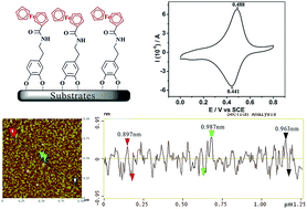
Self-assembly of catecholic ferrocene was studied on different surfaces. The self-assembly kinetics of Fc-terminated (Fc-dopamine) self-assembled monolayers (SAMs) and their stability have been studied and characterized using atomic force microscopy (AFM) and cyclic voltammetry (CV), respectively. AFM images have revealed that the self-assembly process of Fc-dopamine molecules on a mica surface follows the systematic increase of surface coverage with assembly time until a closely packed density is reached. The redox behavior of the Fc-dopamine monolayer in NaClO4 electrolyte solutions was characterized using CV, and the stability of the Fc-dopamine SAMs on an Au surface at different pH values and voltages was evaluated. CV results show that the Fc-dopamine SAMs are stable over a scan voltage range of −0.8–1.0 V under pH values lower than 11, but very rapidly destroyed above pH 11. Finally, the wetting behavior of the Fc-dopamine grafted on rough surfaces is tuned by a redox reaction of the Fc group in SAMs, which exhibits superhydrophobicity with a static water contact angle of 161° on anodized alumina surfaces, and hydrophilicity with a CA of 5° after Fc is oxidized. The work provides useful information for understanding the adhesion and deposition mechanisms of catecholic compounds on substrates.


Introduction

Self-assembled monolayers (SAMs) refer to a single layer of molecules on substrate surfaces, which are molecular assemblies formed spontaneously by chemisorption of molecular constituents from either the vapor or liquid phase and exhibit a high degree of ordered orientation and packing density.1 The attachment of a functional self-assembled monolayer (SAM) onto various substrates via tight covalent bonding is a considerably attractive approach for altering the surface/interface properties, that has potential applications in a variety of fields, such as biological/chemical sensing,2 photolithography,3 photovoltaic devices,4 corrosion protection,5 wetting control and catalysis,6,7 etc. Ferrocene-terminated self-assembled monolayers (Fc-SAMs) are one of the most studied molecular aggregates on metal substrates8 and have attracted considerable interest over the past decade due to the attractive electrochemical characteristics of ferrocene (fast electron-transfer rate, low oxidation potential, and stability).9,10 Its introduction in organic monolayers can also be of interest for the development of devices for electrocatalysis, electroanalysis, biosensing, and so on.11,12 Fc-SAMs have been used extensively as convenient, robust, and well-reproducible surfaces to study the kinetics and thermodynamics of electron transfer processes, the properties of the electrical double layer, and the mechanisms of electron transfer.13–15

Generally, SAM molecules consist of a head group, tail group and functional end group. The common head groups include thiols, silanes, phosphonates, etc.,16 but there are still limitations for present strategies because these anchoring chemistries can only bind to their suitable substrates17 (e.g., thiols to noble metals,18 silanes to silicon,19 and phosph(on)ates to metal oxides20). Furthermore, the stability of these SAMs depends on the strong chemisorption of the “head groups”. So, it is still desirable to look for a universal anchor with excellent stability for wide substrate applicability. Recently, many research groups have reported that catechols and their derived compounds can strongly bind to various inorganic and organic materials, including noble metals, metals, metal oxides, mica, silica, ceramics, etc.21,22 The good adhesion of biomolecules to catecholic compounds23,24 has stimulated our great interest in exploiting catechols as the “head groups” for enhancing interfacial adhesion so as to functionalize the target surfaces.

Compared to the large number of literature reports about ferrocene-terminated thiol- and disulfide-based gold,25 and ferrocene-terminated monolayers covalently bound to silicon surfaces,26 Fc-terminated catecholic molecule-based substrates have not yet been reported up to now. The catecholic group of Fc-dopamine can enable the self-assembly of the molecule on various substrates. Fc can be easily oxidized via chemical and electrochemical methods and the redox behavior of Fc/Fc+ can be studied using cyclic voltammetry (CV). Meanwhile, Fc-dopamine can also be used to control the wettable behavior of the surface smartly using the redox reaction, due to the difference in wettability between Fc and Fc+. Here, we have prepared a biomimetic Fc-dopamine terminated monolayer with a thickness of less than 10 Å and evaluated the stability of the self-assembled monolayers of Fc-dopamine on Au at different pH values and voltages. The self-assembly behavior of the Fc-dopamine monolayer on a mica surface with assembly time was studied and characterized using atomic force microscopy (AFM). Finally, the wettable behavior of the Fc-dopamine based surfaces was investigated and the superhydrophobic and superhydrophilic transformations have been achieved by tuning the valence state of the Fc. This work addresses the issues that are central to our understanding of the assembly and application of catecholic compounds.

Experimental section

Materials

3-Hydroxytyramine hydrochloride (99%) was purchased from Aldrich. Ferrocenecarboxylic acid (98%) was used as received from Alfa Aesar. Dicyclohexylcarbodiimide (DCC, 98%) and 4-dimethylamino-pyridine (DMAP, 99%) were used as received from the Chemical Reagent Co. of J&K Chemical Ltd (Beijing, China). Pyridine (China) was purified by distillation over NaOH. Ultrapure water used in all experiments was obtained from a NANOpure Infinity system from Barnstead/Thermolyne Corporation. Other reagents were used as received. The gold substrates were obtained by sputtering gold on a Si wafer using a K975X turbo evaporator and cleaned using oxygen plasma for 10 s before use.

Experiment

1. Synthesis of the Fc-dopamine compound. A solution of dopamine (1.89 g, 10 mmol), 4-dimethylaminopyridine (DMAP) (61 mg, 0.5 mmol) and ferrocenecarboxylic acid (2.30 g, 10 mmol) in pyridine (30 mL) was degassed for 30 min under Ar flow, and then dicyclohexylcarbodiimide (DCC) (2.27 g, 11 mmol) was added. The reaction mixture was stirred for 24 h at 45 °C under Ar protection. After the reaction finished, the precipitates were filtered off and the crude reaction product was extracted with ethyl acetate (3 × 50 mL). The combined organic extracts were washed with 1 M HCl solution and brine, dried over magnesium sulfate, filtered, and the solvent was evaporated under reduced pressure to give a brown solid. The crude product was purified using silica gel column chromatography to give a yellow solid powder (2.26 g, 6.2 mmol, yield 62%) of the Fc-dopamine compound (Scheme 1).
image file: c5ra03915k-s1.tif
Scheme 1 The synthetic pathway to the Fc-dopamine compound.

1H-NMR (400 MHz, CDCl3), (ppm): 7.90 (s, 0.5H), 6.73 (s, 1H), 6.71 (d, J = 8.4 Hz, 1H), 6.61 (dd, J = 8.0, 1.6 Hz, 1H), 4.74 (t, J = 1.6 Hz, 2H), 4.36 (t, J = 1.6 Hz, 2H), 4.12 (s, 5H), 3.48 (t, J = 7.2 Hz, 2H), 2.75 (t, J = 7.2 Hz, 2H).

13C-NMR (100 MHz, CDCl3), (ppm): 173.6, 146.5, 145.0, 132.3, 121.2, 117.1, 116.6, 77.0, 71.8, 71.0, 69.4, 42.5, 36.3.

HRMS (ESI+): for C19H20FeNO3 (M + H)+: calculated: 366.0787, found: 366.0774.

2. Substrate modification with Fc-dopamine. Au substrates were immersed in a 1 mg mL−1 solution (ultrapure water[thin space (1/6-em)]:[thin space (1/6-em)]ethanol (v/v) = 3[thin space (1/6-em)]:[thin space (1/6-em)]2) of Fc-dopamine in the dark at room temperature for 6–48 h under Ar atmosphere. The modified Au substrates were rinsed by repeated washing with ultrapure water and ethanol, then dried by nitrogen flow. Fc-dopamine SAMs on mica and Al2O3 nanowire substrates27 were performed in the same way.

Characterization

All electrochemical measurements were performed using a three-electrode cell (10 cm3) on a CHI660B electrochemical work station.28 Platinum wire was used as the counter electrode, a saturated calomel electrode was used as the reference electrode, and the Fc-dopamine SAM-modified Au substrate acted as the working electrode (the real surface area of the working electrode was 1 cm × 1 cm). CV measurements were recorded in a 0.1 M NaClO4 solution (at different pH) over the potential range between −1.0 and 1.1 V. Before the electrochemical measurements, the solutions were deoxygenated by bubbling argon gas for 30 min. All electrochemical measurements were carried out inside a homemade Faraday cage at room temperature (25 ± 2 °C) and under a constant flow of argon.

1H (400 MHz) and 13C (100 MHz) Nuclear Magnetic Resonance (NMR) spectra were recorded on a 400 MHz spectrometer (Bruker AM-400) using CDCl3 as the solvent at room temperature. Chemical composition information about the samples was obtained using X-ray photoelectron spectroscopy (XPS), which was carried out on a PHI-5702 multi-functional spectrometer using Al Kα radiation and the binding energies were referenced to the C 1s line at 284.8 eV from adventitious carbon. HRMS spectra were obtained using a Bruker Daltonics microTOF-QII mass spectrometer. The surface topography of the Fc-dopamine monolayer on mica was monitored using atomic force microscopy (AFM) (Nanoscope III, Digital Instruments Co.) with tapping mode in air atmosphere. Sessile water-droplet contact angle (CA) values were acquired using a DSA-100 optical contact-angle meter (Kruss Company, Ltd, Germany) at ambient temperature.

Results and discussion

Scheme 2 describes a self-assembly process of Fc-dopamine molecules onto the surface of substrates. The assembly rule and the structure of the Fc-dopamine monolayer were characterized in detail as follows.
image file: c5ra03915k-s2.tif
Scheme 2 Fc-dopamine molecules forming an ordered monolayer on substrates via self-assembly.

Prior to electrochemical investigation, the self-assembly behavior of the Fc-dopamine SAMs on mica was studied using AFM. In order to validate the immobilization of the Fc-dopamine SAMs on the mica surface, the surface morphology was observed using AFM. As shown in Fig. 1A, the fresh mica displays a very flat and smooth surface favorable for binding functional layers with a root-mean-square (rms) roughness of 0.052 nm for an area of 1 μm2. After modification with Fc-dopamine (48 h), small grains with heights ranging from 0.7 to 1 nm are seen uniformly distributed over the whole mica surface, and the rms roughness of 0.236 nm was obtained from the AFM image in Fig. 1C as a result of the formation of the SAMs. The line profile on the image (Fig. 1D) shows the periodicities of the adsorbed molecules, which indicates that the mean thickness of the Fc-dopamine SAMs is 0.95 nm. The thickness is close to the value expected from the thickness of the dopamine monolayer (0.8 nm).29 Therefore, we can conclude that the Fc-dopamine monolayer was formed onto the mica substrate.


image file: c5ra03915k-f1.tif
Fig. 1 AFM topography images of the bare mica (A), and Fc-dopamine monolayer-modified mica (C), and the corresponding topographic AFM profiles taken along the line on the right (B and D).

Moreover, the assembly time can influence the surface topography of Fc-dopamine SAMs. Different periods of time from 6 h to 48 h were selected to observe the self-assembly process of the Fc-dopamine monolayer. As shown in Fig. 2, after incubation in the Fc-dopamine solution for 24 h, the mica surface appears to be homogeneously covered with Fc-dopamine (rms roughness of 0.248 nm for an area of 1 μm2, Fig. 2D), and the dispersive Fc-dopamine nano-grains (Fig. 2B) gradually link up into a huge archipelago of islands with increased assembly time (Fig. 2D and E). The rms roughness values gradually increased (0.166 nm for 6 h, 0.255 nm for 12 h, 0.248 nm for 24 h, and 0.236 nm for 48 h). Meanwhile, the degree of coverage of Fc-dopamine on the mica surface increases gradually with increasing assembly time. On the basis of these observations in AFM images, the degree of coverage of Fc-dopamine is greatly affected during the initial stage of self-assembly within 24 h (Fig. 2B–D). After 24 h, the surface coverage was nearly unchanged (Fig. 2E), which means that the self-assembly process finished in 24 h and the mica surface was homogeneously covered with the Fc-dopamine monolayers. Moreover, the results have also reflected that the change in the degree of surface coverage with assembly time is merely transverse variation, and the thickness of the Fc-dopamine monolayer was always smaller than 1 nm. So, these AFM images have revealed that the self-assembly process of the Fc-dopamine molecules is dynamic and the grafting density of Fc-dopamine increases with increasing assembly time.


image file: c5ra03915k-f2.tif
Fig. 2 Tapping-mode AFM images (topography) in air, of (A) a bare mica substrate and Fc-dopamine monolayer modified mica at different assembly times: (B) 6 h, (C) 12 h, (D) 24 h, (E) 48 h.

Successful Fc-dopamine modification on the Au surface was first ascertained using XPS. Fig. 3 displays the XPS survey spectra of the bare Au and Fc-dopamine modified Au substrate. XPS analysis of the Fc-dopamine-modified Au surfaces reveals characteristic peaks from the Au substrate itself and from the C 1s, O 1s, N 1s, and Fe 2p core levels of the attached organic molecule. The N 1s core level spectrum of the Fc-dopamine-Au displays a peak at a binding energy of about 399.7 eV, which is attributable to the O[double bond, length as m-dash]C–N species in the monolayer of Fc-dopamine. The Fe 2p spectrum is composed of two peaks at 721.1 and 708.0 eV, corresponding to the Fe 2p3/2 and Fe 2p1/2 signals, respectively. These values are perfectly consistent with the previously published results,9,30 which confirm the attachment of the Fc groups onto the Au substrate.


image file: c5ra03915k-f3.tif
Fig. 3 The XPS survey spectra of the Au substrate (a) and Fc-dopamine modified Au substrate (b).

Ferrocene-terminated monolayer modified substrates have been widely studied as working electrodes in electrochemical measurements.31,32 Herein, the stability of the Fc-dopamine monolayers at different pH was investigated by way of cyclic voltammetry in a solution of 0.1 M NaClO4. Fig. 4 shows the typical cyclic voltammograms of the full-coverage Fc-dopamine SAMs in a 0.1 M NaClO4 solution. The symmetric oxidation and reduction peaks appear, which are attributed to the redox of the ferrocene (Fc)/ferrocenium (Fc+) moieties.33 The peaks centered at 0.47 V (half-wave potential, E1/2) are observed for the oxidation and reduction of the Fc moiety, with a peak separation of 23 mV. This peak position is within the ranges of the redox potentials of the Fc SAMs reported previously.34 In solution, Fc is oxidized at 0.41 and 0.37 V vs. SCE onto platinum and Si (111)–H, respectively. So, the positive shift of about 100 mV observed for bound Fc can be ascribed to the electron-withdrawing character of the underlying amide bonds making the Fc more difficult to be oxidized.9 The electrochemical response of the Fc-dopamine modified Au exhibits symmetric redox waves that are relatively stable in the 0.1 M NaClO4 solution in acid conditions even in pH = 0 solution (Fig. 4B). The Fc-dopamine modified Au also displays the typical cyclic voltammograms of the Fc molecule in 0.1 M NaClO4 alkaline solution (pH < 11). However, these films exhibit irreversible current peaks and instability in the 0.1 M NaClO4 solution with pH = 11 (Fig. 4A). The instability refers to the degradation of Fc-dopamine, which is due to the instability of the catecholic molecule in basic solutions (pH ≥ 11) and then OH–C bond cleavage. These results could also be verified by AFM images, Fig. 5 shows AFM topography of the Fc-dopamine–mica before and after treatment with an acid solution and alkaline solution. With the Fc-dopamine–mica immersed in pH = 3.0 aqueous solution, the AFM topographic images showed nearly no change, but after treatment with the pH 11 aqueous solution for 30 min, some grains were obviously lost due to the degradation of the phenol hydroxy groups of the catechol. All voltammograms in Fig. 4 have shown well-defined surface waves consisting of symmetric redox waves (except at pH 11), which show that the Fc-dopamine modified Au substrate is stable in pH < 11 solutions.


image file: c5ra03915k-f4.tif
Fig. 4 Cyclic voltammograms of the full-coverage Fc-terminated dopamine SAMs on a gold electrode in the 0.1 M NaClO4 solutions at different pH; (A) alkaline solution, (B) acid solution (scan rate = 100 mV s−1, 25 °C).

image file: c5ra03915k-f5.tif
Fig. 5 AFM topography images of the Fc-dopamine monolayer-modified mica (A), following treatment with pH = 3 acid solution (B) and pH = 11 alkaline solution (C).

The surface adsorption coverage of the ferrocenyl group (Γ) can be calculated using Faraday’s law,34

image file: c5ra03915k-t1.tif
where QFc+ is the surface charge density associated with ferrocene oxidation, which can be calculated from the integration of the anodic peak of the cyclic voltammograms, n is the number of electrons involved in the electron-transfer process (n = 1 for the ferrocene/ferricenium couple), F is the Faraday constant, and A is the area of the exposed Fc-dopamine Au electrode. The integration of the oxidation peak is equal to 44.0 μC cm−2 for Fc-dopamine–Au, which affords a ΓFc of 4.56 × 10⁻¹⁰ mol cm−2. The measured ΓFc is consistent with the theoretical coverage of ferrocene calculated from the close packing of ferrocene spheres.35,36 It is noteworthy that the density of oxidizable ferrocene moieties (2.7 molecules per nm2) of the Fc-dopamine–Au is comparable to that of the single-component ferrocene derivative (CH3)3N+C11SAu monolayer reported by Tulpar et al. (2.4 molecules per nm2).37

Fc-dopamine modified Au substrate as the work electrode was performed at different scan voltages to evaluate its electrochemical stability. Fig. 6 also displays the Fc electrochemical signals of the Fc-dopamine–Au in 0.1 M NaClO4 at different scan voltages. Both the presence of a redox peak of the ferrocene/ferrocenium couple and the peak separation of around 20 mV confirm that the Fc-dopamine moieties are immobilized onto the Au electrode surface.38,39 The scan voltage can affect the electrochemical performance of the Fc-dopamine monolayer. Cyclic voltammograms of the Fc-dopamine modified Au substrates were stable over the scan voltage range of 0–1.0 V and exhibited a relatively constant oxidation and reduction peak of the Fc/Fc+. From Fig. 6A, we can see clearly that with scan voltage increasing to 1.1 V, the reduction peak shifted to higher potentials (from 442 mV to 681 mV). The cyclic voltammograms show an irreversible behavior with large peak separation and significant peak currents other than the peak of Fc, indicating that the electron-transfer reaction occurs by an irreversible process. The oxidation peaks of the Fc/Fc+ appear relatively stable at the scan voltage from −0.8 V to 0.8 V (Fig. 6B), whereas the oxidation peak shifted to higher potentials as the scan voltage reduced to −1.0 V. The results have shown that the Fc-dopamine monolayer is unstable under ultra-high or low scan voltage.


image file: c5ra03915k-f6.tif
Fig. 6 Cyclic voltammograms for the Fc-dopamine monolayer on a gold electrode in 0.1 M NaClO4 solution at different scan ranges (scan rate = 100 mV s−1, 25 °C).

Redox of the Fc group in SAMs affects not only the electron transfer behavior but also the wettability behavior of the surface. The change of wettability on the Fc-dopamine SAM surface was tested by modification of alumina nanowires with microscale and nanostructures,27 and thus resulted in extreme wettability. The Fc-dopamine grafting substrate was immersed into an Fe(NO3)3 solution to get the Fc+-dopamine terminated surface. The XPS spectra of the Fc-dopamine modified alumina and Fc+-dopamine on the alumina surface are dominated by signals attributable to carbon, oxygen, and silicon. The appearance of an N 1s signal and Fe 2p peak in the multi-element spectra indicated the existence of Fc-dopamine on alumina nanowires. XPS results also confirm the chemical oxidation of Fc and the formation of Fc+ on the dopamine-Fc monolayers, as depicted in Fig. 7B, prior to oxidation, and the Fe (2p3/2) and Fe (2p1/2) peaks are present at 708.3 eV and 721.5 eV, respectively. After oxidation, both of these peaks shift to 712.1 eV and 724.8 eV, which is consistent with the change in oxidation state from 0 to +1.40 The wettability behavior of the dopamine-Fc surfaces was characterized by contact angle measurements.41 Fig. 7C and D summarize the static CA values of water droplets in contact with Fc-dopamine on the rough alumina nanowire surfaces. The CA of the Fc-dopamine monolayer coated alumina is 161° due to the hydrophobic Fc group on the head group. Once ferrocene (Fc) is oxidized to Fc+, the surface has a lower CA of 5°. The CA of water on the Fc-dopamine modified Au substrates is 65°, and changes to 33° after oxidation.


image file: c5ra03915k-f7.tif
Fig. 7 (A) XPS full survey spectra of the alumina nanowires (curve a), the Fc-dopamine-alumina nanowires before (curve b) and after (curve c) oxidation, (B) the XPS spectra of the Fc-dopamine–alumina nanowires in the Fe (2p) level regions before (curve a) and after (curve b) oxidation reaction. The photos and contact angles of water on the Fc-dopamine modified alumina nanowire surfaces before (C) and after (D) oxidation.

Conclusions

Catecholic Fc can be covalently bound to different substrate surfaces via a biomimetic method, with the ferrocene units as electrochemical probes for verifying the stability of catecholic monolayers. The various aspects of Fc-dopamine monolayer formation, assembly density, surface coverage, wetting properties, and stability have been characterized using contact angle measurements, X-ray photoelectron spectroscopy, cyclic voltammetry, and atomic force microscopy. CV of an Fc-dopamine monolayer functionalized Au electrode has exhibited a well-defined surface wave, all voltammograms have shown that the Fc-dopamine monolayer was stable over the scan range of −0.8–1.0 V, and the Fc-dopamine monolayer was destroyed under a high pH value solution (≥11). Quantitative analysis indicates a high packing density of catecholic-Fc, up to 2.7 molecules per nm2. Moreover, the wettability behavior of the Fc-terminated monolayer can be modulated via the transformation between ferrocene and ferrocenium. A suitable rough substrate of alumina nanowires modified with Fc-dopamine exhibits super hydrophobicity (CAs = 161°), and the ferrocenium-dopamine monolayer based surface turns hydrophilic with a CA of 5°. The attachment of Fc groups to a catecholic molecule opens up interesting opportunities for extending the potential application of Fc-SAMs.

Acknowledgements

This work is financially supported by the National Nature Science Foundation of China (21434009, 21204095, 21125316) and the Western Action Project, Youth Innovation Promotion Association CAS.

Notes and references

  1. J. C. Love, L. A. Estroff, J. K. Kriebel, R. G. Nuzzo and G. M. Whitesides, Chem. Rev., 2005, 105, 1103–1170 CrossRef CAS PubMed.
  2. P. R. Solanki, A. Kaushik, T. Manaka, M. K. Pandey, M. Iwamoto, V. V. Agrawal and B. D. Malhotra, Nanoscale, 2010, 2, 2811–2817 RSC.
  3. J. Y. Huang, D. A. Dahlgren and J. C. Hemminger, Langmuir, 1994, 10, 626–628 CrossRef CAS.
  4. D. Li, M. A. Ratner, T. J. Marks, C. Zhang, J. Yang and G. K. Wong, J. Am. Chem. Soc., 1990, 112, 7389–7390 CrossRef CAS.
  5. P. E. Laibinis and G. M. Whitesides, J. Am. Chem. Soc., 1992, 114, 9022–9028 CrossRef CAS.
  6. J. M. Buriak, Chem. Rev., 2002, 102, 1271–1308 CrossRef CAS PubMed.
  7. A. Ulman, Chem. Rev., 1996, 96, 1533–1554 CrossRef CAS PubMed.
  8. C. E. D. Chidsey, Science, 1991, 251, 919–922 CrossRef CAS PubMed.
  9. Y. Iizumi, H. Suzuki, M. Tange and T. Okazaki, Nanoscale, 2014, 6, 13910–13914 RSC.
  10. D. R. Staveren and N. Metzler-Nolte, Chem. Rev., 2004, 104, 5931–5985 CrossRef PubMed.
  11. B. Yu, H. Y. Hu, D. A. Wang, W. T. S. Huck, F. Zhou and W. M. Liu, J. Mater. Chem., 2009, 19, 8129–8134 RSC.
  12. Y. Song, X. Kang, N. B. Zuckerman, B. Phebus, J. P. Konopelski and S. Chen, Nanoscale, 2011, 3, 1984–1989 RSC.
  13. J. Das, K. Jo, J. W. Lee and H. Yang, Anal. Chem., 2007, 79, 2790–2796 CrossRef CAS PubMed.
  14. N. K. Devaraj, R. A. Decreau, W. Ebina, J. P. Collman and C. E. D. Chidsey, J. Phys. Chem. B, 2006, 110, 15955–15962 CrossRef CAS PubMed.
  15. J. Jalkh, Y. R. Leroux, C. Lagrost and P. Hapiot, J. Phys. Chem. C, 2014, 118, 28640–28646 CAS.
  16. J. J. Gooding and S. Ciampi, Chem. Soc. Rev., 2011, 40, 2704–2718 RSC.
  17. S. H. Hsu, D. N. Reinhoudt, J. Huskens and A. H. Velders, J. Mater. Chem., 2011, 21, 2428–2444 RSC.
  18. M. A. Mezour, I. I. Perepichka, O. Ivasenko, R. B. Lennox and D. F. Perepichka, Nanoscale, 2015, 7, 5014–5022 RSC.
  19. M. K. Chaudhury and G. M. Whitesides, Science, 1992, 255, 1230–1232 CAS.
  20. S. Tosatti, R. Michel, M. Textor and N. D. Spencer, Langmuir, 2002, 18, 3537–3548 CrossRef CAS.
  21. H. Lee, S. M. Dellatore, W. M. Miller and P. B. Messersmith, Science, 2007, 318, 426–430 CrossRef CAS PubMed.
  22. Q. Ye, F. Zhou and W. M. Liu, Chem. Soc. Rev., 2011, 40, 4244–4258 RSC.
  23. J. P. Park, I. T. Song, J. Lee, J. H. Ryu, Y. Lee and H. Lee, Chem. Mater., 2015, 27, 105–111 CrossRef CAS.
  24. Q. Ye, X. L. Wang, S. B. Li and F. Zhou, Macromolecules, 2010, 43, 5554–5560 CrossRef CAS.
  25. T. Auletta, F. C. J. M. Veggel and D. N. Reinhoudt, Langmuir, 2002, 18, 1288–1293 CrossRef CAS.
  26. B. Fabre, Acc. Chem. Res., 2010, 43, 1509–1518 CrossRef CAS PubMed.
  27. W. C. Wu, X. L. Wang, D. A. Wang, M. Chen, F. Zhou, W. M. Liu and Q. J. Xue, Chem. Commun., 2009, 1043–1045 RSC.
  28. F. Zhou, H. Y. Hu, B. Yu, V. L. Osborne, W. T. S. Huck and W. M. Liu, Anal. Chem., 2007, 79, 176–182 CrossRef CAS PubMed.
  29. Y. Liu, B. Yu, J. C. Hao and F. Zhou, J. Colloid Interface Sci., 2011, 362, 127–134 CrossRef CAS PubMed.
  30. C. M. Woodbridge, D. L. Pugmire, R. C. Johnson, N. M. Boag and M. A. Langell, J. Phys. Chem. B, 2000, 104, 3085–3093 CrossRef CAS.
  31. L. L. Norman and A. Badia, J. Am. Chem. Soc., 2009, 131, 2328–2337 CrossRef CAS PubMed.
  32. J. Zhang, Y. Zhao, C. G. Yuan, L. N. Ji, X. D. Yu, F. B. Wang, K. Wang and X. H. Xia, Langmuir, 2014, 30, 10127–10132 CrossRef CAS PubMed.
  33. G. Valincius, G. Niaura, B. Kazakeviciene, Z. Talaikyte, M. Kazemekaite, E. Butkus and V. Razumas, Langmuir, 2004, 20, 6631–6638 CrossRef CAS PubMed.
  34. L. L. Norman and A. Badia, Langmuir, 2007, 23, 10198–10208 CrossRef CAS PubMed.
  35. C. E. D. Chidsey, C. R. Bertozzi, T. M. Putvinski and A. M. Mujsce, J. Am. Chem. Soc., 1990, 112, 4301–4306 CrossRef CAS.
  36. T. Kondo, T. Kanai and K. Uosaki, Langmuir, 2001, 17, 6317–6324 CrossRef CAS.
  37. A. Tulpar and W. A. Ducker, J. Phys. Chem. B, 2004, 108, 1667–1676 CrossRef CAS.
  38. L. Y. S. Lee, T. C. Sutherland, S. Rucareanu and R. B. Lennox, Langmuir, 2006, 22, 4438–4444 CrossRef CAS PubMed.
  39. K. Uosaki, Y. Sato and H. Kita, Langmuir, 1991, 7, 1510–1514 CrossRef CAS.
  40. J. A. M. Dinglasan, M. Bailey, J. B. Park and A. Dhirani, J. Am. Chem. Soc., 2004, 126, 6491–6497 CrossRef CAS PubMed.
  41. X. M. Wang, Z. Gershman, A. B. Kharitonov, E. Katz and I. Willner, Langmuir, 2003, 19, 5413–5420 CrossRef CAS.

This journal is © The Royal Society of Chemistry 2015
Click here to see how this site uses Cookies. View our privacy policy here.