Versatility of divinylsulfone supports permits the tuning of CALB properties during its immobilization

Jose C. S. dos Santosab, Nazzoly Ruedaac, Alfredo Sanchezd, Reynaldo Villalongad, Luciana R. B. Gonçalvesb and Roberto Fernandez-Lafuente*a
aDepartamento de Biocatálisis, Instituto de Catálisis-CSIC, ICP-CSIC, C/ Marie Curie 2, Campus UAM-CSIC, Cantoblanco, 28049 Madrid, Spain. E-mail: rfl@icp.csic.es
bDepartamento de Engenharia Química, Universidade Federal do Ceará, Campus do Pici, CEP 60455-760, Fortaleza, CE, Brazil
cEscuela de Química, Grupo de Investigación en Bioquímica y Microbiología (GIBIM), Universidad Industrial de Santander, Edificio Camilo Torres 210, Bucaramanga, Colombia
dDepartment of Analytical Chemistry, Faculty of Chemistry, Complutense University of Madrid, 28040 Madrid, Spain

Received 3rd March 2015 , Accepted 7th April 2015

First published on 14th April 2015


Abstract

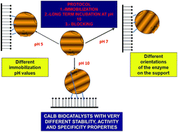
The lipase B from C. antarctica (CALB) has been immobilized on divinylsulfone (DVS) activated agarose beads under different conditions (pH 5–10). In the presence of 0.3% Triton X-100, the immobilization rate was rapid at pH 10 and the slowest one was at pH 5. Incubation at pH 10 for 72 h of the immobilized enzymes before blocking of the support with ethylenediamine permitted improvement of the enzyme stability. Enzyme features (activity, stability, specificity versus different substrates, effect of the pH on enzyme properties) were quite different on the different CALB preparations, suggesting the different orientation of the enzyme. The alkaline incubation produced an increase in enzyme activity with some substrates, and some of the DVS-CALB preparations exhibited a higher specific activity than the octyl-preparations. The indirect fluorescence spectrum of the different immobilized preparations confirmed that different structures of the CALB molecules were generated after immobilization.

1. Introduction

Lipases are the most utilized enzymes in biocatalysis1–6 due to their wide substrate specificity, high stability under a broad range of conditions and reaction media (aqueous, organic solvent, neoteric solvents)7–11 and broad range of reactions (e.g., hydrolysis, esterifications, aminations, acydolysis, transesterifications,1,6 and also other promiscuous reactions like perhydrolysis or C–C bond synthesis)12–14 that they are able to catalyze.

Moreover, lipase properties, including selectivity, specificity and activity are very easily modulated by almost any change in the enzyme or in the reaction media (including genetic manipulation,15,16 medium engineering,17 physico-chemical modification of the enzyme surface by polymers or small reagents,18–20 or via immobilization21–24). This is due to the flexibility of their active center, which is a consequence of the conformational changes that the lipases suffer during catalysis, involving the movement of an oligopeptide chain (lid or flat) that usually isolates the active center of lipases from the medium.25–28 The open form of the lipases becomes strongly adsorbed to their natural substrates (drops of oils) or any other hydrophobic surface, becoming stabilized.4,29,30

Enzyme immobilization is a prerequisite for most industrial processes, as a way to easily recover and reuse these relatively expensive biocatalysts and to avoid product contamination.31–36 Thus, the coupling of enzyme immobilization to the improvement of other enzyme features seems to be a very adequate goal in biocatalyst design, and in fact it has been reported improvement in enzyme stability, activity, selectivity, etc. upon immobilization.22,23,37–40

The tuning of lipase catalytic properties via immobilization is based on involving different regions of the enzyme on the interaction with the support and on the control of the support–enzyme interaction degree;22–24 this may generate different nano-environments on the enzyme surroundings, may distort the regions involved in the immobilization, or may just avoid some movements during the opening/closing conformational changes. This has been achieved by using different immobilization protocols, which involve different enzyme moieties in the immobilization.22–24 However, in some cases a versatile support may permit to immobilize an enzyme by different orientations by controlling the immobilization conditions.24 This is the case of heterofunctional supports, such as glutaraldehyde. This support has been used to give 4 different preparations of a lipase just by altering the ionic strength or adding detergents during immobilization.41,42

Divinylsulfone activated supports have been used for the successful immobilization of some proteins.43–51 Recently, activated divinylsulfone agarose beads have been described as a suitable support to stabilize enzymes via multipoint covalent attachment.52 The reactive group is very stable in a broad range of pH values (from 5 to 10), capable of reacting with primary and secondary amines, hydroxyl, phenyl, thiol and imidazol groups.52 However, the reactivity of each enzyme group versus the vinylsulfone support differed greatly, and also was greatly influenced by the pH value.52 At pH 10, the Lys residues are only slightly less reactive that Cys or His (the most reactive ones), while at pH 5 event he reactivity of the Tyr overpassed the reactivity of Lys residues.52 Thus, altering the immobilization conditions, it is possible to immobilize an enzyme via different orientations on supports activated with divinylsulfone.52 The further long time incubation at alkaline pH value permitted to increase the number of enzyme–support linkages, increasing the enzyme rigidity.52

In this paper, we show the results obtained in the immobilization under different conditions of the most popular lipase, the lipase B from Candida antarctica,53,54 on agarose beads activated with divinylsulfone with the objective of checking the possibility of using the features of this support to alter the catalytic properties of lipases. To this goal, the hydrolytic activity versus structurally different substrates and the stability of the immobilized enzyme under different conditions will be studied. Finally, we will try to correlate the changes in enzyme function after immobilization on the same support but following different protocols with changes in the lipase structure for the first time in the literature.

2. Materials and methods

2.1. Materials

Lipase B from Candida antarctica (CALB) was kindly donated by Novozymes (Spain), p-nitrophenyl butyrate (p-NPB), divinylsulfone (DVS), Triton X-100, ethylenediamine (EDA), 8-anilino-1-naphthalenesulfonic acid (ANS), 2-mercaptoethanol, methyl mandelate, methyl phenylacetate and ethyl hexanoate were from Sigma Chemical Co. (St. Louis, MO, USA). Octyl sepharose beads 4BCL and cyanogen bromide Sepharose beads 4BCL (CNBr) were from GE Healthcare. All reagents and solvents were of analytical grade.

All experiments were performed by triplicate and the results are reported as the mean of this value and the standard deviation (usually under 10%).

2.2. Standard determination of enzyme activity

This assay was performed by measuring the increase in absorbance at 348 nm produced by the released p-nitrophenol in the hydrolysis of 0.4 mM p-NPB in 50 mM sodium phosphate at pH 7.0 and 25 °C (ε = 5150 M−1 cm−1 under these conditions). To start the reaction, 50–100 μL of lipase solution or suspension were added to 2.50 mL of substrate solution. One unit of activity (U) was defined as the amount of enzyme that hydrolyzes 1 μmol of p-NPB per minute under the conditions previously described. Protein concentration was determined using Bradford's method,55 bovine serum albumin was used as the reference.

In the studies of the effects of pH on the enzyme activity, the protocol was similar but the buffer in the measurements was changed according to the pH value: sodium acetate at pH 5, sodium phosphate at pH 6–8 and sodium borate at pH 9–10. At 25 °C, all the preparations remained fully active after incubation for several hours at any of these pH values.

2.3. Immobilization of CALB on octyl Sepharose beads

Lipase CALB was immobilized on octyl Sepharose beads at low ionic strength as previously described.56 A volume of 1.6 mL of commercial enzyme (containing 6.9 mg mL−1 of protein) was diluted in 88.4 mL of 5 mM sodium phosphate at pH 7, maintaining a 1/10 support–enzyme solution ratio, (w/v) for 60 min. Suspension and supernatant samples were withdrawn for evaluation of immobilization through enzymatic activity measurement as described above. This immobilization strategy also permitted the purification of lipases from contaminant esterases.56

2.4. Immobilization of CALB on CNBr-agarose beads

Immobilization of CALB on CNBr-agarose beads was performed following a protocol previously described for this enzyme.57 A volume of 1 mL of commercial CALB was diluted in 99 mL of 5 mM sodium phosphate at pH 7. Then, 6 g of wet CNBr support was added. After 90 min at 4 °C under stirring at 250 rpm, around 56% of lipase became immobilized on the support. The enzyme–support reaction was ended by incubating the support with 1 M ethanolamine at pH 8 for 2 h. Finally, the immobilized preparation was washed with abundant distilled water.

2.5. Immobilization of CALB on DVS-agarose beads

2.5.1. Preparation of DVS-agarose beads. 1.5 mL divinylsulfone was added to 40 mL of 333 mM sodium carbonate at pH 12.5 and stirred until the mixture becomes homogeneous, then 2 g of agarose beads was added and left under gentle agitation for 35 minutes.52 Finally, the activated support was washed with an excess of distilled water and stored at 4 °C.
2.5.2. Immobilization of CALB on DVS-agarose beads. A 10 g portion of support was suspended in 100 mL of solutions of CALB (maximum protein concentration was 1 mg mL−1) at 25 °C using 10 mM of different buffers (sodium acetate at pH 5, sodium phosphate at pH 7 or sodium carbonate at pH 10). In some instances, Triton X-100 was added. In some cases, the immobilized lipase preparations were filtered and a portion of the derivatives was incubated in 100 mL of 100 mM bicarbonate at pH 10.0 and 25 °C for 72 h. As an enzyme–support reaction end-point, all the immobilized biocatalysts were incubated in 1 M EDA at pH 10 and 25 °C for 24 h to block the remaining reactive groups on the support (this was the optimal blocking reagent using chymotrypsin and this support).52 Finally, the immobilized preparation was washed with an excess of distilled water and stored at 4 °C.

2.6. Thermal inactivation of different CALB immobilized preparations

To check the stability of the different enzyme derivatives, 1 g of immobilized enzyme was suspended in 5 mL of 50 mM sodium acetate at pH 5, sodium phosphate at pH 7 or sodium carbonate at pH 9 and at different temperatures. Periodically, samples were withdrawn and the activity was measured using pNPB. Half-lives were calculated from the observed inactivation courses.

2.7. Stability assays in the presence of dioxane

Enzyme preparations were incubated in mixtures of 70% dioxane/30% 100 mM Tris buffer at pH 7 and at different temperatures to proceed with their inactivation. Periodically, samples were withdrawn and the activity was measured using p-NPB as described above. Half-lives were calculated from the observed inactivation courses. The acetonitrile presented in the measurement samples had no significant effect on enzyme activity determination experiments.

2.8. Hydrolysis of methyl mandelate

200 mg of the immobilized preparations was added to 2 mL of 50 mM substrate in 100 mM sodium acetate at pH 5, 100 mM sodium phosphate at pH 7 or 100 mM sodium carbonate at pH 8.5 and 25 °C under continuous stirring. The conversion degree was analyzed by RP-HPLC (Spectra Physic SP 100 coupled with an UV detector Spectra Physic SP 8450) using a Kromasil C18 (15 cm × 0.46 cm) column. Samples (20 μL) were injected and eluted at a flow rate of 1.0 mL min−1 using acetonitrile/10 mM ammonium acetate (35[thin space (1/6-em)]:[thin space (1/6-em)]65, v/v) at pH 2.8 as mobile phase and UV detection was performed at 230 nm. The acid has a retention time of 2.5 minutes while the ester has a retention time of 10 minutes. One unit of enzyme activity was defined as the amount of enzyme necessary to produce 1 μmol of mandelic acid per minute under the conditions described above. Activity was determined by triplicate with a conversion ranging 20–30%, and data are given as average values.

2.9. Hydrolysis of methyl phenylacetate

200 mg of the immobilized preparations were added to 2 mL of 5 mM substrate in 100 mM buffer containing 50% CH3CN. The buffers were sodium acetate at pH 5, sodium phosphate at pH 7 and sodium bicarbonate at pH 8.5. All experiments were carried out at 25 °C under continuous stirring. The conversion degrees were analyzed by RP-HPLC (Spectra PhysicSP 100 coupled with an UV detector Spectra Physic SP 8450) using a Kromasil C18 (15 cm × 0.46 cm) column. Samples (20 μL) were injected and eluted at a flow rate of 1.0 mL min−1 using a mixture of acetonitrile: 10 mM ammonium acetate aqueous solution (35[thin space (1/6-em)]:[thin space (1/6-em)]65, v/v) and pH 2.8, as mobile phase and UV detection was performed at 230 nm. The acid has a retention time of 3 minutes while the ester has a retention time of 12 minutes. One unit of enzyme activity was defined as the amount of enzyme necessary to produce 1 μmol of phenyl acetic acid per minute under the conditions described above. The activity was determined by triplicate with a maximum conversion of 20–30%, and data are given as average values.

2.10. Hydrolysis of ethyl hexanoate

Enzyme activity was determined by using ethyl hexanoate; 200 mg of the immobilized preparations were added to 2 mL of 25 mM substrate in 50 mM buffer containing 50% CH3CN. The buffer was sodium acetate at pH 5, sodium phosphate at pH 7 and sodium bicarbonate at pH 8.5. All experiments were carried out at 25 °C under continuous stirring. The conversion degree was analyzed by RP-HPLC (Spectra Physic SP 100 coupled with an UV detector Spectra Physic SP 8450) using a Kromasil C18 (15 cm × 0.46 cm) column. Samples (20 μL) were injected and eluted at a flow rate of 1.0 mL min−1 using acetonitrile/10 mM ammonium acetate aqueous solution (50[thin space (1/6-em)]:[thin space (1/6-em)]50, v/v) and pH 3.2 as mobile phase and UV detection was performed at 208 nm. Hexanoic acid has a retention time of 3.4 minutes while the ester has a retention time of 14.2 minutes. One unit of enzyme activity was defined as the amount of enzyme necessary to produce 1 μmol of hexanoic acid per minute under the conditions described above. Activity was determined by triplicate with a maximum conversion of 20–30%, and data are given as average values.

2.11. Fluorescence studies of the different immobilized enzyme preparations

The immobilized enzyme preparations (150 mg) were mixed with 15 mL of 13.5 μM 8-anilino-1-naphthalenesulfonic acid (ANS) solution in 10 mM Tris–HCl buffer, pH 7.0. The mixtures were incubated at 25 °C during 1 h under magnetic stirring. The samples were centrifuged and the emission fluorescence spectra of the supernatant solutions were recorded after excitation at 360 nm by using a Cary Eclipse Spectrophotometer (Varian).58

3. Results

3.1. Immobilization of CALB on divinylsulfone activated agarose beads at different pH values

Fig. 1 shows the immobilization courses of CALB at pH 5, 7 and 10. It should be remarked that free CALB remained fully active under all assayed conditions (not shown results). Surprisingly, the immobilization was very rapid in all cases, even though at pH 5 the reactivity of most nucleophilic groups of a protein versus vinylsulfone should be quite reduced.52 Furthermore, an increase in enzyme activity after immobilization was appreciated, approximately 50% in the 3 cases.
image file: c5ra03798k-f1.tif
Fig. 1 Immobilization courses of CALB at pH 5, 7 and 10 on DVS-agarose. Experimental conditions are detailed in Section 2. Circles, solid black line: suspension pH 5; circles, solid dash line: supernatant pH 5; square, solid black line: suspension pH 7; square, dash line: supernatant pH 7; triangles, solid black line: pH10 suspension; triangles, solid dash line: supernatant pH 10.

These facts could be explained if the enzyme was immobilized via another mechanism, such as physical adsorption. This could be, for example, the interfacial activation of the lipase in the fairly hydrophobic divinylsulfone layer on the agarose surface.56 This hydrophobicity feature of the support was not detected using chymotrypsin.52 Fig. 2 shows the structure of the activating group.45,51 This group is moderately hydrophobic, so that a dense layer of this group may enable interfacial activation of the enzyme.56 To check if any physical adsorption could be the cause of the immobilization of CALB, the reactive groups in the support were blocked by incubation with 2-mercaptoethanol or destroyed by incubation at pH 12 and 50 °C.52 These unreactive supports were incubated in the presence of CALB and even though the effects on enzyme activity were not identical, the immobilization rates remained pH independent and were very similar to those of the activated support (results not shown). After these treatments, it has been described that aminoacids cannot immobilize on the support, because their chemical reactivity has been destroyed, and the immobilization of the enzyme confirmed that the covalent attachment was not the first step in the immobilization of CALB on DVS activated agarose in the previous experiments.


image file: c5ra03798k-f2.tif
Fig. 2 Structure of the activated support.

3.2. Effect of Triton X-100 on the immobilization of CALB on divinylsulfone support beads

A detergent is able to desorb the enzyme from a hydrophobic support, even a very hydrophobic one, and may be used to prevent the lipase immobilization via interfacial activation.59,60 By progressively adding Triton X-100 to the DVS-support and the lipase suspension, it was possible to reduce the adsorption of the enzyme on the inactivated support (Fig. 3). Using 0.3% detergent, CALB did not immobilize on any of the inactivated supports. These results confirmed that the immobilization on this support could be founded on the interfacial activation of CALB on the fairly hydrophobic surface of the support. In fact, if the enzymes adsorbed on the reactive (neither blocked nor incubated at pH 12) DVS support at pH 5 or 7 were incubated in the presence of detergent just after immobilization, more than 80% of the enzyme released from the support. When this experiment was performed on the preparation at pH 10, less than 10% of the immobilized enzyme was released, showing that most of the enzyme was covalently attached to the support (although it is not clear which one is the first step of the immobilization; covalent attachment or interfacial activation; at least a 10% of the enzyme molecules is not covalently immobilized after 3 hours but it is already immobilized).
image file: c5ra03798k-f3.tif
Fig. 3 Effect of Tritón X-100 on the immobilization of CALB on inactivated DVS-supports. The support was incubated 24 h in 0.1 M NaOH to destroy the vinylsulfone groups. Experimental conditions are detailed in Section 2. Circles, solid black line: supernatant without Tritón X-100; squares, solid black line: supernatant with 0.05% Tritón X-100; triangles, solid black line: supernatant with 0.15% Tritón X-100; rhombus, solid black line: supernatant with 0.3% Tritón X-100.

Thus, a new batch of CALB immobilizations on DVS-agarose was carried out at pH 5, 7 and 10, but in the presence of enough detergent to prevent lipase adsorption on the inactive DVS support (0.3% Triton X-100) (Fig. 4). Immobilization was relatively rapid at pH 10 (full immobilization after 3 h). At pH 7, immobilization was slower (70% after 24 h) and even slower still at pH 5 (under 30% after 24 h). These results fitted better with the expected chemical reactivity of the enzyme groups at different pH values versus the DVS activated support.52


image file: c5ra03798k-f4.tif
Fig. 4 Immobilization courses of CALB on DVS-supports in presence of 0.3% Triton at pH 5, 7 and 10. Experimental conditions are detailed in Section 2. Circles, solid black line: suspension pH5; circles, solid dash line: supernatant pH 5; square, solid black line: suspension pH 7; square, dash line: supernatant pH 7; triangles, solid black line: pH 10 suspension; triangles, solid dash line: supernatant pH 10.

Looking at the activity, the immobilization at pH 10 produced an increase in enzyme activity (around 30%) while at the other pH values, the activity slightly decreased after immobilization. This higher activity at pH 10 is curious, as it may not be due to a lower intensity of the enzyme–support reaction.52

To enhance immobilization yields, a ratio of 1 g of support to 3 mL of enzyme suspension was used. Under these conditions CALB immobilization was almost complete even at pH 5 after 24 h (results not shown).

3.3. Effect of the long term incubation at alkaline pH value on enzyme activity

After immobilization, and in order to favor the multipoint covalent immobilization, the three immobilized CALB biocatalysts (immobilized at pH 5, 7 or 10) were incubated at pH 10 for 72 h, after washing the detergent. Results are shown in Fig. 5.
image file: c5ra03798k-f5.tif
Fig. 5 Effect of the long term incubation at pH 10 value on enzyme activity on CALB immobilized on DVS agarose at different pH values: Experimental conditions are detailed in Section 2. Circles, solid black line: pH 5; square, solid black line: pH 7; triangles, solid black line: pH 10.

The preparation immobilized at pH 10 increased the activity for 48 h, and later kept that value constant (near 170%).

The enzyme immobilized at pH 7 suffered an increase in the activity during the alkaline incubation (around 220%), and this effect was even more relevant if the enzyme had been immobilized at pH 5 (over 250%). The most active preparations were those incubated at pH 10 in all cases (Table 1), even though under these conditions a higher enzyme–support chemical reaction should occur. This increase in enzyme activity upon incubation at alkaline pH values could be explained as a function of enzyme distortions caused by the enzyme–support reaction that, in this case, presented positive effects on enzyme activity.

Table 1 Activities of the different CALB preparations versus p-NPB. DVS-CALB was blocked using EDA. Activity was determined at pH 7 and 25 °C as indicated in Section 2. Activity is given in μmol of substrate hydrolyzed per minute and mg of immobilized enzyme. The preparation of the biocatalyst is in Section 2
Biocatalysts Activity
DVS-pH 5-EDA 7.79 ± 1.7
DVS-pH 5/pH 10-EDA 22.3 ± 2.2
DVS-pH 7-EDA 20.44 ± 2.9
DVS-pH 7/pH 10-EDA 27.15 ± 2.5
DVS-pH 10 (2 h) EDA 23.79 ± 1.91
DVS-pH 10 (72 h)-EDA 32.15 ± 1.94
Octyl 16.92 ± 2.16
CNBr 5.90 ± 1.17


In order to compare the enzyme properties after immobilization, CALB was also immobilized on octyl agarose and CNBr agarose. The enzyme immobilized on octyl agarose (results not shown) presented less than 60% of the activity of the enzyme immobilized on DVS support and incubated at pH 10. It should be considered that the small lid of CALB makes that the activity of the enzyme is not significantly increased after immobilization on octyl agarose (around a 10%). The enzyme immobilized on CNBr agarose did not significantly alter its activity (Table 1).

3.4. Characterization of the immobilized biocatalysts

The 6 new covalent preparations have been compared against each other and also with the two standard immobilization protocols, CALB immobilized on CNBr- and octyl-Sepharose.
3.4.1. Activity/pH versus pNPB. Table 1 shows the activities of the 8 preparations under standard conditions after blockage. The hyperactivation caused by the alkaline incubation at pH 10 is clearly visualized, the enzyme immobilized at pH 5 started with 2 fold less activity than the enzyme immobilized at pH 10, but after alkaline incubation, the higher increase on enzyme activity permitted to almost equilibrate the observed activities. All of them (except the enzyme just immobilized at pH 5) are more active than the octyl preparation, which is also slightly more active than the CNBr preparation.

Fig. 6 shows the enzyme activity/pH profile using the different immobilized samples. The main difference is found when comparing the enzymes immobilized on different supports. The enzyme immobilized on CNBr-Sepharose presented the maximum of activity at pH 7, with a sharp decrease at either alkaline or acidic pH values (activity was around 40% at pH 5 and 10). Using octyl agarose as support, the maximum activity was found at pH 8, and the decrease in activity at acidic and alkaline pH values is milder (55% at pH 5 and 70% at pH 10). The enzyme immobilized on DVS support under different conditions presented the maximum activity at the highest pH used in the study (pH 10), and only slight differences were found on the immobilization pH or long term incubation at alkaline pH value. The enzyme immobilized at pH 5 showed an 18% or 25% of the maximum activity at pH 5, for the non-incubated or long term incubated enzyme preparations respectively. Both enzyme preparations immobilized at pH 7 exhibited 40% of the maximum activity at pH 5, while the preparations immobilized at pH 10 showed around 30% of this activity.


image file: c5ra03798k-f6.tif
Fig. 6 Effect of the pH on the activity versus pNPB of the different CALB preparations. Experimental conditions are detailed in Section 2. Circles, solid black line: pH 5; gray circles, solid gray line: pH 5–10; squares, solid black line: pH 7; grays squares, solid gray line: pH 7–10; triangles, solid black line: pH10; gray triangles, solid gray line: pH 10–10. stars, solid black line: octyl; gray stars, solid gray line: CNBr.
3.4.2. Thermal stability under different conditions at different pH values. Table 2 shows the half-lives of the different CALB preparations under different inactivation conditions. We only show the results obtained in the temperature where the inactivations have a rate that permitted to obtain reliable results in a reasonable time.
Table 2 Half-lives (expressed in minutes) of the different CALB preparation under different inactivation conditions. Experiments were performed as described in Section 2. * The enzyme retained full activity during the inactivation assay
CALB preparation Inactivation conditions
pH 5, 55 °C pH 7, 55 °C pH 9, 55 °C 70% dioxane, 25 °C, pH 7
DVS-pH 5-EDA 4.5 ± 0.3 3 ± 0.3 4.5 ± 0.3 5 ± 0.3
DVS-pH 5–pH 10-EDA 35 ± 1.2 10 ± 1.1 33 ± 1.9 5.3 ± 0.7
DVS-pH 7-EDA 33 ± 1.0 33 ± 2.2 27 ± 2.1 1.5 ± 0.2
VS-pH 7–pH 10-EDA 60 ± 2.4 60 ± 3.3 60 ± 3.2 7.3 ± 0.3
DVS-pH 10-EDA 32 ± 1.3 4 ± 0.2 4.2 ± 0.4 1.7 ± 0.2
DVS-pH 10–pH 10-EDA 46 ± 2.2 25 ± 1.2 25 ± 1.2 2.6 ± 0.4
Octyl 240 (100%)* 240 (100%)* 30 ± 2.1 0.17 ± 0.02
CNBr 45 ± 3.3 24 ± 2.3 4.6 ± 0.3 0.21 ± 0.02


The most stable preparation was that obtained using octyl-agarose when the inactivations were performed at pH 5 or 7. The just immobilized DVS preparations were far less stable, but their stabilities improved after long-term incubation to favor multipoint covalent attachment, becoming more stable than the CNBr-CALB in all cases.

If the inactivation was performed at pH 5, the alkaline incubation increased the half live from 4.5 to 35 minutes for the enzyme immobilized at pH 5, if the immobilization was performed at pH 7, the stability increased to a lower extent, from 33 to 60 minutes. The value of the half live of the enzyme immobilized at pH 10 went from 32 to 46 minutes after the long term incubation, a value lower than that obtained with the immobilization at pH 7 and incubated at pH 10.

The pattern was somehow similar looking at the inactivations carried out at pH 7, the enzyme immobilized at pH 7 and incubated at alkaline conditions was the most stable one, followed by the enzyme immobilized at pH 10 and the enzyme immobilized at pH 5.

At pH 9, the situation varied. The enzyme immobilized at pH 7 presented a stability similar to that of the octyl, and the alkaline incubation of this preparation permitted to double the half-life. The stabilities of the enzymes immobilized at pH 5 or 10 were quite similar, both after immobilization and after long term alkaline incubation before blocking. In both cases, the stability became similar to that of the octyl-CALB after the alkaline incubation. It may be likely that at pH 9 the cause of the inactivation is a conformational change in another area of the enzyme or just a chemical modification of some groups, this can explain the significant qualitative change in the stability of the different preparations.

Considering that in all cases the support was the same for the DVS immobilized enzymes, and that the long term incubation of 3 days should permit a similar reaction between the enzyme and the support, the differences on enzyme stability must be related to populations of enzyme molecules having different orientations, with different relevance for enzyme stability61,62 or different density of groups able to react with the support, giving differences in the final intensity of the multipoint covalent attachment.22

The high thermostability of the lipases adsorbed on hydrophobic supports has been previously described.63 These preparations are much more stable than the glyoxyl agarose-CALB, and this was explained by the very stable conformation that the open form of the adsorbed lipases presented,64 and the moderate amount of nucleophilic groups that many lipases presented in its surface make complex a very intense multipoint covalent attachment (e.g., CALB has 9 Lys plus the Leu 1, all of them exposed to the medium).65,57

3.4.3. Solvent stability. In opposition to the results obtained during thermal inactivations, Table 2 shows that in all cases the DVS preparations were by far more stable that the octyl or CNBr-Sepharose immobilized enzymes when they were incubated in the presence of 70% dioxane. Analyzing the DVS preparations blocked just after immobilization, the most stable biocatalyst was that prepared at pH 5 (half live of 5 minutes), being the stability of the enzymes immobilized at pH 7 and 10 very similar (1.5–1.7 minutes). However, after the long term incubation the enzyme immobilized at pH 7 greatly improved the stability (to more than 7 minutes), while the enzyme immobilized at pH 5 maintained its stability practically unaltered after alkaline incubation and the enzyme immobilized at pH 10 improved its the stability by only 50%.

The low stability of CALB immobilized on octyl-agarose in the presence of dioxane may be related to the enzyme desorption caused by the presence of this very high cosolvent concentration, the free enzyme is rapidly inactivated under these drastic conditions.52,66

The different stability of the enzymes immobilized at different pH value on DVS activated supports, where after long term alkaline incubation the only difference may be the enzyme orientation, suggests that the inactivation of CALB follows a different route on different inactivation conditions, some protein regions are more relevant on the stability at certain conditions, while some other areas may be more relevant on other experimental conditions.61,62,67

3.4.4. Activity versus different esters. Immobilization has been reported to alter enzyme specificity and the influence of the pH on the activity, if enzyme orientation on the support or the intensity of the enzyme–support interaction is different.22,24 Thus, differences in enzyme specificity or influence on activity/pH curve upon different immobilization protocols can reinforce the idea on a different enzyme orientation on the support surface. Three different substrates have been used at 3 different pH values: esters formed by an aliphatic acid (ethyl hexanoate), one aromatic acid (phenylacetate) or one aromatic and chiral one (mandelic acid) and the results are resumed on Table 3.
Table 3 Activity of different CALB preparations versus different substrates at different pH values. Experimental details may be found in Section 2. MM, methyl mandelate; MPA, methyl phenylacetate; EH, ethyl hexanoate. The activity is given in μmol of substrate hydrolyzed per minute and mg of immobilized enzyme
CALB preparations MM/pH 5 MM/pH 7 MM/pH 8.5 MPA/pH 5 MPA/pH 7 MPA/pH 8.5 EH/pH 5 EH/pH 7 EH/pH 8.5
Octyl 16.45 ± 0.8 55.00 ± 2.8 41.07 ± 2.1 14.02 ± 0.7 24.27 ± 1.2 19.17 ± 1 450.00 ± 23 300.00 ± 15 273.44 ± 14
CNBr 28.25 ± 1.1 124.15 ± 5.0 85.61 ± 3.4 15.32 ± 0.6 30.54 ± 1.2 19.00 ± 0.8 627.85 ± 25 436.65 ± 17 197.44 ± 8
DVS-pH 5 11.61 ± 0.6 58.78 ± 2.9 23.28 ± 1.2 6.97 ± 0.3 3.94 ± 0.2 2.39 ± 0.1 200.89 ± 10 139.18 ± 7 30.97 ± 2
DVS-pH 5–pH 10 39.6 ± 0.9 82.32 ± 1.1 35.08 ± 1.8 2.86 ± 0.1 1.38 ± 0.1 1.95 ± 0.1 74.40 ± 4 194.20 ± 10 50.22 ± 3
DVS-pH 7 52.17 ± 2.6 57.07 ± 2.9 52.41 ± 2.6 22.47 ± 1.1 25.64 ± 1.3 5.43 ± 0.3 760.87 ± 38 456.52 ± 23 188.52 ± 9
DVS-pH 7–pH 10 56.13 ± 2 78.80 ± 3.9 86.43 ± 1.9 9.34 ± 0.5 9.29 ± 0.5 3.36 ± 0.2 217.39 ± 11 157.07 ± 8 142.66 ± 7
DVS-pH 10 12.50 ± 0.6 67.92 ± 3.5 51.07 ± 2.6 18.65 ± 0.9 17.59 ± 0.9 8.13 ± 0.4 425.00 ± 21 191.25 ± 10 24.38 ± 1
DVS-pH 10–pH 10 8.35 ± 0.4 69.00 ± 3.5 29.11 ± 1.5 6.47 ± 0.3 6.68 ± 0.3 2.71 ± 0.1 62.50 ± 3 41.25 ± 2 38.53 ± 2


Using ethyl hexanoate, results are quite diverse depending on the biocatalyst. The highest activity was usually found at pH 5, except for the preparation immobilized at pH 5 and then incubated at alkaline pH, where the maximum activity was found at pH 7. The enzyme just immobilized at pH 7 on DVS was the most active one at pH 5 and pH 7 while at pH 8.5 the most active one was the octyl-Sepharose preparation. The long term incubation at alkaline pH of the DVS preparations usually decreased the enzyme activity, mainly at pH 5. The enzyme immobilized at pH 5 is the one with the most drastic change after alkaline incubation, with a shift in the maximum activity at pH 7 (becoming more active than the enzyme just immobilized at pH 5 under these conditions, that is, alkaline incubation produced an hyper-activation at pH 7). On the other hand, the enzyme immobilized at pH 5 and at pH 10 improved the activity after alkaline incubation if the activity was determined at pH 8.5. In general, the effect of the change of the pH in the activity determination presented a more drastic effect on DVS preparations without long term alkaline incubation (e.g., from 425 U mg−1 to 24 U mg−1 using the enzyme immobilized at pH 10) than in octyl or CNBr preparations (activity at pH 8.5 was around 60% and 30% than that at pH 5, respectively). Long term incubation at alkaline pH reduced this effect of the pH on DVS-CALB activity.

Using methyl phenylacetate, at pH 5 the most active preparations are two DVS preparations, those just immobilized at pH 7 (22.5 U mg−1) and pH 10 (18.7 U mg−1). At pH 8.5, octyl and CNBr CALB preparations presented the highest activity, while at pH 7 the most active preparations were CNBr and DVS immobilized at pH 7. The lowest activity for all preparations immobilized on DVS was that found at pH 8.5, except for the enzyme immobilized at pH 5 on DVS and submitted to alkaline incubation that have the minimum activity at pH 7. The highest activity depended on the immobilization protocol. The DVS preparations immobilized at pH 5 had a clear maximum at pH 5; while both preparations immobilized at the other two pH values have not a clear maximum (similar activities are detected at pH 5 and 7). Octyl and CNBr CALB had a clear maximum at pH 7. Long term alkaline incubation decreased enzyme activity in all cases, but the intensity of this effect depended on the immobilization pH and activity determination pH.

Using mandelic ester, new changes were found. The most active preparations at pH 5 were both preparations immobilized at pH 7 on DVS, at pH 7 the most active preparations were the CNBr preparation and the enzyme immobilized at pH 5 or pH 7 and long term submitted to alkaline incubation before blocking. At pH 8.5, the most active biocatalysts were those immobilized at pH 7 and long term incubated and the CNBr preparation. There are examples where the highest activity was found at pH 7 (octyl, CNBr, both DVS immobilized at pH 5 and both immobilized at pH 10). The enzyme immobilized at pH 7 has not a clear maximum activity, and after incubation this optimum is clearly at pH 8.5. The long term incubation of the DVS preparations used to have a positive effect on enzyme activity, except when the enzyme was immobilized at pH 10, where the alkaline incubation decreased the enzyme activity when measured at pH 5 or 8.5, while having almost no effect at pH 7.

Thus, CALB immobilized following different protocols on DVS-activated supports (different immobilization pH values, long term incubation or not under alkaline conditions) presented very different enzyme specificity and very different response to changes on environmental conditions, confirming that the different preparations have different orientation and/or degree of enzyme–support interaction.68

3.4.5. Evaluation of the structure of different CALB immobilized preparations. The influence of the different immobilization strategies on the 3D conformation of the enzyme was determined by using the ANS-binding fluorescence assays. ANS is a hydrophobic fluorescent dye that strongly binds the clusters from hydrophobic amino acid side chains in β-sheet conformations of proteins.59 Usually, a great density of those hydrophobic clusters is well protected from the solvent in native enzymes due to the rigid packing of the globular protein conformation. Accordingly, a decrease in the fluorescence intensity of the ANS dye can be attributed to its binding to the exposed hydrophobic regions in partially unfolded proteins.

Fig. 7 shows the fluorescence emission spectra of the biocatalysts prepared through different immobilization protocols. In comparison with the raw support (line a), the fluorescence intensity of ANS decreased after incubation with all immobilized lipase preparations. This fact can be ascribed to the presence of the enzyme molecules on the support surface, and thus, to the binding of ANS molecules to the exposed hydrophobic clusters in these proteins. On the other hand, the results obtained using the biocatalyst prepared by immobilization at pH 5 and further incubation at 10 (line c) showed the lowest fluorescence signal, much lower than using the enzyme immobilized at pH 5 (line b). This result suggested that the immobilization approach based on two consecutive incubation steps at pH 5 and 10 leads to protein conformations with partially exposed hydrophobic β-sheet clusters, and accordingly, more prone to bind the hydrophobic ASN molecules, than when the enzyme is just immobilized at pH 5 and them blocked. That is, alkaline incubation produced conformational changes on the enzyme that led to the exposition of more hydrophobic groups to the medium.


image file: c5ra03798k-f7.tif
Fig. 7 Spectra of ANS incubated in the presence of different DVS immobilized CALB. Experimental conditions are detailed in Section 2. Line (a) blocked DVS-support; line (b) DVS-CALB-pH 5, line (c) DVS-CALB pH 5 + 72 h at pH 10, line (d) DVS-CALB pH 10; line (e) DVS-CALB pH 10 + 72 h at pH 10.

When the enzyme is immobilized at pH 10 (line d), the effect of the further alkaline incubation is in the opposite direction (line e), the fluorescence signal increased after the alkaline incubation, less hydrophobic groups are partially exposed suggesting a more rigid and compact structure. Again, the changes in enzyme properties could be correlated to conformational changes.

Moreover, it is clear that the difference in the exposition of protein hydrophobic groups of the enzyme immobilized at pH 5 and that immobilized at pH 10, in both cases after 72 h of incubation at pH 10 before support blocking is quite significant, with much higher exposition using the enzyme immobilized at pH 5 and incubated at pH 10. The results may be explained by the implication of different areas of the enzyme in the multipoint covalent attachment. This produced fully different effects on the enzyme structure (making more compact one and more relaxed the other), The effects on the exposition of the hydrophobic groups surrounding the active of the lipase (the small lid and adjacent areas) may be also considered. These differences may explain the drastic changes of enzyme properties when immobilized at different pH values discussed along this paper, and suggest that the areas reacting with the support for those 72 h could be different.

4. Conclusions

Immobilization of CALB on DVS-supports under different conditions permits to have covalently immobilized preparations exhibiting very different properties. The change in the immobilization pH permits to alter the enzyme specificity, activity and stability, while further incubation under alkaline conditions (described as a way to improve the enzyme support reaction)52 also produced changes in enzyme features. The indirect determination of the ANS incubated enzyme fluorescence showed that the different enzyme derivatives have different structures.

Thus, DVS activated supports may be a potent way to tuning lipase properties via immobilization. The DVS activation of supports compatible with organic media may increase the range of reactions where the biocatalysts may be used and provide new data on the different behavior of CALB immobilized on different supports.

Acknowledgements

We gratefully recognize the support from the MINECO of Spanish Government, by the grant CTQ2013-41507-R. The predoctoral fellowships for Ms Rueda (Colciencias, Colombian Goberment) and Mr dos Santos (CNPq, Brazil) are also recognized. The authors wish to thank Mr Ramiro Martínez (Novozymes, Spain) for kindly supplying the enzymes used in this research. The help and comments from Dr Ángel Berenguer (Instituto de Materiales, Universidad de Alicante) are kindly acknowledged.

References

  1. K. E. Jaeger and T. Eggert, Curr. Opin. Biotechnol., 2002, 13, 390–397 CrossRef CAS.
  2. R. Sharma, Y. Chisti and U. C. Banerjee, Biotechnol. Adv., 2001, 19, 627–662 CrossRef CAS.
  3. K. E. Jaeger and M. T. Reetz, Trends Biotechnol., 1998, 16, 396–403 CrossRef CAS.
  4. R. D. Schmid and R. Verger, Analysis, 1998, 37, 1608–1633 Search PubMed.
  5. A. Pandey, S. Benjamin, C. R. Soccol, P. Nigam, N. Krieger and V. T. Soccol, Biotechnol. Appl. Biochem., 1999, 29(pt 2), 119–131 CAS.
  6. J. Zhang, H. Shi, D. Wu, Z. Xing, A. Zhang, Y. Yang and Q. Li, Process Biochem., 2014, 49, 797–806 CrossRef CAS PubMed.
  7. P. Adlercreutz, Chem. Soc. Rev., 2013, 42, 6406–6436 RSC.
  8. P. Lozano, J. M. Bernal, G. Sánchez-Gómez, G. López-López and M. Vaultier, Energy Environ. Sci., 2013, 6, 1328 CAS.
  9. P. Lozano, J. M. Bernal and A. Navarro, Green Chem., 2012, 14, 3026 RSC.
  10. P. Lozano, J. M. Bernal and M. Vaultier, Fuel, 2011, 90, 3461–3467 CrossRef CAS PubMed.
  11. Y. Fan and J. Qian, J. Mol. Catal. B: Enzym., 2010, 66, 1–7 CrossRef CAS PubMed.
  12. C. Li, X.-W. Feng, N. Wang, Y.-J. Zhou and X.-Q. Yu, Green Chem., 2008, 10, 616 RSC.
  13. E. Busto, V. Gotor-Fernández and V. Gotor, Chem. Soc. Rev., 2010, 39, 4504–4523 RSC.
  14. M. Kapoor and M. N. Gupta, Process Biochem., 2012, 47, 555–569 CrossRef CAS PubMed.
  15. N. J. Turner, Nat. Chem. Biol., 2009, 5, 567–573 CrossRef CAS PubMed.
  16. U. T. Bornscheuer and M. Pohl, Curr. Opin. Chem. Biol., 2001, 5, 137–143 CrossRef CAS.
  17. J. M. Palomo, G. Fernandez-Lorente, C. Mateo, C. Ortiz, R. Fernandez-Lafuente and J. M. Guisan, Enzyme Microb. Technol., 2002, 31, 775–783 CrossRef CAS.
  18. R. C. Rodrigues, Á. Berenguer-Murcia and R. Fernandez-Lafuente, Adv. Synth. Catal., 2011, 353, 2216–2238 CrossRef CAS PubMed.
  19. C. Garcia-Galan, J. C. S. dos Santos, O. Barbosa, R. Torres, E. B. Pereira, V. C. Corberan, L. R. B. Gonçalves, R. Fernandez-Lafuente and L. R. B. Goncalves, Process Biochem., 2014, 49, 604–616 CrossRef CAS PubMed.
  20. J. C. S. dos Santos, C. Garcia-Galan, R. C. Rodrigues, H. B. de Sant'Ana, L. R. B. Gonçalves and R. Fernandez-Lafuente, Enzyme Microb. Technol., 2014, 60, 1–8 CrossRef CAS PubMed.
  21. C. Garcia-Galan, Á. Berenguer-Murcia, R. Fernandez-Lafuente and R. C. Rodrigues, Adv. Synth. Catal., 2011, 353, 2885–2904 CrossRef CAS PubMed.
  22. K. Hernandez and R. Fernandez-Lafuente, Enzyme Microb. Technol., 2011, 48, 107–122 CrossRef CAS PubMed.
  23. R. C. Rodrigues, C. Ortiz, Á. Berenguer-Murcia, R. Torres and R. Fernández-Lafuente, Chem. Soc. Rev., 2013, 42, 6290–6307 RSC.
  24. O. Barbosa, R. Torres, C. Ortiz, A. Berenguer-Murcia, R. C. Rodrigues and R. Fernandez-Lafuente, Biomacromolecules, 2013, 14, 2433–2462 CrossRef CAS PubMed.
  25. A. M. Brzozowski, U. Derewenda, Z. S. Derewenda, G. G. Dodson, D. M. Lawson, J. P. Turkenburg, F. Bjorkling, B. Huge-Jensen, S. A. Patkar and L. Thim, Nature, 1991, 351, 491–494 CrossRef CAS PubMed.
  26. K. K. Kim, H. K. Song, D. H. Shin, K. Y. Hwang and S. W. Suh, Structure, 1997, 5, 173–185 CrossRef CAS.
  27. K. E. Jaeger, S. Ransac, H. B. Koch, F. Ferrato and B. W. Dijkstra, FEBS Lett., 1993, 332, 143–149 CrossRef CAS.
  28. M. Cygler and J. D. Schrag, Biochim. Biophys. Acta, 1999, 1441, 205–214 CrossRef CAS.
  29. R. Verger, Trends Biotechnol., 1997, 15, 32–38 CrossRef CAS.
  30. C. Cambillau, S. Longhi, A. Nicolas and C. Martinez, Curr. Opin. Struct. Biol., 1996, 6, 449–455 CrossRef CAS.
  31. I. Chibata, T. Tosa and T. Sato, J. Mol. Catal., 1986, 37, 1–24 CrossRef CAS.
  32. W. Hartmeier, Trends Biotechnol., 1985, 3, 149–153 CrossRef CAS.
  33. E. Katchalski-Katzir, Trends Biotechnol., 1993, 11, 471–478 CrossRef CAS.
  34. J. F. Kennedy, E. H. M. Melo and K. Jumel, Chem. Eng. Prog., 1990, 86, 81–89 CAS.
  35. A. M. Klibanov, Science, 1983, 219, 722–727 CAS.
  36. D. Brady and J. Jordaan, Biotechnol. Lett., 2009, 31, 1639–1650 CrossRef CAS PubMed.
  37. R. Fernandez-Lafuente, Enzyme Microb. Technol., 2009, 45, 405–418 CrossRef CAS PubMed.
  38. C. Mateo, J. M. Palomo, G. Fernandez-Lorente, J. M. Guisan and R. Fernandez-Lafuente, Enzyme Microb. Technol., 2007, 40, 1451–1463 CrossRef CAS PubMed.
  39. M. Petkar, A. Lali, P. Caimi and M. Daminati, J. Mol. Catal. B: Enzym., 2006, 39, 83–90 CrossRef CAS PubMed.
  40. U. Guzik, K. Hupert-Kocurek and D. Wojcieszyńska, Molecules, 2014, 19, 8995–9018 CrossRef PubMed.
  41. O. Barbosa, R. Torres, C. Ortiz and R. Fernandez-Lafuente, Process Biochem., 2012, 47, 1220–1227 CrossRef CAS PubMed.
  42. O. Barbosa, C. Ortiz, Á. Berenguer-Murcia, R. Torres, R. C. Rodrigues and R. Fernandez-Lafuente, RSC Adv., 2014, 4, 1583 RSC.
  43. F. J. Lopez-Jaramillo, M. Ortega-Munõz, A. Megia-Fernandez, F. Hernandez-Mateo and F. Santoyo-Gonzalez, Bioconjugate Chem., 2012, 23, 846–855 CrossRef CAS PubMed.
  44. P. Prikryl, J. Lenfeld, D. Horak, M. Ticha and Z. Kucerova, Appl. Biochem. Biotechnol., 2012, 168, 295–305 CrossRef CAS PubMed.
  45. M. Ortega-Muñoz, J. Morales-Sanfrutos, A. Megia-Fernandez, F. J. Lopez-Jaramillo, F. Hernandez-Mateo and F. Santoyo-Gonzalez, J. Mater. Chem., 2010, 20, 7189 RSC.
  46. J. C. Begara-Morales, F. J. López-Jaramillo, B. Sánchez-Calvo, A. Carreras, M. Ortega-Muñoz, F. Santoyo-González, F. J. Corpas and J. B. Barroso, BMC Plant Biol., 2013, 13, 61 CrossRef CAS PubMed.
  47. A. L. Medina-Castillo, J. Morales-Sanfrutos, A. Megia-Fernandez, J. F. Fernandez-Sanchez, F. Santoyo-Gonzalez and A. Fernandez-Gutierrez, J. Polym. Sci., Part A: Polym. Chem., 2012, 50, 3944–3953 CrossRef CAS PubMed.
  48. K. Labus, A. Turek, J. Liesiene and J. Bryjak, Biochem. Eng. J., 2011, 56, 232–240 CrossRef CAS PubMed.
  49. J. Bryjak, J. Liesiene and B. N. Kolarz, Colloids Surf., B, 2008, 61, 66–74 CrossRef CAS PubMed.
  50. M. D. Bale Oenick, S. J. Danielson, J. L. Daiss, M. W. Sunderberg and R. C. Sutton, Ann. Biol. Clin., 1990, 48, 651–654 CAS.
  51. J. Morales-Sanfrutos, J. Lopez-Jaramillo, M. Ortega-Muñoz, A. Megia-Fernandez, F. Perez-Balderas, F. Hernandez-Mateo and F. Santoyo-Gonzalez, Org. Biomol. Chem., 2010, 8, 667–675 CAS.
  52. J. C. S. dos Santos, N. Rueda, O. Barbosa, J. F. Fernández-Sánchez, A. L. Medina-Castillo, T. Ramón-Márquez, M. C. Arias-Martos, M. C. Millán-Linares, J. Pedroche, M. del, M. Yust, L. R. B. Gonçalves and R. Fernandez-Lafuente, RSC Adv., 2015, 5, 20639–20649 RSC.
  53. E. M. Anderson, K. M. Larsson and O. Kirk, Biocatal. Biotransform., 1998, 16, 181–204 CrossRef CAS.
  54. V. Gotor-Fernández, E. Busto and V. Gotor, Adv. Synth. Catal., 2006, 348, 797–812 CrossRef PubMed.
  55. M. M. Bradford, Anal. Biochem., 1976, 72, 248–254 CrossRef CAS.
  56. R. Fernandez-Lafuente, P. Armisén, P. Sabuquillo, G. Fernández-Lorente and J. M. Guisán, Chem. Phys. Lipids, 1998, 93, 185–197 CrossRef CAS.
  57. O. Barbosa, M. Ruiz, C. Ortiz, M. Fernández, R. Torres and R. Fernandez-Lafuente, Process Biochem., 2012, 47, 867–876 CrossRef CAS PubMed.
  58. G. V Semisotnov, N. A. Rodionova, O. I. Razgulyaev, V. N. Uversky, A. F. Gripas' and R. I. Gilmanshin, Biopolymers, 1991, 31, 119–128 CrossRef PubMed.
  59. K. Hernandez, C. Garcia-Galan and R. Fernandez-Lafuente, Enzyme Microb. Technol., 2011, 49, 72–78 CrossRef CAS PubMed.
  60. C. Garcia-Galan, O. Barbosa, K. Hernandez, J. C. S. dos Santos, R. C. Rodrigues and R. Fernandez-Lafuente, Molecules, 2014, 19, 7629–7645 CrossRef PubMed.
  61. J. Mansfeld and R. Ulbrich-Hofmann, Biotechnol. Appl. Biochem., 2000, 32(pt 3), 189–195 CrossRef CAS.
  62. J. Mansfeld, G. Vriend, B. Van Den Burg, V. G. H. Eijsink and R. Ulbrich-Hofmann, Biochemistry, 1999, 38, 8240–8245 CrossRef CAS PubMed.
  63. J. M. Palomo, G. Muñoz, G. Fernández-Lorente, C. Mateo, R. Fernández-Lafuente and J. M. Guisán, J. Mol. Catal. B: Enzym., 2002, 19–20, 279–286 CrossRef CAS.
  64. G. H. Peters, O. H. Olsen, A. Svendsen and R. C. Wade, Biophys. J., 1996, 71, 119–129 CrossRef CAS.
  65. M. Galvis, O. Barbosa, M. Ruiz, J. Cruz, C. Ortiz, R. Torres and R. Fernandez-Lafuente, Process Biochem., 2012, 47, 2373–2378 CrossRef CAS PubMed.
  66. G. Fernandez-Lorente, M. Filice, D. Lopez-Vela, C. Pizarro, L. Wilson, L. Betancor, Y. Avila and J. M. Guisan, J. Am. Oil Chem. Soc., 2010, 88, 801–807 CrossRef.
  67. V. Grazú, F. López-Gallego, T. Montes, O. Abian, R. González, J. A. Hermoso, J. L. García, C. Mateo and J. M. Guisán, Process Biochem., 2010, 45, 390–398 CrossRef PubMed.
  68. J. M. Palomo, G. Fernández-Lorente, C. Mateo, M. Fuentes, R. Fernández-Lafuente and J. M. Guisan, Tetrahedron: Asymmetry, 2002, 13, 1337–1345 CrossRef CAS.

This journal is © The Royal Society of Chemistry 2015
Click here to see how this site uses Cookies. View our privacy policy here.