Giacomo Prampolini*a,
Ivo Cacelliab and
Alessandro Ferrettia
aIstituto di Chimica dei Composti OrganoMetallici (ICCOM-CNR), Area della Ricerca, via G. Moruzzi 1, I-56124 Pisa, Italy. E-mail: giacomo.prampolini@pi.iccom.cnr.it
bDipartimento di Chimica e Chimica Industriale, Universitá di Pisa, via G. Moruzzi 3, I-56124 Pisa, Italy
First published on 7th April 2015
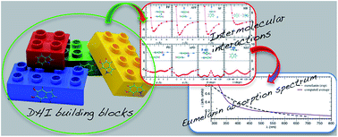
The non-covalent interactions between pairs of the smallest eumelanins building blocks, 5,6-dihydroxy-indole (DHI) and its redox derivatives, are subjected to a systematic theoretical investigation, elucidating their nature and commenting on some of their possible effects on the layered structure of eumelanin. An accurate yet feasible protocol, based on second order perturbation theory, was set up and validated herein, and thereafter used to sample the intermolecular potential energy surfaces of several DHI related dimers. From the analysis of the resulting local minima, the crucial role of stacking interactions is assessed, evidencing strong effects on the geometrical arrangement of the dimer. Furthermore, the absorption spectra of the considered dimers in their most stable arrangements are computed and discussed in relation to the well known eumelanin broadband features. The present findings may help in elucidating several eumelanin features, supporting the recently proposed geometrical order/disorder model (Chen et al., Nat. Commun. 2014, 5, 3859).
Notwithstanding the many theoretical17–38 and experimental2,5,13,33,39–50 studies, the structure–property relationships underpinning the above mentioned multipurpose applications are still far from clearly understood, and the patterns of supramolecular assembly or even the chemical nature of eumelanin constituting molecules are still under debate.7,35,36 The reason for this has been clearly pointed out in a very recent review by D’Ischia et al.,7 who traced back the difficulties in establishing a detailed picture of eumelanin structure to the fact that “unlike the vast majority of natural pigments, eumelanins cannot be described in terms of a single well-defined structure, and it is not possible to provide an accurate picture beyond a statistical description of main units and functional groups”. More specifically, since the original amorphous semiconductor model39 was definitively abandoned in light of experimental evidence of a certain degree of supramolecular structure,40,41,43,49,51,52 many of eumelanin’s peculiar characteristics (and in particular the broadband absorption spectra) were explained through the so-called chemical disorder model,3,4,27 where eumelanins are constituted by a plethora of distinct species, which may differ in structure, redox state or geometrical arrangement. In this scenario, most of the resulting eumelanin properties can be rationalized as a consequence of such disorder: the broadband absorption, for instance, might be interpreted as a superposition of the spectra of the multiple constituents of eumelanin. The main drawback of the chemical disorder model is undoubtedly the neglect of both the non-covalent interactions occurring among the various eumelanin constituents and the effect that the supramolecular motifs that originate from these interactions might have on the optical and mechanical properties of eumelanin, as reported in many investigations.18,23,36–38,48 Very recently, Chen et al.36,38 have complemented the chemical disorder model with the geometrical order/disorder picture, where the existence of stacked layers containing a number of eumelanin forming units (protomolecules) and the conformational and configurational disorder within and among them is also taken into account (see also Scheme 3 in ref. 7). Despite the elusive nature of this description, several points seem now to be shared by the scientific community:1–7 (i) the basic2 unit components (see also Fig. 1) of natural and synthetic eumelanins are 5,6-dihydroxy-indole-2-carboxylic acid (DHICA), 5,6-dihydroxy-indole (DHI), its redox form indole-quinone (IQ) and its tautomer quinone-imine (MQ); (ii) the above units are linked through covalent C–C bonds into small oligomers (protomolecules), ranging from tetramers to octamers, although a monomeric-based picture has also been recently proposed;34 (iii) to date, the most probable and investigated oligomeric structures are those of a cyclic, porphyrin-like planar tetramer and linear or cross linked oligomers. It might be interesting to note that the former structures were mainly deduced from computational results,26 whereas the latter were hypothesized, based on experimental measurements;46,47 (iv) whatever their structure, the protomolecules are arranged into stacked layers, with a typical separation of 3–4 Å; (v) the dimensions of these stacked aggregates are nanometric and no long-range orientational or positional order was found among the layers. Additionally, it is most probable that, within each layer, protomolecules can adopt a variety of geometrical conformations and/or redox states.
![]() | ||
Fig. 1 Monomeric species involved in eumelanins2,7 that are considered in the present work: 5,6-dihydroxy-indole (DHI), its redox forms semi-quinone (SQ) and indole-quinone (IQ) and its tautomer quinone-imine (MQ). |
Based on these few points, it appears that it is now of fundamental importance to deeply understand and characterize the non-covalent interactions among protomolecules that are responsible for both the formation of the stacked layers and the tertiary structure arising from their spatial disposition. In this context, the potential of a computational approach can be exploited to unravel the inherent complexity of the supramolecular structure of eumelanin. Surprisingly, to our knowledge, only a few attempts to thoroughly investigate non-covalent interactions in eumelanins have been reported to date in the literature. Indeed, apart from two recent works, where quantum mechanical (QM) methods, such as density functional theory (DFT)31 and its tight binding extension combined with the self-consistent charge technique (SCC-DFTB),35 were employed, most of the computational studies dealing with interactions between protomolecules were based on empirical, transferable force-fields (FFs),23,36,38 whose accuracy in handling non-covalent forces has been recently questioned.53–55 Moreover, all previous studies dealt only with a few spatial arrangements of the protomolecules. On the contrary, considering the crucial role of the non-covalent interactions that may occur between eumelanin building blocks, the effect of their relative orientation as well as the possible balance between van der Waals (stacked) and hydrogen bond (HB) forces should be also taken into account. Therefore, a more systematic though accurate investigation appears to be necessary.
The first step towards a deeper comprehension of the structural motifs of protomolecules consists of devising a computational method able to accurately investigate the interactions between pairs of the smallest eumelanin building blocks, i.e. DHI and its redox (IQ) and tautomeric (MQ) forms. This is the main objective of the present work. Considering the aromatic character of all species considered, and the presence of one or more –OH functional groups, it is not unlikely that the most favorable arrangements of such pairs will be driven by a delicate balance between stacked (π–π) and HB interactions. The most accurate and reliable QM method to handle such non-covalent forces is probably the coupled cluster technique, evaluated at complete basis set (CCSD(T)@cbs), often referred to as the gold standard of quantum chemistry.56 Unfortunately, the prohibitive computational cost of this technique rules out its adoption in sampling a large number of non-covalent dimer conformations. Computational convenience would suggest to turn to DFT dispersion corrected calculations, but the accuracy of the results is known to depend on the choice of the functional and its empirical correction.57,58 In a rather time-consuming procedure, some purposely computed CCSD(T)@cbs data could be used as a reference to select the most accurate empirical dispersion correction,57,59–61 although it has been recently reported that the performances of a given functional may vary dramatically with the target dimer and even with its spatial arrangement.62,63
In this scenario, an alterative approach was adopted in our group, exploiting the combination of the second order Möller–Plesset perturbation theory (MP2) with purposely modified basis sets (therefore named64 MP2mod). Very recently, the MP2mod method was successfully applied by us64,65 to a non-covalent dimer, similar to those encountered in eumelanins, namely quinhydrone. As a matter of fact, MP2mod results obtained for quinhydrone agreed well with reference high-level CCSD(T)@cbs data, whereas, thanks to the reduced basis set dimensions, the observed computational times were remarkably reduced with respect to the other investigated methods.64 Given the similarities (also recently pointed out in ref. 34) between quinhydrone’s component monomers and the eumelanin building blocks considered here, MP2mod is expected to give good performances in this case also. Nonetheless, for a more robust assessment of the accuracy of the method, it is here again validated against CCSD(T)@cbs data, purposely computed on a few selected arrangements. Subsequently, MP2mod is employed to calculate the interaction energy of different non-covalent pairs arising from the combination of the considered eumelanin minimal building blocks. Finally, the effect of the resulting dimer conformations on the shape of the absorption spectrum is also considered and discussed in some detail.
The interaction potential energy surface (IPES) of each investigated pair was first sampled by computing the interaction energy in several dimer arrangements, constructed without altering the internal (B3LYP/cc-pVTZ) geometry of each monomer. In this case, the interaction (ΔEinter) and binding (ΔEbind) energies between monomers A and B have the same expression:
ΔEinter = EAB − (E0A + E0B) = ΔEbind | (1) |
Next, several local minima of each considered IPES were further explored by optimizing the dimer geometries corresponding to the most representative arrangements found in the previous sampling. In this case, since the monomer geometries may change during the full optimization, ΔEinter and ΔEbind may differ, i.e.
ΔEinter = EAB − (EA + EB) | (2) |
ΔEbind = EAB − (E0A + E0B) = ΔEinter + (ΔEA + ΔEB) | (3) |
ΔEM = EM − E0M; M = A, B | (4) |
For each investigated AB dimer geometry, the computation of the interaction energies was performed at two different levels of theory.
(i) For selected arrangements (either obtained by assembling the two rigid monomers or through dimer optimization) ΔEinter was computed according to eqn (2), at the CCSD(T)@cbs56 level, following the procedure employed by us for quinhydrone,64 which is reported in detail in the ESI.†
(ii) All considered dimer pairs were investigated through the MP2mod method,64 computing both ΔEinter and ΔEbind according to eqn (2) and (3), respectively. All calculations were performed with 6-31G*(αC, αO, αN), where αC, αO and αN are the modified polarization exponents for carbon, oxygen, and nitrogen atoms, respectively. The first two exponents, namely 0.25 and 0.44, were transferred from the ones optimized for quinhydrone,64 whereas αN was optimized in this work through the EXOPT64,66,67 procedure against the reference CCSD(T)@cbs data obtained for pyrrole, as detailed in the ESI† and in the next section.
Finally, the absorption vertical transition energies for the first 25 excited states were obtained for eumelanin related monomers and dimers at the TD-DFT level, using the B3LYP functional with the aug-cc-pVDZ basis set. Absorption spectra were subsequently obtained by broadening each transition with a Gaussian function with an half-width at half maximum of 0.33 eV. All calculations were performed with the Gaussian09 package.68
As far as o-hydroquinone derivatives are concerned, the polarization exponents for carbon (0.25) and oxygen (0.44) atoms were transferred from previous work performed on a very similar dimer (quinhydrone).64 To test this assumption, o-benzoquinone/o-hydroquinone pairs were prepared in three different geometries (see Fig. A of the ESI†): face-to-face (FF), antiparallel face-to-face (AFF) and hydrogen bonded (HB). The intermolecular distance between the centers of mass of the monomers was varied stepwise in the 2.8–7.0 Å range, and the interaction energy was computed for each of the resulting dimer arrangements at the MP2mod/6-31G*(0.25, 0.44) level. From the computed curves, reported in detail in Fig. B of the ESI,† two main observations may be made. First, as could be expected, it is clear from the difference between the MP2mod energies and their HF contributions that the dispersion forces routed in the dynamic electron correlation dominate the stacked wells (FF and AFF), and play only a minor (but not negligible) role in the H-bonded (HB) dimers. As a consequence, the FF and AFF arrangements are much more favorable than the HB one.
Starting from the most stable AFF geometry, a complete energy optimization was carried out at the MP2mod level. The final arrangement (dimer I) is shown in the left panel of Fig. 2. The interaction energy ΔEinter of this geometry has been computed according to eqn (2) at both the MP2mod and CCSD(T) levels, as reported in Table 1. For a more extended validation, a second optimization was also carried out at the MP2mod level, starting with the HB minimum conformation. As could be expected from the comparison of the AFF and HB curves (ESI, Fig. B†), the HB dimer optimization ends up in a stacked conformation (dimer II), shown in the right panel of Fig. 2. In contrast with dimer I, in dimer II one hydroquinone hydrogen is found out of the aromatic plane, allowing for the formation of two weak hydrogen bonds, which concur to further stabilize the stacked geometry. MP2mod and CCSD(T) interaction energies for dimer II were then computed, and are compared to each other in Table 1. For both dimers, the agreement between MP2mod and CCSD(T) reference values is rather good, with a ΔEinter difference less than 0.5 kcal mol−1, and the adoption of the αC and αO exponents to handle o-hydroquinone derivatives appears reliable.
![]() | ||
Fig. 2 Optimized geometries of the o-benzoquinone/o-hydroquinone pair computed at the MP2mod level, starting from the AFF (dimer I, left) and HB (dimer II, right) arrangements, displayed in Fig. A of the ESI.† Hydrogen bonds in dimer II are highlighted with green dashed lines. |
Dimer | Method | Basis set | ΔEinter (kcal mol−1) | ΔEbind (kcal mol−1) |
---|---|---|---|---|
Dimer I | CCSD(T) | cbs | −5.34 | — |
Dimer I | MP2mod | 6-31G*(0.25, 0.44) | −5.80 | −5.35 |
Dimer II | CCSD(T) | cbs | −9.54 | — |
Dimer II | MP2mod | 6-31G*(0.25, 0.44) | −9.91 | −7.04 |
Since no α exponent was available for the nitrogen atom in pyrrole, αN was optimized following the procedure adopted for quinhydrone in ref. 64, and discussed in some detail in the ESI.† The optimized polarization exponent for the nitrogen atom resulting from the EXOPT minimization is 0.37, a value not far from that found for the oxygen atom in hydroquinone (0.44).64 The pyrrole IPES was scanned according to selected cross sections, described in Fig. C of the ESI.† From looking at Fig. D in the ESI,† the MP2mod results match well with the scenario that emerged from previous calculations performed on the pyrrole dimer and reported in the literature (see ref. 69 and references therein). Indeed, the stacked face-to-face (FF) arrangements are found not to be stable, whereas the antiparallel displaced one shows a significant well (∼3.5 kcal mol−1) at rather short ring–ring distances (∼3 Å). More importantly, the most favorable conformations (>5 kcal mol−1) among the four investigated arrangements are those where a hydrogen bond between the –NH group of one monomer and the π cloud of the other is established. To further check the reliability of the MP2mod protocol, two additional dimer optimizations were performed, starting from the two most favorable geometries (T-shaped (TS) and APD, see Fig. C and D in ESI†). The final structures, labeled dimer I and II, are reported in Fig. 3. In the first case, the two monomers are found in a T-shaped like arrangement, where the hydrogen of on the –NH group strongly interacts with the π cloud of the neighboring monomer, the distance with the aromatic plane being around 2 Å. However, because the HF contribution is only slightly attractive (−0.5 kcal mol−1), dispersion forces still play the major role in dimer stabilization. As far as dimer II is concerned, the planar rings are found to be in a stacked conformation, though displaced by ∼2 Å and with the NH bonds pointing in opposite directions. The interaction energies were computed for both dimers at the MP2mod level with the optimized 6-31G*(0.25, 0.37) basis set, finding ΔEinter = −5.41 and −5.97 for dimer I and II, respectively. Despite these values being well within the interaction energy range (−4.0 to 6.3 kcal mol−1) found with previous high level calculations,69 the many local minima expected for the pyrrole IPES do not allow for a direct comparison. To better assess the reliability of MP2mod, the ΔEinter was computed for dimer I and II also at the CCSD(T)@cbs level, and the results are compared with their MP2mod counterparts in Table 2. It appears that the agreement with the reference values is remarkable, with the differences between the reference and MP2mod values being 0.1 kcal mol−1 and 0.5 kcal mol−1for dimer I and II geometries, respectively.
![]() | ||
Fig. 3 Optimized geometries of the pyrrole dimer computed at the MP2mod level, starting from the TS (dimer I, left) and APD (dimer II, right) arrangements, displayed in Fig. C and D of the ESI.† |
Dimer | Method | Basis set | ΔEinter (kcal mol−1) | ΔEbind (kcal mol−1) |
---|---|---|---|---|
Dimer I | CCSD(T) | cbs | −5.51 | — |
Dimer I | MP2mod | 6-31G*(0.25, 0.37) | −5.41 | −5.35 |
Dimer II | CCSD(T) | cbs | −6.52 | — |
Dimer II | MP2mod | 6-31G*(0.25, 0.37) | −5.97 | −4.77 |
Exploiting the exponent reliability found for both the o-quinone and pyrrole dimer cases, the 6-31G*(0.25, 0.44, 0.37) basis set was implemented, and the MP2mod procedure was applied in the calculations of the interaction energy between pairs of the eumelanin-related monomers displayed in Fig. 1.
DHI/IQ pairs were initially placed in eight different arrangements, devised to consider all types of intermolecular interactions (stacking, H–π, hydrogen bond, etc.) that could arise between such non-covalent dimers, i.e. stacked face-to-face (FF), anti-parallel face-to-face (AFF), side-to-face (SF), hydrogen bonded (HB), parallel displaced (PD), anti-parallel displaced (APD), stacked rotated (ST) and not stacked rotated (NST). For each arrangement, a number of dimer geometries were built by moving one monomer with respect to another along one IPES coordinate (either a distance or an angle), without changing the internal geometries of the monomer. All considered configurations and the chosen displacement coordinates are displayed in the insets of Fig. 4. Finally, for each arrangement, the IPES cross sections, also displayed in Fig. 4, were obtained at the MP2mod/6-31G*(0.25, 0.44, 0.37) level by computing the interaction energy of the proper set of configurations.
The first general observation that arises by looking at the resulting curves is that the DHI and IQ monomers show rather strong non-covalent interactions, which remarkably depend on their relative orientation, and span a ΔEinter range between −10 and −3 kcal mol−1. Notwithstanding the dominant stacking interactions, the most stable conformers are found when the molecular planes are twisted (ST, −9.51 kcal mol−1) or displaced along the plane direction (APD, −9.28 kcal mol−1). In all cases, an antiparallel arrangement of the CO dipoles is preferred to a parallel one. It is also worth noticing that in all the stacked configurations considered, the inter-ring distance corresponding to the curve minimum is found between 3.4 to 3.6 Å, a value in good agreement with the range (3–4 Å) proposed7,36,40,41 for the eumelanin inter-layer distance. Interestingly, while the lack of stacking interactions causes the HB configurations to be the least stable, the side-to-face like pairs (SF, −7.95 kcal mol−1 and NST, −8.10 kcal mol−1) also show a non-negligible interaction, although the distance between the centers of mass of the monomers is around 5 Å. As a consequence, even in the (most likely) hypothesis that eumelanin protomolecules consist of DHI related oligomers, the latter side-to-face arrangements between the building blocks of two neighboring oligomers cannot be ruled out, as they could be realized by a rotation around the C–C bond connecting two protomolecules moieties, if the gain in non-covalent energy is more than the one needed for the dihedral torsion. However, a full investigation of the subtle balance of the interaction energy between oligomer building blocks and the torsional barriers driving the internal structure of the protomolecules is beyond the aims of the present work, but will be the main topic of a future investigation currently in progress in our group.
![]() | ||
Fig. 5 Optimized geometries of the DHI/IQ pair, starting from selected minima of the APD (dimer A), HB (dimer B), NST (dimer C) and ST (dimer D) curves (see Fig. 4). The geometrical parameters reported in Table 3 are highlighted with blue and green dashed lines. |
Dimer | a (Å) | b(b′) (Å) | ΔEinterCCSD(T) (kcal mol−1) | ΔEinterMP2mod (kcal mol−1) | ΔEbindMP2mod (kcal mol−1) |
---|---|---|---|---|---|
A | 2.82 | 2.11 | −11.4 | −12.4 | −10.0 |
B | 2.80 | 1.92 | −14.4 | −15.6 | −10.7 |
C | 2.95 | 2.25 (2.54) | −11.0 | −12.4 | −10.1 |
D | 2.95 | 1.92 | −14.3 | −14.8 | −12.1 |
CPU time (hours) | ∼15![]() |
∼1 |
The comparison between the MP2mod values and those obtained using the CCSD(T)@cbs method is rather good, with a standard deviation of ∼1 kcal mol−1, i.e. less than 10% of the total ΔEinter value. It is worth noticing that the accuracy obtained with the MP2mod protocol is even more appreciable if one considers the computational times, reported in the last row of Table 3, that underline the feasibility of MP2mod calculations, even on a large number of dimer arrangements.
Once the reliability of MP2mod has been finally assessed, a closer look at the four minimum energy conformations found using MP2mod optimizations can help in understanding the type and strength of the non-covalent interactions established between DHI and IQ units. First, a general feature shared by the four pairs is the stacked arrangement of the two monomers, which is a clear indication of the fundamental role of dispersion forces. Indeed, the two molecular planes are in all cases very close, the inter-plane distance being slightly less than 3 Å. Another specific characteristic of the DHI/IQ pair is the formation of a hydrogen bond between the two units, in addition to the intramolecular one found in the DHI monomer for all A–D conformations. The intermolecular H-bond is formed through an out-of-plane rotation of one hydrogen of the –OH group around the C–OH bond. For dimers B and D, this rotation is very effective, as the intermolecular H-bond distance is found to be below 2 Å. It is worth noticing that the same kind of torsion also takes place in the quinhydrone dimer,64 and is involved in both dimer stability and electron and proton transfers between the redox pair in solution.65 Finally, by comparing the ΔEinterMP2mod and ΔEbindMP2mod values reported in Table 3 for all investigated dimers, it appears that the distortion energy [ΔEDHI + ΔEIQ] reported in eqn (4) ranges from 2 to 5 kcal mol−1, depending on dimer conformation. However, the former contribution (ΔEDHI) is more than 75% of the total distortion energy, indicating that larger distortions from the monomer equilibrium geometries are located in the DHI species, and essentially consist of the aforementioned dihedral rotation.
The starting conformations employed in the previous MP2mod optimizations, carried out on the DHI/IQ pair (that eventually led to the A–D dimers displayed in Fig. 5), were employed as starting points also for the other considered dimers, after proper addition/removal of hydrogen atoms. The final dimer arrangements (labeled E to T) are displayed in Fig. 6. By looking at these optimized conformers, and at the geometrical and energy data reported in Table 4 for all pairs, some general conclusions may be drawn.
![]() | ||
Fig. 6 From top to bottom, MP2mod-optimized geometries of the DHI/DHI (E–H), DHI/MQ (I–L), IQ/MQ (M–P) and DHI/SQ (Q–T) pairs, starting from the same initial conformations (i.e. antiparallel displaced (APD), hydrogen bonded (HB), not stacked (NST) and stacked (ST)) used in the optimization of DHI/IQ shown in Fig. 5, as indicated in the top panel. The geometrical parameters reported in Table 4 are highlighted with blue and green dashed lines. |
Dimer | a (Å) | b(b′) (Å) | ΔEinter (kcal mol−1) | ΔEbind (kcal mol−1) |
---|---|---|---|---|
DHI/DHI | ||||
E | 3.00 | 2.21 | −11.4 | −7.8 |
F | 3.10 | 2.04 | −6.9 | −4.5 |
G | 2.95 | 1.95 | −15.7 | −10.7 |
H | 2.92 | 2.16 (2.17) | −15.8 | −11.3 |
![]() |
||||
DHI/MQ | ||||
I | 2.72 | 2.00 | −21.9 | −14.3 |
J | 2.90 | 1.85 (2.90) | −10.5 | −8.0 |
K | 2.70 | 1.66 | −11.4 | −6.4 |
L | 2.70 | 2.02 | −18.5 | −13.8 |
![]() |
||||
IQ/MQ | ||||
M | 3.20 | — | −14.0 | −6.3 |
N | 2.80 | 2.51 | −11.8 | −7.1 |
O | 2.85 | — | −6.7 | −4.8 |
P | — | 2.56 | −5.9 | −5.7 |
![]() |
||||
DHI/SQ | ||||
Q | 3.14 | 2.56 | −6.8 | −4.9 |
R | — | 1.91 | −3.3 | −3.1 |
S | 2.87 | 2.92 | −11.4 | −7.2 |
T | — | 2.30 | −3.9 | −3.5 |
The first observation is that, as already noticed for the DHI/IQ pair, most of the considered optimizations end up in a stacked arrangement, independently of the starting conformation. More precisely, after optimization, only P, R and T dimers are found to not be in a stacked-like arrangement, but it should be mentioned that in all three cases the starting conformation was also not stacked. However, by looking at the ΔEbind values reported in Table 4, it appears that they are among the less stable conformers. On the contrary, dimers B, F, J and N all start from a side-by-side, H-bonded conformer (see Fig. 4, top left panel), but, during optimization, the molecular planes rotate around the intermolecular H-bond(s) and the two moieties are eventually found in an almost aligned, stacked geometry. By looking at the IQ/MQ pair, where only one or no H-bonds can be formed, it is clear that the stabilization of the stacked arrangements is ensured essentially by dispersion forces, which may contribute up to ∼8 kcal mol−1 to the pair stability. The formation of one or more H-bonds, of course, favors the stability of the dimer with an additional gain in binding energy that can be quantified, depending on the case considered, to be between 2 and 4 kcal mol−1. Furthermore, when H-bonds are formed between the two moieties, the two monomers tend to orient their long axes in an almost parallel (or antiparallel) disposition, whereas, in their absence, the molecular planes are found to be mostly twisted with respect to each other. However, by looking at the binding energies reported in Tables 3 and 4, it seems that there is no systematic trend in the strength of the non-covalent interaction exhibited by different types of monomers pairs, although DHI/IQ, DHI/DHI and DHI/MQ dimers are on average more stable than those involving IQ/MQ and, in particular, DHI/SQ.
To investigate the effects of the afore-discussed dimers on the absorption properties of eumelanin, TD-DFT calculations were performed on both the isolated monomers and their optimized combinations. The accuracy of the chosen functional (B3LYP) was validated (see Fig. E in the ESI†) by comparing the computed absorption spectrum of the DHI monomer with the available experimental data.42 The absorption spectra of all monomers considered in this work have been computed and reported in the top panel of Fig. 7. By looking at the resulting spectral lines, it appears that the differences in the redox form (DHI vs. SQ, vs. IQ) and in the geometrical arrangement of their constituent atoms (MQ vs. IQ) may cause significant shifts in the position of the strong absorption peak in the UV region and/or changes in intensity in the broad band in the visible range. In agreement with the chemical disorder model, when the separate monomer signals are averaged (see light green line in the bottom panel of Fig. 7), a broad band is formed in the UV region, whose intensity gradually increases with decreasing wavelength, as expected for eumelanin.2,3 On the contrary, not all of the visible region is covered by the average absorption band, and small or null absorption intensities are found in the 350–550 nm range, confirming that the eumelanin broadband absorption, which covers monotonically (see top panel of Fig. 10) the whole UV/visible spectrum, cannot simply originate from the superposition of the absorption lines of the non-interacting monomers.
![]() | ||
Fig. 7 Top panel: TD-DFT absorption spectra of the monomer species displayed in Fig. 1: DHI (green line), IQ (blue), SQ (orange) and MQ (magenta). Bottom panel: average of all monomer spectra (light green line) and average (red line) of the absorption spectra computed for DHI/IQ dimers A–D, displayed in Fig. 5. |
In Fig. 8 and 9 the absorption spectra obtained for the considered non-covalent dimer arrangements A to T are displayed. From looking at Fig. 8, where only the spectra relative to the DHI/IQ pair are displayed, it is evident that the formation of the stacked complex, and the consequent excitonic coupling, causes the appearance of a much more diffuse absorption band, which in some cases (dimer C) almost covers the whole visible region or presents maxima (dimer A) at wavelengths around 400–450 nm, where the separate monomers do not absorb at all. This last observation is also evidenced in Fig. 7, where the absorption band of the DHI/IQ dimer, averaged over the four considered minima (A–D), is directly compared to the one arising from the monomeric species.
![]() | ||
Fig. 8 TD-DFT absorption spectra of the DHI/IQ dimers, in the optimized conformations displayed in Fig. 5: A (black line), B (cyan), C (magenta) and D (blue). The spectra resulting from the superimposition of the former band shapes is reported with a solid red line. |
![]() | ||
Fig. 9 TD-DFT absorption spectra of the DHI related pairs in the optimized conformations displayed in Fig. 6. |
A more complete picture can be achieved by also taking into account the absorption due to the other dimer configurations E–T (see Fig. 9), built by assembling different monomer pairs. Similarly to what was previously found for the DHI/IQ dimer, all the DHI/MQ and IQ/MQ complexes (central panels of Fig. 9) show rather diffuse bands, whose position and intensity strongly depend upon the dimer arrangement. In particular, dimer M, formed from the IQ and MQ monomers, shows an intense band centered around 400 nm, a region not covered when considering all the isolated monomers. Conversely, dimers E–H and Q–T are characterized by a much reduced dependency upon the relative orientation of their constituent monomers, all bands being centered in the high energy region (∼100 nm).
Finally, it is interesting to superimpose all the computed signals, either originating from monomers or non-covalent dimers, and compare the resulting curve with the experimental3 absorption band shape of real eumelanin. The outcomes of both these operations are displayed in Fig. 10. As shown in the upper panel, the computed curve matches well with most of the characteristics of the experimental signal, i.e. the monotonic increase towards lower wavelengths and the broad band covering a great part of the investigated spectrum. Nonetheless, it is important to stress that the computed average displayed in Fig. 10 neglects the contributions to the spectral signal due to chemical heterogeneity, which comes into play when covalent combinations of different building blocks (e.g. tetramers) and their relevant redox states are also taken into account. This however goes beyond the aims of the present study, and is the object of investigations currently in progress in our group.
![]() | ||
Fig. 10 Top: experimental3 absorption spectrum of eumelanin. Bottom: absorption spectrum computed in this work by averaging the TD spectra obtained for monomers and their combinations in the discussed optimized conformations. |
Exploiting the computational feasibility of the tuned basis set, the IPES of several dimer pairs was investigated. From the information retrieved, some general conclusions on the type and extent of the most likely non-covalent interactions characterizing the structure of eumelanin may be drawn. First of all, the stacking interactions are dominant, even if some hydrogen bonds can occur in the considered pairs. The latter indeed reinforce dimer stability but are not able to alter the preferred stacked disposition. More specifically, most of the investigated building block pairs are found with their molecular planes in parallel arrangements, though often twisted or slightly displaced. The computed interaction energies are found to be remarkably dependent on the relative orientation of the monomer, spanning a rather wide range of values from 5 to 12 kcal mol−1. It might be thus speculated that such strong stacked interactions, sometimes complemented by non-negligible hydrogen bonds, should be able to promote the layered structure hypothesized for eumelanin by many authors. As a matter of fact, the extended IPES sampling carried out herein revealed that for the most stable local minima the distance between two stacked rings of the considered building blocks is found to be between 3.0 and 3.5 Å, in agreement with the 3–4 Å range proposed7,36,40,41 for eumelanin inter-layer distance.
Once the presence of closely stacked aromatic planes was confirmed by calculations, its consequences in the optical properties of the resulting non-covalent dimers were investigated. As a matter of fact, considering the rather small values found for the inter-ring separation, it can be hypothesized that the π clouds that characterize each of the two aromatic moieties will realistically overlap, hence affecting the frontier orbitals involved in the electronic transition. Such supposition was confirmed by observing, for most of the considered pairs, the appearance of a broad band upon dimer formation, whose position and intensity remarkably depend on dimer type and spatial arrangement. On the contrary, when the intermolecular interactions are completely neglected and the absorption spectrum is built as a superimposition of all the signals arising from the isolated monomers, a negligible absorption intensity is found in the 350–550 nm range, in contrast with the well known broad band features of real eumelanin. On the other hand, when the contribution of the stacked non-covalent dimers conformations is included in the calculation of the spectrum, the averaged computed signal shows two specific features that characterize the experimental absorption of eumelanin: a broad band that covers practically the whole investigated spectrum and a monotonic increase towards higher energies.
Despite the fact that these results seem to suggest that many of the considered configurations do effectively take place in eumelanin structure, the covalent bonds that may connect the investigated building blocks to form small protomolecules (as tetrameric structures) should be taken into account. Indeed, work in this direction is currently in progress in our group. In this context, the validation of a reliable and effective method to compute the non-covalent interactions in eumelanin, taking place either between the simple building blocks investigated herein or between larger DHI based oligomers, paves the way to a further step in the comprehension of eumelanin supramolecular motifs.
It should be thus pointed out that this is the first part of a multi-step work, which will hopefully lead us toward an accurate large scale simulation of a model eumelanin material. Indeed, in the present work the interaction between the minimal building blocks was investigated and the proposed computational method reliably validated: a second step will consist of extending the investigation to the interaction between larger oligomers (protomolecules). This could be done, for instance, by resorting to the idea that the interaction energy between two large protomolecules (for instance two DHI tetramers) can be expressed as a sum of the interactions between all pairs of their building blocks.70 In this hypothesis, the interaction energy between two protomolecules would strongly depend on the relative distances and orientations of their building blocks. However, unlike the case investigated here, the intramolecular flexibility of the involved oligomers should be also accounted for, since the constraints imposed by the structure of each protomolecule could realistically alter the distribution of geometrical conformations of all possible building blocks pairs. Beside the dimensions of the protomolecules, a further obstacle to such an approach is that the same level of accuracy is required to handle intramolecular flexibility and intermolecular interactions, if a correct balance is to be achieved. Therefore, the next step of this work will consist of applying to different kinds of protomolecules a number computational protocols, developed in our group70–73 to reliably investigate van der Waals dimers of large dimensions.
Finally, an accurate sampling of the six dimensional interaction potential energy surface (IPES) of the building block pairs considered here could be also exploited to validate the performances of the FFs adopted in previous studies or employed as a reference to parameterize a specifically tailored FF.66,67,74,75 Once both the achieved intra- and inter-molecular FF descriptions have been validated, the last step of this project will consist of a large-scale simulation on different eumelanin models, based on different combinations of DHI building blocks.
Footnote |
† Electronic supplementary information (ESI) available. See DOI: 10.1039/c5ra03773e |
This journal is © The Royal Society of Chemistry 2015 |