Structural, dielectric and magnetic investigations on Al3+ substituted Zn-ferrospinels

Naveen Kumari*, Vinod Kumar and S. K. Singh
Physics Department, DCR University of Science and Technology, Murthal-131039, India. E-mail: naveenmalik6@yahoo.co.in

Received 3rd March 2015 , Accepted 20th April 2015

First published on 20th April 2015


Abstract

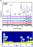
A series of ZnAlxFe2−xO4 (0.1 ≤ x ≤ 0.5) ferrospinels has been prepared by chemical co-precipitation method to understand the effect of Al3+ substitution on the structural, dielectric and magnetic response of ZnFe2O4 nanoparticles. X-ray diffraction (XRD) and transmission electron microscopy (TEM) images confirmed the nano size formation of particles. The lattice parameter (a), X-ray density (ρx) and bulk density (ρm) were found to decrease with increasing inclusion of Al3+ ions. AC conductivity (σac) measurements as a function of temperature show that the samples behave like semiconductors. Decrease in the hopping conduction between Fe2+ ↔ Fe3+ ions at octahedral site is observed with increasing inclusion of Al3+ ions. The Nyquist plots of the prepared materials reveal the inherent phenomenon involved in conduction mechanism of Al3+ substituted ZnFe2O4 ferrites. The magnetization studies revealed that magnetic moment (ηB) showed decreasing trend with increase in substitution of Al3+, its value decreases from 0.56 (for x = 0.1) to 0.34 (for x = 0.5). The Ms values decrease from 13.29 emu g−1 for x = 0.1 to 8.42 emu g−1 for x = 0.5. DM (magnetic particle size) was found to be less than the particle size calculated from TEM micrographs due to presence of magnetically dead layer on the surface of particle. Squareness (S) values infer that particles interact by magnetostatic interactions. The MH loop of all the samples is narrow with low value of coercivity and retentivity; indicates the superparamagnetic nature of prepared nanoparticles.


1. Introduction

Ferrospinels have attracted considerable attention due to their technological importance in different applications such as high frequency devices, biomedicine, catalysis, sensors, microwave absorbers, magnetic drug delivery and magnetic refrigerators.1,2 Spinel ferrites with general formula AB2O4 are widely used in many electronic and magnetic devices because of their high magnetic permeability and low magnetic loss.3 Moreover, advanced applications often require materials with magnetic properties that can be deliberately tuned by external control parameters.4,5 The wide range of applications for ferrites is attributed to their resistivity, low eddy current losses, high Curie temperature, magnetocrystalline anisotropy, reasonable cost and excellent chemical stability.6 In nanocrystalline phase magnetic, structural and electrical properties of ferrites change drastically in comparison to their bulk counterpart. This change in properties is also strongly dependent on preparation techniques, preparation conditions, site chosen for substitution and composition. Various methods (ceramic, sol–gel, co-precipitation, hydrothermal, auto-combustion,7–9 etc.) have been reported to generate nanosize materials. The ceramic method involves certain disadvantages including particle size inhomogeneity, high sintering temperatures, impurity introduced during grinding process.10 Although chemical methods like co-precipitation and sol–gel are not economic for large scale production.11 In the last decade, co-precipitation method has been extensively used for ferrites synthesis with metal nitrates or metal chlorides as cation sources, ammonia as precipitating agent and oleic acid as a surfactant to protect particles from atmospheric oxygen. The co-precipitation method produces powders of homogeneous chemical composition, fine particle size, high yield, low preparation temperature and high chemical stability. It has been demonstrated by several experimental results that the A-site Zn2+ ions substitution is an effective method for tuning the physical properties of Fe3O4.5 Substitution of Fe3+ by Al3+ on B-site in ferrite nanoparticles modifies the structural, electrical and magnetic properties of ferrites.12 The Al3+ substituted ferrites find a wide range of practical applications where minimum electrical and magnetic losses are required.13 Several reports are available on Al3+ substituted Li ferrites,14 Al3+ substituted Ni–Zn ferrites15 and Al3+ substituted Mg–Mn–Ni ferrites12 etc. where various structural, dielectric and magnetic properties have been studied. However detailed reports are not available for Al3+ substituted ZnFe2O4 ferrite nanoparticles prepared via chemical co-precipitation method. In the present paper we have studied the effect of Al3+ substitution on various structural, dielectric and magnetic parameters at different frequencies and temperatures using XRD, Impedance analyzer and Vibrating sample magnetometer respectively. Evaluation of dielectric and magnetic parameters i.e. low loss, high resistivity and superparamagnetic nature of particles provides the information regarding the usefulness of materials in various practical applications where minimum core losses are required. Porosity of materials was found to be >70% making these materials useful for sensing applications.

2. Experimental

2.1 Materials & sample preparation

A series of ZnAlxFe2−xO4 (x = 0.1, 0.2, 0.3, 0.4 and 0.5) samples was prepared by chemical co-precipitation method. The materials required for synthesis viz. ZnCl2, Al (NO3)3·9H2O and Fe(NO3)3·9H2O were procured from E-Merck, Germany and were used without further purification. Metal salts taken in required stoichiometric ratio were then dissolved in a required amount of double distilled water and their homogeneous solutions were prepared using magnetic stirring. Oleic acid was added to avoid agglomeration of particles and to protect particles from atmospheric oxidation. Analytical grade ammonia (NH3) solution was added drop by drop under constant stirring so as the pH of the solution attains value equal to 8 at which the precipitation of ferrites takes place. Required solution was then repeatedly washed with de-ionized water to remove unwanted salt residues and finally dried at 100 °C to remove remaining water contents.16 Dried samples were then powdered using pestle and mortar for XRD, TEM and VSM analysis. The powdered samples were then pressed using hydraulic press under equal pressure conditions in the form of pallet of diameter 13 mm and were used for dielectric analysis.

2.2 Characterization techniques

X-ray diffraction study of powdered samples annealed at 523 K was carried out with a PANalytical X'Pert PRO X-ray diffractometer with CuKα radiation (λ = 1.5406 Å) in the 2θ range of 20–70° and with a scan rate of 2° per minute. Transmission electron microscopy studies were carried out from high resolution transmission electron microscopy analysis (HRTEM, Technai G2 200 kV). Dielectric measurements were carried out using a HIOKI IM 3570 impedance analyzer in frequency range 1 kHz to 5 MHz and temperature range 300 K to 523 K. The MH loops were recorded at room temperature with maximum applied field up to 15 kOe.

3. Results and discussion

3.1 Structural analysis

Fig. 1(a) shows the X-ray diffraction pattern of developed ZnAlxFe2−xO4 (x = 0.1, 0.2, 0.3, 0.4 and 0.5) nanostructured particles. The XRD data reveals that all the composition exhibit single phase of prepared ferrites having space group Fd3m. Presence of diffraction planes (220), (311), (422), (611) and (440) in the diffraction pattern confirms the formation of cubic spinel ferrite structure. In all the observed diffraction peaks a slight shift towards higher angle is being observed, indicating a little decrease in the unit cell of samples with increasing Al3+ content which shows that Al3+ ions have been incorporated into the spinel structure.17,18 The average crystalline size (D) (Table 1) of the particles was estimated using well known Debye–Scherrer formula.19
 
image file: c5ra03745j-t1.tif(1)
here λ is the wavelength of Cu-Kα radiation (λ = 1.5406 Å), β is FWHM in radians.

image file: c5ra03745j-f1.tif
Fig. 1 (a) X-ray diffraction pattern of ZnAlxFe2−xO4 (x = 0.1, 0.2, 0.3, 0.4 and 0.5) for all samples calcined at 523 K. (b) Typical EDAX spectrum for ZnAl0.5Fe1.5O4 sample.
Table 1 Crystalline size (D), lattice constant (a), X-ray density (ρx), measured density (ρm), porosity (P), radii of tetrahedral and octahedral sites rA and rB respectively, tetrahedral bond length (dAL), octahedral bond length (dBL), jump length of A-site (LA) and jump length of B-site (LB) for ZnAlxFe2−xO4 (x = 0.1, 0.2, 0.3, 0.4 and 0.5)
x 0.1 0.2 0.3 0.4 0.5
a (Å) 8.396 8.385 8.373 8.346 8.342
D (nm) 29.0 28.5 27.8 27.0 25.6
ρx (g cm−3) 5.343 5.301 5.258 5.243 5.184
ρm (g cm−3) 1.53 1.47 1.46 1.43 1.39
P 0.683 0.711 0.723 0.725 0.729
rA (Å) 0.4979 0.4954 0.4929 0.4870 0.4862
rB (Å) 0.7792 0.7762 0.7733 0.7661 0.7656
dAL (Å) 1.8179 1.8154 1.8129 1.8070 1.8062
dBL (Å) 2.0992 2.0962 2.0933 2.0866 2.0856
LA (Å) 3.6359 3.6308 3.6258 3.6141 3.6124
LB (Å) 2.9687 2.9645 2.9604 2.9509 2.9495


Present investigation shows that crystallite size of samples is in nanoscale range and decreases continuously with increasing inclusion of Al3+ ions concentration.20

Lattice constant (a) has been calculated by d-spacing using the relation:

 
image file: c5ra03745j-t2.tif(2)
where (h, k, l) are the miller indices. As shown in Table 1 lattice constant was found to decrease linearly with increasing substitution of Fe3+ by Al3+ obeying Vegard's law.20 The decrease in lattice constant is attributed to the fact that the ionic radii of Al3+ ∼(0.54 Å) ions is smaller than that of Fe3+ ∼(0.67 Å) ions.

X-ray density (ρx), apparent density (ρm) and porosity (P) as shown in (Table 1) has been calculated using following standard relations shown in eqn (3)–(5) respectively.

 
image file: c5ra03745j-t3.tif(3)
M is molecular weight of sample, N is Avogadro's number
 
image file: c5ra03745j-t4.tif(4)
where m is mass, r is radius, h is height of sample for cylindrical pallets of the samples.
 
image file: c5ra03745j-t5.tif(5)

Both X-ray density and apparent density show decreasing trend with increasing content of Al3+ ions as density and atomic weight of Aluminium atoms (2.702 g cm−3 and 26.98154 g) is less than that of iron atoms (7.86 g cm−3 and 55.845 g).21 The X-ray density is higher than apparent density. This may be due to the existence of pores in the samples, which depend upon sintering conditions (apparent density calculations were executed using pellets made by applying 25 N m−2 pressure for 5 minutes to the powdered samples. Pellets so formed were sintered at 200 °C for 1 hour). In addition, the porosity shows increasing trend with increasing Al3+ ion content, which is due to lower density of Al3+ ions, revealing that aluminium enhances the disorder of spinel ferrite system. Relatively large porosity values for nanosize samples and low temperature sintered ferrites are commonly observed.22

For cubic spinel structures X-ray parameters viz. A-site radii (rA), B-site radii (rB),23 tetrahedral bond length (dAL), octahedral bond length (dBL),24 jump length of A-site (LA) and jump length of B-site (LB)25 as shown in (Table 1) were calculated using values of lattice constant (a) and oxygen positional parameter (u) from the eqn (6)–(11).

 
image file: c5ra03745j-t6.tif(6)
here u = 3/8 and R0 is oxygen ion radius26,27
 
rB = (0.625 − u)aR0 (7)
 
image file: c5ra03745j-t7.tif(8)
 
image file: c5ra03745j-t8.tif(9)
 
image file: c5ra03745j-t9.tif(10)
 
image file: c5ra03745j-t10.tif(11)

It is observed that rA, rB, dAL and dBL decrease with increasing substitution of Fe3+ by Al3+ ions content which may be due to the increasing substitution of the smaller ionic radii Al3+ ∼(0.54 Å) ions at octahedral site (B-site) instead of Fe3+ ∼(0.67 Å) ions. The decreasing trend of LA and LB with increasing Al3+ content is due to decrease in distance between magnetic ions by the substitution of smaller Al3+ at octahedral site.28 From Table 1, it is also observed that LA > LB which indicates that the electron hopping between ions at A and B sites is less probable than that between B and B sites.

Fig. 1(b) shows the typical EDAX spectrum of the composition ZnAl0.5Fe1.5O4. The spectrum marks the presence of Zn, Al, Fe and O which further confirms the formation of pure ZnAl0.5Fe1.5O4. Fig. 2 shows the TEM micrographs of ZnAlxFe2−xO4 (x = 0.1, 0.3, and 0.5) samples. It is observed from TEM images that the particles are little agglomerated. The agglomeration can be attributed to magnetic interaction arising among ferrite nanoparticles. The average particle sizes for x = 0.1, x = 0.3 and x = 0.5 samples are ∼31 nm, ∼29 nm and ∼26 nm respectively.


image file: c5ra03745j-f2.tif
Fig. 2 TEM micrograph at different Al3+ ion content: (a) x = 0.1; (b) x = 0.3 (c) x = 0.5 calcined at 523 K.

3.2 Temperature and frequency dependent dielectric properties

Various dielectric parameters i.e. the real (ε′) and imaginary (ε′′) parts of dielectric constant, dielectric loss tangent (tan[thin space (1/6-em)]δ) and ac conductivity (σac) were calculated using standard relations of eqn (12)–(14) respectively.
 
image file: c5ra03745j-t11.tif(12)
where C is capacitance in farad (F), A is cross-sectional area of pallet in m2, t is the thickness of the pallet in mm and ε0 is constant of permittivity in free space
 
ε′′ = ε[thin space (1/6-em)]tan[thin space (1/6-em)]δ (13)
 
σac = 2πε0[thin space (1/6-em)]tan[thin space (1/6-em)]δ (14)
where f is frequency of alternating applied field in Hz.
3.2.1 Real (ε′) and imaginary (ε′′) part of dielectric constant. Fig. 3 and 4 show the variation of real and imaginary part of dielectric constant of nano ZnAlxFe2−xO4 (x = 0.1, 0.2, 0.3, 0.4 and 0.5) ferrite samples with frequency from 1 kHz to 5 MHz at 523 K. It can be observed that all the compositions exhibit dielectric dispersion where both real and imaginary part of dielectric constant decreases rapidly with frequency in low frequency region while it behaves almost frequency independent in high frequency region. This type of decrease can be explained on the basis of Koop's theory29 in accordance with Maxwell–Wagner two layer model for in-homogeneous structure30 as during the preparation of ferrites in poly-crystalline form the formation of highly conducting grains with thin layers of poorly conducting grain boundaries take place. Thus they behave as heterogeneous dielectric materials. According to Maxwell–Wagner model, structure of ferrite materials are supposed to be consisted of highly conducting phases (grains) in insulating matrix (poorly conducting grain boundaries).31,32 At lower frequencies grain boundaries are more effective and as frequency increases highly conducting grains come in action due to which dielectric constant decreases. Further analysis of variation of ε′ and ε′′ shows that both of these dielectric parameters have high value at lower frequency and its value becomes so small that it becomes frequency independent at higher frequencies which can be explained on the basis of space charge polarization31 which is produced due to presence of high conducting phases (grains) in between thin poorly conducting grain boundaries. Electron exchange between Fe2+ ↔ Fe3+ results in the local displacement of electrons in the direction of applied field that determines the polarization in ferrites. On the application of alternating electric field as the electrons reach the poorly conducting grain boundary they pile up there and cause space charge polarization due to which the dielectric constant is high at low frequencies and as frequency increases polarization decreases. According to classical polarization mechanism, as frequency increases mobility of electrons in between ferrous and ferric ions decreases because of which surface charge polarization contribution effect decreases and gets eliminated.32–34 At sufficiently higher frequencies of external applied field electronic movements are not able to keep pace with rapidly changing ac field as charge carriers require finite time to change their orientation in response to the applied electric field.35 It is because of the predominance of species like Fe2+ ions, oxygen vacancies, grain boundary defects, interfacial dislocation pile ups, voids, etc.30,36 Also, the dielectric constant decreases with increasing substitution of Fe3+ ions by Al3+ ions because of decreasing availability of ferrous and ferric ions at octahedral site which are preferentially occupied by Al3+ ions.
image file: c5ra03745j-f3.tif
Fig. 3 Variation of real part of dielectric constant (ε′) of ZnAlxFe2−xO4 (x = 0.1, 0.2, 0.3, 0.4 and 0.5) with frequency at 523 K (inset: variation of dielectric constant (ε′) with temperature for ZnAlxFe2−xO4 (x = 0.1) at 10 kHz, 100 kHz, 5 MHz).

image file: c5ra03745j-f4.tif
Fig. 4 Variation of imaginary part of dielectric constant (ε′′) of ZnAlxFe2−xO4 (x = 0.1, 0.2, 0.3, 0.4 and 0.5) with frequency at 523 K. (inset: variation of imaginary part of dielectric constant (ε′′) with temperature for ZnAlxFe2−xO4 (x = 0.1) at 1 kHz, 100 kHz, 5 MHz).

Inset of Fig. 3 and 4 show the temperature dependence of ε′ and ε′′ at 10 kHz, 100 kHz and 1 MHz for x = 0.1. Analysis shows that there is increase in the value of ε′ and ε′′ with increase in temperature. This increase an in dielectric constant could be because of increase in drift mobility of charge carriers as electron hoping between Fe2+ (ferrous) and Fe3+ (ferric) ions present at octahedral site is thermally enhanced with increasing temperature which causes local displacement in the direction of applied electric field which in turn enhance their contribution to the space charge polarization.37 This leads to increase in the value of ε′ and ε′′. It is observed that, this increase is quite significant at lower frequencies and as frequency increases the increase in dielectric constant (ε′) becomes quite insignificant. It is known that polarization of ferrite materials, in general, is due to interfacial, dipolar, electronic and ionic polarization.38 Dipolar and interfacial polarization are known to play dominant role at lower frequencies and both are temperature dependent.39 At higher frequencies electronic and ionic polarization are main contributors and do not depend significantly upon temperature.38 So an increase in the value of ε′ and ε′′ with temperature at lower frequencies arises from the combined effect of dipolar and interfacial polarizations. At high frequencies the variation of ε′ and ε′′ with temperature is insignificant due the dominant effect of ionic and electronic polarization. Dielectric constant of these ferrites is sufficiently low which is probably due to low sintering temperature during preparation. Sintering at lower temperature results in reducing the possibility of ions to exist in different valance states and ultimately reduces the probability of electron hopping.

3.2.2 Dielectric loss (tan[thin space (1/6-em)]δ). The variation of dielectric loss with frequency was studied at 523 K and is depicted in Fig. 5. It is clear that dielectric loss shows normal dielectric behavior for all the samples. Again the dielectric loss decreases with increase in frequency at low frequencies and becomes almost frequency independent at higher frequencies. Conduction in ferrites is due to hopping between ions of same element at octahedral site. When the frequency of applied ac electric field is much smaller than the hopping frequency of electrons between ferrous and ferric ions at octahedral site, electrons follow the field and loss is maximum. At higher frequencies electron exchange between Fe2+ and Fe3+ ions can't keep pace with applied ac electric field which causes a decrease in contribution of space charge polarization and we observe a decrease in dielectric loss. The high value of dielectric loss at low frequencies is due to high resistivity of grain boundaries which are more effective at lower frequencies. Due to high resistivity of grain boundaries more energy is required for electron exchange between ferrous (Fe2+) and ferric (Fe3+) ions, which corresponds to maximum energy loss. On the other hand with increase in frequency small energy is sufficient for electron exchange, which corresponds to less energy loss.40,41 It is also observed that tan[thin space (1/6-em)]δ of prepared nano particles depends upon the composition. It decreases with increasing inclusion of Al3+ ions. Inset of Fig. 5 shows the temperature dependence of tan[thin space (1/6-em)]δ at 10 kHz, 100 kHz and 1 MHz for x = 0.1. It is observed that there is an increase in the values of tan[thin space (1/6-em)]δ with increase in temperature. It is also observed that, this increase is quite significant at lower frequencies and as frequency increases this increase in dielectric loss becomes quite insignificant which can be explained on the same basis as in the case of dielectric constant.
image file: c5ra03745j-f5.tif
Fig. 5 Variation of dielectric loss (tan[thin space (1/6-em)]δ) of ZnAlxFe2−xO4 (x = 0.1, 0.2, 0.3, 0.4 and 0.5) with frequency at 523 K (inset: variation of dielectric loss (tan[thin space (1/6-em)]δ) with temperature for ZnAlxFe2−xO4 (x = 0.1) at 10 kHz, 100 kHz, 5 MHz).
3.2.3 AC conductivity (σac). Fig. 6 shows the frequency dependence of ac conductivity (σac) at 523 K for all the prepared samples. Analysis shows that the total conductivity almost remains constant in lower frequency region, slowly increases in middle frequency region and shows dispersion for higher frequency region which is in accordance with eqn (15).
 
σtot = σ0(T) + σ(ω,T) = σ0(T) + s (15)
where first part is dc conductivity and is due to band conduction which is frequency independent part42 and second part is ac conductivity due to hopping mechanism among ions of same element which are present in more than one valance state. ‘B’ and ‘s’ are constants which depend on both temperature and composition; s is a dimensionless quantity having values between 0 and 1, when s = 0 conduction is dc conduction, but for s ≤ 1, the conduction is ac conduction. ‘B’ has the dimensions of electrical conductivity. The conductivity of samples increases with increasing frequency which is universal dielectric behavior and can be explained on the basis of hopping model. In the low frequency region, grain boundaries are more effective with high resistance due to which we obtain constant plateau region (σdc). At higher frequencies, the increase in conductivity is due to increased hopping of charge carriers between Fe2+/Fe3+ ions at octahedral site and also due to grain effect.43 With the increase in substitution of Fe3+ by Al3+ ions conductivity decreases due to its stable oxidation state of Al3+ ion which do not participate in conduction and also limits the conduction between Fe3+ and Fe2+ ions as Al3+ ion preferentially occupy octahedral site. Inset of Fig. 6 represents the variation of σac for x = 0.1 with temperature at different frequencies i.e. 1 kHz, 100 kHz and 1 MHz. It is observed that σac increases with increase in temperature which may be due to increased hopping of charge carriers and increase in grain size as a result of which number of grain boundaries decreases with increasing temperature.44

image file: c5ra03745j-f6.tif
Fig. 6 Variation of ac conductivity (σac) of ZnAlxFe2−xO4 (x = 0.1, 0.2, 0.3, 0.4 and 0.5) with frequency at 523 K (inset: variation of ac conductivity (σac) with temperature for ZnAlxFe2−xO4 (x = 0.1) at 10 kHz, 100 kHz, 5 MHz).
3.2.4 Impedance spectroscopy. Impedance measurements were carried out in the frequency range of 1 kHz to 5 MHz at 523 K. The impedance spectroscopy helps us to distinguish the effect of grains and grain boundaries because both of them have different relaxation time. Impedance spectroscopy (Cole–Cole) is studied by plotting real part (Z′) with imaginary (Z′′) part of impedance. Fig. 7(a) shows impedance spectroscopy measurements for ZnAlxFe2−xO4 (x = 0.1, 0.2, 0.3, 0.4 and 0.5) at 523 K. Analysis of plots shows that the diameter of semicircle increases with increasing Al3+ substitution which means impedance increases which supports the decrease in conductivity and increase in relaxation time. Variation of Cole–Cole plots also supports the variation in dielectric parameters and ac conductivity. After extrapolating the Cole–Cole plots what we observe is that all these semicircles merge and terminate at Z′ (real) axis at higher frequency side, this indicates the presence of bulk resistance. Also, grain boundary resistance is there but it is very small as no second semicircle is obtained.45,46 The values of Rg (grain resistance), Cg (grain capacitance), τg (relaxation time) are calculated for ZnAlxFe2−xO4 (x = 0.1, 0.2, 0.3, 0.4 and 0.5) and are reported in Table 2. Cg (grain capacitance) is calculated using frequency peaks of semicircle arcs at maximum Z′ = −Z′′.
 
image file: c5ra03745j-t12.tif(16)
 
image file: c5ra03745j-t13.tif(17)

image file: c5ra03745j-f7.tif
Fig. 7 (a) Cole–Cole plot for ZnAlxFe2−xO4 (x = 0.1, 0.2, 0.3, 0.4 and 0.5) at 523 K. (b) Cole–Cole plot for composition ZnAlxFe2−xO4 (x = 0.1) at different temperatures i.e. 423 K, 473 K, 523 K).
Table 2 Relaxation time (τg), grain resistance (Rg) and grain capacitance (Cg) for ZnAlxFe2−xO4 (x = 0.1, 0.2, 0.3, 0.4 and 0.5) at 523 K
x 0.1 0.2 0.3 0.4 0.5
τg (μs) 2.35 2.56 2.95 73.3 134
Rg (kΩ) 108.00 170.45 217.79 6496.38 10[thin space (1/6-em)]826.01
Cg (pF) 21.75 15.02 13.50 11.29 12.37


Fig. 7(b) shows Cole–Cole plots of ZnAlxFe2−xO4 (x = 0.1) sample at different temperatures i.e. 423 K, 473 K and 523 K. Analysis shows that the diameter of semicircle decreases with increasing temperature which means impedance decreases that support the increase in conductivity and decrease in relaxation time. The values of Rg, Cg, τg calculated at different temperatures are reported in Table 3.

Table 3 Relaxation time (τg), grain resistance (Rg) and grain capacitance (Cg) for ZnAlxFe2−xO4 (x = 0.1) at different temperatures i.e. 423 K, 473 K, 523 K
T (K) τg (μs) Rg (kΩ) Cg (pF)
523 2.35 108.00 21.75
473 7.19 308.72 23.29
423 17.0 711.69 23.88


4. Magnetic analysis

Fig. 8(a) shows the variation of the magnetization (M) as a function of applied magnetic field (H) for ZnAlxFe2−xO4 (x = 0.1, 0.2, 0.3, 0.4, 0.5) ferrite samples measured at room temperature. Variation in various magnetic parameters i.e. saturation magnetization (Ms), remanence magnetization (Mr), coercivity (Hc), anisotropy constant (K), squareness (S) [represented as a measure of how square the loop is and is a dimensionless quantity between 0 and 1] and magnetic moment (ηB) as a function of substitution of Fe3+ by Al3+ ions is shown in Table 4. The Ms, Mr and Hc values gradually decreases with increasing inclusion of Al3+. The Ms values decrease from 13.29 emu g−1 for x = 0.1 to 8.42 emu g−1 for x = 0.5. This may be due to the fact that Fe3+ ions (magnetic moment 5 μB) are replaced by lesser magnetic Al3+ (magnetic moment 0 μB) ions at the octahedral B-sites of ferrite sublattice.47 It is observed that the magnetic moment decreases with Al3+ substitution which may also be due to substitution of lesser magnetic Al3+ ions which have strong site preference for octahedral B-site.48 From the hysteresis loops S is derived to determine whether the intergrain exchange exists or not.49 Stoner and Wohlfarth have reported S = 0.5 for randomly oriented non-interacting particles, while S < 0.5, particles interact by magnetostatic interaction.50 In this study, for all samples, S values are less than 0.5 indicating that particles interact by magnetostatic interactions. Upper limit of magnetic particle size (Dm) was calculated from MH loop using the relation:51,52
 
image file: c5ra03745j-t14.tif(18)
where kB is Boltzmann's constant, T is measurement temperature, χi is initial magnetic susceptibility (χi = (dM/dH)H→0), ρ is density and Ms is saturation magnetization of sample. DM was found to be less than the particle size calculated from TEM micrographs due to presence of magnetically dead layer on the surface of particle. As the surface layer of dimensions nearly half the unit cell dimensions do not contribute towards magnetization and thus magnetic particle size comes out to be less than the particle size calculated from electron microscopy data. Low values of retentivity and coercivity reveals that Al3+ substituted Zn-ferrite particles are superparamagnetic in nature. Fig. 8(b) shows the typical hysteresis loop of the sample with composition ZnAl0.1Fe1.9O4 annealed at two different temperatures (523 and 773 K) along with those of as obtained sample. Similar type of behavior was observed for other compositions also. It is observed from Fig. 8(b) that Ms value increases with increase in annealing temperature possibly due to the fact that increase in annealing temperature results in an increase in particle size. Consequently, a decrease in the amount of super-paramagnetic particles occurs which allows an increase in overall magnetization.53,54

image file: c5ra03745j-f8.tif
Fig. 8 (a) MH curve of ZnAlxFe2−xO4 (x = 0.1, 0.2, 0.3, 0.4 and 0.5). (b) MH curve of ZnAlxFe2−xO4 (x = 0.1) in as obtained form and annealed at 523 K and 773 K.
Table 4 Particle size (DT) calculated from TEM micrograph, particle size (DM) calculated from hysteresis loop, saturation magnetization (Ms), remanance (Mr), coercivity (Hc), anisotropy constant (K), squareness (S) and magnetic moment (ηB)
Parameters x = 0.1 x = 0.2 x = 0.3 x = 0.4 x = 0.5
DM (nm) 21.6 21.3 21.5 25.3 26.9
Ms (emu g−1) 13.29 12.70 12.5 10.11 8.42
Mr (emu g−1) 1.128 0.778 0.655 0.385 0.115
Hc (Oe) 88.00 75.27 62.73 53.11 41.35
K (erg per Oe) 1218.26 995.75 812.88 559.31 362.67
S 0.084 0.061 0.052 0.038 0.014
ηB (μB) 0.566 0.535 0.517 0.415 0.341


5. Conclusions

In the present study, single phase ferrospinels ZnAlxFe2−xO4 (x = 0.1, 0.2, 0.3, 0.4 and 0.5) were successfully synthesized by chemical co-precipitation method. The structure and nano size of prepared samples were confirmed using XRD method and TEM images. Lattice parameter (a), X-ray density (ρx), apparent density (ρm) has been found to decrease with Al3+ substitution which is explained on the basis of smaller ionic radii and density of Al3+ ion. However, increasing trend in porosity was attributed to the substitution of Fe3+ by Al3+ ions, thereby, making all samples porous. The dielectric parameters show normal dielectric behavior with frequency and temperature which is explained on the basis of Koop's theory in accordance with Maxwell–Wagner two layer model by taking surface charges into account. The dielectric properties i.e. real and imaginary part of dielectric constant (ε′ & ε′′), dielectric loss (tan[thin space (1/6-em)]δ) and ac conductivity (σac) decrease with Al3+ ion substitution. The area under the semicircle of Cole–Cole plots increases with increasing inclusion of Al3+ ions which also support the decrease in conductivity. It has been observed that as temperature increases, the area under the semicircle of Cole–Cole plots decreases, which represents the tendency of better conductivity of samples. Low value of dielectric loss (tan[thin space (1/6-em)]δ) and high resistivity obtained in these ferrites is suitable for devices where low eddy current losses are required in low as well as high frequency region. Saturation magnetization was found to be decreasing with increasing Al3+ substitution which was due to less magnetic moment of Al3+ ions as compared to the magnetic moment of Fe3+ ions. Values of coercivity and retentivity were found to be very small due to super-paramagnetic nature of particles. Squareness (S) values revealed that particles interact by magnetostatic interactions.

Acknowledgements

The authors are greatly thankful to UGC, New Delhi (India) for providing financial support under Major Research Project (F. no. 39-500/2011 (SR)) at department of physics, DCR University of science and technology, Murthal, Haryana, India. Co-ordinator CIL, DCRUSTM is also acknowledged for providing the Impedance Analyzer facility. Sincere acknowledgement is expressed to Director SAIF (IIT Chennai) for providing VSM facility.

References

  1. S. Odenbach, Ferrofluids, Springer, Berlin, Heidlberg, 2002 Search PubMed.
  2. S. Adarsh, H. Hiroshi and A. Masanori, Synthesis and applications of magnetic nanoparticles for biorecognition and point of care medical diagnostics, Nanotechnology, 2010, 21, 442001 CrossRef PubMed.
  3. L. Hao, Y. Zhao, Q. Jiao and P. Chen, Synthesis of zinc–nickel ferrite nanorods and their magnetic properties, RSC Adv., 2014, 4, 15650 RSC.
  4. D. Venkateshvaran, M. Althammer, A. Nielsen, S. Geprägs, M. S. Ramchandra Rao, S. T. B. goennenwein, M. Opel and R. Gross, Epitaxial ZnxFe3−xO4 thin films: A spintronic material with tunable electrical and magnetic properties, Phys. Rev. B: Condens. Matter Mater. Phys., 2009, 74, 134405 CrossRef.
  5. Y. H. Cheng, L. Y. Li, W. H. Wang, H. Liu, S. W. Ren, X. Y. Cui and R. K. Zheng, Tunable electrical and magnetic properties of half-metallic ZnxFe3−xO4 from first principles, Phys. Chem. Chem. Phys., 2011, 13, 21243 RSC.
  6. A. M. Wahba, N. A. Ali and M. M. Eltabey, Effect of Al-ion substitution on structural and magnetic properties of Co–Ni ferrites nanoparticles prepared via citrate precursor method, Mater. Chem. Phys., 2014, 146, 224 CrossRef CAS PubMed.
  7. S. E. Shirsath, R. H. Kadam, S. M. Patange, M. L. Mane, A. Ghasemi and A. Morisako, Enhanced magnetic properties of Dy3+ substituted Ni–Cu–Zn ferrite nanoparticles, Appl. Phys. Lett., 2012, 100, 042407 CrossRef PubMed.
  8. J.-M. Li, X.-L. Zeng and Z.-A. Xu, Partial cationic inversion-induced magnetic hardening of densely packed 23 nm-sized nanocrystallite-interacting nickel ferrite electrospun nanowires, Appl. Phys. Lett., 2013, 103, 232410 CrossRef PubMed.
  9. H. W. Wang and S. C. Kung, Crystallization of nanosized Ni–Zn ferrite powders prepared by hydrothermal method, J. Magn. Magn. Mater., 2004, 270, 230 CrossRef CAS PubMed.
  10. A. R. Bueno, M. L. Gregori and M. C. S. Nobrega, Effect of Mn substitution on the microstructure and magnetic properties of Ni0.50−xZn0.50−xMn2xFe2O4 ferrite prepared by the citrate–nitrate precursor method, Mater. Chem. Phys., 2007, 105, 229 CrossRef CAS PubMed.
  11. R. V. Mangalaraja, S. Ananthakmr, P. Manohara, F. D. Gnanama and M. Awano, Characterization of Mn0.8Zn0.2Fe2O4 synthesized by flash combustion technique, Mater. Sci. Eng., A, 2004, 367, 301 CrossRef PubMed.
  12. S. Verma, J. Chand and M. Singh, Structural and electrical properties of Al3+ ions doped nanocrystalline Mg0.2Mn0.5Ni0.3AlyFe2−yO4 ferrites synthesized by citrate precursor method, J. Alloys Compd., 2014, 587, 763 CrossRef CAS PubMed.
  13. M. Kisielewski, A. Maziewski, M. Tekielak, J. Ferre, S. Lemerle, V. Mathet and C. Choppert, Magnetic anisotropy and magnetization reversal processes in Pt/Co/Pt films, J. Magn. Magn. Mater., 2003, 260, 231 CrossRef CAS.
  14. M. A. Dar, K. M. Batoo, V. Verma, W. A. Siddiqui and R. K. Kotnala, Synthesis and characterization of nano-sized pure and Al-doped lithium ferrite having high value of dielectric constant, J. Alloys Compd., 2010, 493, 553 CrossRef CAS PubMed.
  15. M. Hasim, Alimuddin, S. Kumar, S. Ali, B. H. Koo, H. Chung and R. Kumar, Structural, magnetic and electrical properties of Al3+ substituted Ni–Zn ferrite nanoparticles, J. Alloys Compd., 2012, 511, 107 CrossRef PubMed.
  16. N. Kumari, V. Kumar and S. K. Singh, Synthesis, structural and dielectric properties of Cr3+ substituted Fe3O4 nano-particles, Ceram. Int., 2014, 40, 12199 CrossRef CAS PubMed.
  17. J. A. Toledo, M. A. Valenzuela, P. Bosch, H. Armendariz, A. Montoya, N. Nava and A. Vazquez, Effect of Al3+ introduction into hydrothermally prepared ZnFe2O4, Appl. Catal., A, 2000, 198, 235 CrossRef CAS.
  18. N. Kumari, V. Kumar and S. K. Singh, Dielectric study of Al3+ transition metal doped Fe3O4 ferrite nanoparticles, Int. J. Mod. Phys. B, 2014, 28, 1450193 CrossRef CAS.
  19. B. D. Cullity, Elements of X-ray Diffraction, Addision-Wesely, Reading, MA, USA, 1978 Search PubMed.
  20. C. G. Whinfrey, D. W. Eckart and A. Tauber, Preparation and X-Ray Diffraction Data1 for Some Rare Earth Stannates, J. Am. Chem. Soc., 1960, 82, 2695 CrossRef CAS.
  21. M. A. Dar, K. M. Batoo, V. Verma, W. A. Siddiqui and R. K. Kotnala, Synthesis and characterization of nano-sized pure and Al-doped lithium ferrite having high value of dielectric constant, J. Alloys Compd., 2010, 493, 553 CrossRef CAS PubMed.
  22. M. Hasim, Alimuddin, S. Kumar, B. H. Koo, S. E. Shirsath, E. M. Mohammed, J. Shah, R. K. Kotnala, H. K. Choi, H. Chung and R. Kumar, Structural, electrical and magnetic properties of Co–Cu ferrite nanoparticles, J. Alloys Compd., 2012, 518, 11 CrossRef PubMed.
  23. J. Smit and H. P. J. Wijn, Ferrites-Physical Properties of Ferromagnetic Oxides in Relation to Their Technical Applications, N.V. Philips Gloeilampenfabrienken, Eindhoven, 1959 Search PubMed.
  24. M. A. Amer, 57Fe Mössbauer, Infrared and X-Ray Studies of the System Zn1−xCuxCr0.8Fe1.2O4, Phys. Status Solidi A, 2000, 181, 539 CrossRef CAS.
  25. A. Globus, H. Pascard and V. Cagan, Distance between Magnetic Ions and Fundamental Properties in Ferrites, J. Phys., Colloq., 1977, 38, 163 Search PubMed.
  26. M. F. Al-Hilli, S. Li and K. S. Kassim, Microstructure, electrical properties and Hall coefficient of europium-doped Li–Ni ferrites, Mater. Sci. Eng., B, 2009, 158, 1 CrossRef CAS PubMed.
  27. K. J. Standley, Oxide Magnetic Materials, Oxford University Press, 2nd edn, 1972, p. 28 Search PubMed.
  28. S. Verma, J. Chand and M. Singh, Structural and electrical properties of Al3+ ions doped nanocrystalline Mg0.2Mn0.5Ni0.3AlyFe2−yO4 ferrites synthesized by citrate precursor method, J. Alloys Compd., 2014, 587, 763 CrossRef CAS PubMed.
  29. C. G. Koops, On the dispersion of resistivity and dielectric constant of some semiconductors at audiofrequencies, Phys. Rev., 1951, 83, 121 CrossRef CAS.
  30. K. W. Wagner and E. L. Heilman, The distribution of relaxation times in typical dielectrics, Ann. Phys., 1993, 40, 818 Search PubMed.
  31. A. Rana, O. P. Thakur and V. Kumar, Effect of Gd3+ substitution on dielectric properties of nano cobalt ferrite, Mater. Lett., 2011, 65, 3191 CrossRef CAS PubMed.
  32. S. M. Patange, S. E. Shirsath, K. S. Lohar, S. S. Jadhav, N. Kulkarni and K. M. Jadhav, Electrical and switching properties of NiAlxFe2−xO4 ferrites synthesized by chemical method, Phys. B, 2011, 406, 663 CrossRef CAS PubMed.
  33. A. M. M. Farea, S. Kumar, K. M. Batoo, A. Yousef and C. G. Lee, Alimuddin, Structure and Electrical Properties of Co0.5CdxFe2.5−xO4 Ferrites, J. Alloys Compd., 2008, 464, 361 CrossRef CAS PubMed.
  34. M. Tan, Y. Köseoğlu, F. Alan and E. Sentürk, Overlapping large polaron tunneling conductivity and giant dielectric constant in Ni0.5Zn0.5Fe1.5Cr0.5O4 nanoparticles (NPs), J. Alloys Compd., 2011, 509, 9399 CrossRef CAS PubMed.
  35. V. R. Murthy and J. Sobhananadri, Dielectric properties of some nickel–zinc ferrites at radio frequency, Phys. Status Solidi A, 1977, 36, K133 CrossRef PubMed.
  36. J. C. Maxwell, Electricity and Magnetism, Oxford Universitry Press, New York, 1973, vol. 2 Search PubMed.
  37. N. Kumari, V. Kumar, S. Khasa and S. K. Singh, Structural and dielectric investigations on ferrite nanoparticles prepared by chemical co-precipitation method, J. Adv. Phys., 2014, 3(2), 111 CrossRef PubMed.
  38. N. Singh, A. Agarwal and S. Sanghi, Dielectric relaxation, conductivity behavior and magnetic properties of Mg substituted Zn–Li ferrites, Curr. Appl. Phys., 2011, 11, 783 CrossRef PubMed.
  39. K. P. Thummer, H. H. Joshi and R. G. Kulkarni, Electrical and dielectric properties of zinc substituted magnesium rich manganese ferrites, J. Mater. Sci. Lett., 1999, 18, 1529 CrossRef CAS.
  40. M. R. Bhandare, H. V. Jamadar, A. T. Pathan, B. K. Chougule and A. M. Shaikh, The effects of thermal treatment on the physical properties of Sr2FeMo1−xMxO6 perovskite with M = W, Ta and x ≤ 0.3, J. Alloys Compd., 2011, 509, 113 CrossRef PubMed.
  41. D. M. Jnaneshwara, D. N. Avadhani, B. D. Prasad, B. M. Nagabhushana, H. Nagabhushana, S. C. Sharma, S. C. Prashantha and C. Shivakumara, Effect of zinc substitution on the nanocobalt ferrite powders for nanoelectronic devices, J. Alloys Compd., 2014, 587, 50 CrossRef CAS PubMed.
  42. A. M. Abo El Ata, M. K. El Nirma, S. M. Attia, D. El Kony and A. H. Al-Hammadi, Studies of AC electrical conductivity and initial magnetic permeability of rare-earth-substituted Li–Co ferrites, J. Magn. Magn. Mater., 2006, 297, 33 CrossRef CAS PubMed.
  43. H. Bottger and V. V. Bryksin, Hopping Conduction in Solids, Akademie-Verlag, Berlin, 1985 Search PubMed.
  44. R. S. Devan, Y. D. Kolekar and B. K. Chougule, Effect of cobalt substitution on the properties of nickel–copper ferrite, J. Phys.: Condens. Matter, 2006, 18, 9809 CrossRef CAS.
  45. B. Ramesh, S. Ramesh, R. V. Kumar and M. L. Rao, AC impedance studies on LiFe5−xMnxO8 ferrites, J. Alloys Compd., 2013, 513, 289 CrossRef PubMed.
  46. N. Kumari, V. Kumar and S. K. Singh, Effect of Cr3+ substitution on properties of nano-ZnFe2O4, J. Alloys Compd., 2015, 622, 628 CrossRef CAS PubMed.
  47. U. N. Trivedi, K. B. Modi and H. H. Joshi, Magnetic Behaviour of the Oxide Spinels: Li0.5Fe2.5−2xAlxCrxO4, Pramana, 2002, 58, 1031 CrossRef CAS.
  48. J. Smith and H. P. J. Wijn, Ferrites, Wiely, New York, 1959 Search PubMed.
  49. Z. L. Wang, Y. Liu and Z. Zhang, Handbook of Nanophase and Nanostructured Materials (Vol. III: Materials Systems and Applications I), Kluwer Academic/Plenun Publishers, USA, 2003 Search PubMed.
  50. E. C. Stoner and E. P. Wohlfarth, A Mechanism of Magnetic Hysteresis in Heterogeneous Alloys, Philos. Trans. R. Soc. London, Ser. A, 1948, 240, 599 CrossRef.
  51. S. Giri, S. Samanta, S. Maji, S. Ganguli and A. Bhumik, Magnetic properties of α-Fe2O3 nanoparticle synthesized by a new hydrothermal method, J. Magn. Magn. Mater., 2005, 285, 296 CrossRef CAS PubMed.
  52. N. Kumari, V. Kumar, S. Khasa and S. K. Singh, Chemical synthesis and magnetic investigations on Cr3+ substituted Zn-ferrite superparamagnetic nano-particles, Ceram. Int., 2015, 41, 1907 CrossRef CAS PubMed.
  53. B. S. Chauhan, R. Kumar, K. M. Jadhav and M. Singh, Magnetic study of substituted Mg–Mn ferrites synthesized by citrate precursor method, J. Magn. Magn. Mater., 2004, 283, 71 CrossRef CAS PubMed.
  54. M. J. Iqbal, M. N. Ashiq, P. H. Gomez, J. M. M. Munoj and C. T. Cabrera, Influence of annealing temperature and doping rate on the magnetic properties of Zr–Mn substituted Sr-hexaferrite nanoparticles, J. Alloys Compd., 2010, 500, 113 CrossRef CAS PubMed.

This journal is © The Royal Society of Chemistry 2015