Shu-Min Hsu,
Jui-Wen Chang,
Fang-Yi Wu,
Yu-Chun Lin,
Tsung-Sheng Lai,
Hsun Cheng and
Hsin-Chieh Lin*
Department of Materials Science and Engineering, National Chiao Tung University, Hsinchu, 300, Taiwan, Republic of China. E-mail: hclin45@nctu.edu.tw; Fax: +886-3-5724727; Tel: +886-3-5731949
First published on 1st April 2015
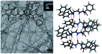
We report a new aromatic-capped peptide amphiphile which is able to form a supramolecular hydrogel under neutral pH. This hydrogel can also be obtained by enzymatic transformation from the hydrogelator precursor. The newly discovered hydrogel has excellent biocompatibility for four different cell lines, thus making it a potentially useful scaffolding material for biomedical applications.
The synthesis of the PFB-YF is shown in Scheme 1, the PFB-YF was synthesized by the solid-phase peptide synthesis (SPPS, see ESI† for details). The PFB-YF formed a self-supporting gel in water at 1 wt% (18.64 mM) under neutral pH. As shown in Fig. 1a, the appearance of the hydrogel is transparent and quite stable up to 43 °C, at which temperature the gel-to-sol transition began to occur. The microscopic nanostructures of the hydrogel at 1 wt% was measured by transmission electron microscopy (TEM). The TEM image of a air-dried sample of PFB-YF gel revealed the formation of nanofibers with uniform diameters of 9.6 ± 2 nm (Fig. 1a). From the TEM image, it is unclear whether the formation of nanofibers results from the sample drying or whether the water evaporation influences the process of the aggregation. To examine this, we measured the TEM image of a 1 wt% freeze-dried gel of PFB-YF and it showed fibrous nanostructures with 8.2 ± 2 nm in diameter which is similar to that of air-dried sample (Fig. S1†). Furthermore, we studied the microstructures of air-dried gels with different concentrations of 1 wt%, 2 wt% and 5 wt% (Fig. 1, S2 and S3†). From the TEM images, we clearly observed nanofibrous structures obtained from these gels and the average diameter for the 5 wt% gel was 11.9 ± 2 nm which is slightly larger than that of 1 wt% gel. These results indicate the change in evaporation rate of water or concentration of the PFB-YF has small effect on the formation of self-assembled nanostructures. Therefore, we conclude that the self-assembled nanofibers are formed through hydrogelation of PFB-YF molecules and the physical crosslinking of these nanofibers could entangle to trap water within the gel. Furthermore, hydrogelation of PFB-YpF at 18.6 mM was performed by using 200 U of the ALP in PBS buffer at 37 °C for 12 h resulting in the formation of the hydrogel with a fibrous network observed by TEM (Fig. 1b). During the enzymatic transformation, these molecules self-assemble to form long and uniform nanofibers with average widths of 8.5 ± 2 which is consistent with that of PFB-YF gel at 1 wt%. These results imply the success of enzymatic transformation of PFB-YpF in PBS buffer.
The viscoelastic properties of the PFB-YF gel were measured using oscillatory rheology. The mechanical data for a PFB-YF gel was collected in Fig. 1c where the storage modulus (G′) is found to be higher than the loss modulus (G′′), indicating the PFB-YF gel is an elastic material rather than a viscous one. The rheological characterization of the gel obtained from enzymatic transformation of PFB-YpF indicated that the G′ is higher than G′′ and these values have the same order of magnitude compared with those of PFB-YF gel (Fig. 1c). We further explored the kinetics of enzymatic transformation of the PFB-YpF. In Fig. 1d, we conducted the time-dependent rheology study of PFB-YpF. After adding the ALP in PFB-YpF solution for 52 min, we found the increase of G′ is faster than G′′ which indicates the hydrogelator precursor turn into hydrogelator PFB-YF. Furthermore, the sol–gel transition of PFB-YpF was also examined. We observed that PFB-YpF solution turns into a stable hydrogel within 3 h after the addition of ALP (200 U). It is interesting to note that a unstable gel formed within 1 h which is consistent with the time point for sharply increase of G′ in the time-dependent rheology study. These results suggested that the efficient enzymatic transformation of PFB-YpF occurred within 3 h after addition of ALP.21,24
To further examine the intermolecular interactions in the assemblies, we investigate the concentration-dependent and solvent effects of PFB-YF at pH 7.0 by using UV-Vis absorption, circular dichroism (CD), fluorescence emission and FT-IR. The concentration-dependent absorption spectra of the PFB-YF in water are given in Fig. 2, where absorption bands of π–π* transitions of the aromatic rings at ca. 260 nm (240–300 nm). The concentration-dependent CD spectra revealed a negative part at 260 nm (240–300 nm) when the concentrations are higher than 5 mM (Fig. S4†). The concentration-dependent emission spectra of the PFB-YF were measured with an excitation wavelength of 260 nm, as shown in Fig. 2, the emission bands of the dilute solutions are centered at ca. 303 nm (50 and 500 μM), and the emission maxima shift to ca. 310 nm with the relatively higher emission intensities for the concentrations higher than 5 mM. Notably, new emission bands at 380 nm (350–450 nm) were observed at the concentrations of 10, 15 and 20 mM, thus indicating the strong π–π interactions may occur in the gel state of PFB-YF.
Fig. 2 Concentration-dependent UV-Vis absorption (left) and fluorescence emission (right) spectra of PFB-YF in water. |
We now turn to the impact of solvents on the self-assembly of PFB-YF at 1 wt%. Because the PFB-YF is soluble in polar organic solvents, the spectroscopic characterization of PFB-YF at 1 wt% in methanol was compared with that in water. The absorption peak shows a slightly blue shift (ca. 5 nm) and the enhanced CD signal at 240–300 nm is detected upon transitioning from methanol to water (Fig. S5†). As displayed in Fig. 3a, a red-shift (ca. 10 nm) of the emission maximum occurs upon assembly in water, and the emission band at 380 nm is relatively prominent compared to that in methanol. These results indicate the π–π interactions of the hydrogelators are essential in the assemblies. FT-IR spectrum of the PFB-YF gel displayed two new well-defined amide I bands centered at 1632 and 1681 cm−1 compared with those in homogeneous methanol solutions (Fig. 3b), which suggested β-sheet-like structures formed in the self-assembled nanofibers.18,25–30
Fig. 3 (a) Normalized UV-Vis absorption (left) and normalized fluorescence emission (right) and (b) FT-IR spectra of PFB-YF at 1 wt% in water (black) and in methanol (red). |
Combining the results obtained from these spectroscopic characterization it was possible to propose a reasonable molecular model for the arrangement of the PFB-YF molecules within the self-assembled nanostructures. As mentioned above, the peptide moiety of the molecule should pack in a β-sheet arrangement and the aromatic rings should form π stack in the assemblies. Fig. 4 shows the possible packing model for PFB-YF, they were stacked in an β-sheet structure with the hydrogen bonding between the peptides. In addition, the π–π interactions in the assemblies can be explained by the packing of pentafluorophenyl, phenol and phenyl groups of the PFB-YF molecules within this model.
Since the PFB-YF gel formed under neutral pH, we further study the biocompatibility of the hydrogelator. It has been reported that the biocompatibility of aromatic-capped peptide hydrogelators can be preliminarily evaluated in the concentration range of 10–500 μM.17,31 Accordingly, the biocompatibility tests of PFB-YF and precursor PFB-YpF were examined using MTT assay.32 As a potential material for tissue engineering and drug delivery, we examined the viability of CTX TNA2, WS1, HeLa and MCF-7 cells. As shown in Fig. 5, after being incubated the four cells with the material (10–500 μM) for 48 h, cells that were grown in liquid medium showed the excellent survival ratios of 95%, 94%, 97% and 88% respectively, at 500 μM. In Fig. 5, the experiments revealed that the 50% inhibition (IC50) are all higher than 500 μM after 48 h. These observations indicate that PFB-YF is relatively biocompatible compared with previously reported cases of PFB-capped dipeptides.17 In addition, the IC50 values of the hydrogelator precursor PFB-YpF for the four cell lines were all higher than 500 μM (Fig. S6–S9†).
In summary, we report a new chemical structure of PFB-capped peptide amphiphile which forms a supramolecular hydrogel under neutral pH. More importantly, this self-assembled hydrogel can also be obtained by enzymatic transformation of PFB-YpF in PBS buffer under physiological condition. On the basis of the spectroscopic characterization and computational calculations, the hydrogen-bonding and aromatic–aromatic interactions might be the major driving force behind the self-assembly of PFB-YF. In addition, the PFB-YF hydrogel has excellent biocompatibility for four different cell lines, thus making it a potentially useful scaffolding material for biomedical applications.
Footnote |
† Electronic supplementary information (ESI) available: Experimental details, Fig. S1–S10 and additional information. See DOI: 10.1039/c5ra03290c |
This journal is © The Royal Society of Chemistry 2015 |