Rapid microwave assisted hydrothermal synthesis of porous α-Fe2O3 nanostructures as stable and high capacity negative electrode for lithium and sodium ion batteries

B. Nageswara Raoa, P. Ramesh Kumara, O. Padmaraja, M. Venkateswarlub and N. Satyanarayana*a
aDepartment of Physics, Pondicherry University, Pondicherry-605014, India. E-mail: nallanis2011@gmail.com; Tel: +91-413-2654404
bR&D, Amara Raja Batteries, Tirupati-517520, India

Received 20th February 2015 , Accepted 7th April 2015

First published on 7th April 2015


Abstract

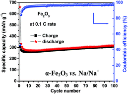
Hematite porous α-Fe2O3 nanostructures were prepared within 60 minutes by a rapid microwave assisted hydrothermal process. X-ray diffraction (XRD) and Fourier transform infrared (FTIR) spectroscopy studies confirm the phase and structural co-ordination of α-Fe2O3, respectively. Scanning electron microscopy (SEM) images reveal the formation of well defined uniform shaped α-Fe2O3 nanospheres. The transmission electron microscopy (TEM) image shows the porous nature of the α-Fe2O3 nanospheres with an interconnected monocrystallites structure. Swagelok type lithium and sodium batteries were fabricated using the developed porous α-Fe2O3 nanostructures as a negative electrode material. As a negative electrode material in Li-ion cells, the porous α-Fe2O3 nanostructures deliver a second cycle discharge capacity of 1000 mA h g−1 at a 0.1 C rate. At a higher current density of 5 C, the porous α-Fe2O3 nanostructures exhibit a specific capacity of 264 mA h g−1. In the case of Na-ion batteries, the porous α-Fe2O3 nanostructures as negative electrode exhibit a reversible capacity of 300 mA h g−1 with excellent cycleability and coulombic efficiency at 0.1 C up to 100 cycles. The observed excellent electrochemical performance is attributed to the porous nature and uniform shape of the α-Fe2O3 nanostructures that are composed of interconnected monocrystallites. Importantly, the architecture of α-Fe2O3 nanostructures, comprising interconnected monocrystallites could be developed within a short time by a rapid microwave synthesis process and it can be applied to develop different morphological nanostructures for better energy storage device applications.


1. Introduction

Lithium ion batteries (LIBs) with high energy densities and high voltages have become the main rechargeable power source for portable electronic devices and, more recently, for electric vehicles (EVs) and hybrid electric vehicles (HEVs).1 However, the scarcity of lithium resources in the Earth's crust and high processing cost have limited their large scale applications.2,3 In this regard, sodium ion batteries (SIBs) are found to be an attractive alternative to LIBs because of the high abundance and low cost of sodium.4,5 Furthermore, the similarities between Li+ and Na+ intercalation reactions favour the use of analogous materials, which could match both the applications.1 The ionic radius of Na+ (0.95 Å) is almost 1.6 times larger than that of Li+ (0.60 Å), which undoubtly restricts the development of electrode materials for SIBs. In recent years, many positive electrode materials have been investigated for both LIBs and SIBs.6–11 So far, a few varieties of materials, as a viable negative electrode, have been developed and studied for LIBs and SIBs. Graphite usage as negative electrode in LIBs has limitations, such as low Li storage capability (372 mA h g−1) and safety problems due to lithium plating on its surface.12 Also, it is not suitable as negative electrode in SIBs, because of its inability to intercalate Na atoms, dendrites formation and safety issues.13,14 Thus, the development of new negative electrode materials with high storage capacity and power density has become an urgent task to meet the ever growing performance demand. Recently, metal oxides (NiO, CuO, Mn2O3, Co3O4, TiO2, α-Fe2O3, etc.) have received great interest as negative electrode materials in LIBs and SIBs,1,15–19 because of their improved electrochemical performance. Hematite α-Fe2O3 has attracted much interest as negative electrode material in both LIBs and SIBs, because of its low cost, high stability under ambient conditions, non-toxicity, high resistivity towards corrosion and environmentally benign.20 The performance of α-Fe2O3 as negative electrode in secondary batteries strongly depends on the structure, particle size and morphology.21,22 Recently, nanostructured porous materials have paid much attention as negative electrodes in LIBs because, the nanosize particles provide shorter diffusion lengths for ions and high surface area contact with electrolyte for electron transfer, leading to an increased rate capability, while the porous nature provides facile strain relaxation during charge–discharge reactions, leading to an improved cycleability.23,24 So, research efforts have been focused for the development of nanostructured porous materials with different morphologies, such as nanospheres, nanorods, nanowires, nanofibers, etc.25–29

Several innovative methods have been developed for the synthesis of nanostructured materials, such as sol–gel, quasi-emulsion-templated, electrostatic spray deposition, hydrothermal, etc.30–35 Sun et al. have reported the electrochemical performance of mesoporous nanostructured α-Fe2O3 obtained by soft template method. The prepared mesoporous α-Fe2O3 delivered a higher specific capacity.36 As reported by D. Chen et al., α-Fe2O3 nanoparticles prepared by a simple wet chemistry route showed good electrochemical performance.26 However, longer reaction times, nonuniform distribution of particles and low yield are recognized as the limitations of above preparation methods. Recently, microwave assisted hydrothermal/solvothermal method has been recognized as a potential technique to overcome the above mentioned limitations. Microwave synthesis has many advantages: (i) shorter reaction times (it can reduce the reaction times from several hours to minutes), (ii) low temperature synthesis, (iii) environmentally friendly and energy saving, (iv) high productivity and efficiency, (v) uniform distribution of particles, (vi) better reproducibility and large scalability, (vii) excellent control over experimental parameters (provides better understanding of the influence of synthesis conditions on nucleation and growth mechanism), (viii) its a versatile approach and can be used effectively and efficiently to develop nanocrystalline materials with different morphologies.37–39 A. V. Murugan et al. have reported the microwave assisted hydrothermal and solvothermal synthesis of LiFePO4 nanorods as positive electrode material for Li ion batteries.40 T. Muraliganth et al. group could prepare nanostructured Li2MSiO4 (M = Fe, Mn) positive electrodes for Li ion batteries by facile microwave assisted solvothermal process.41 More recently, nanostructured metal oxides (Co3O4/graphene, Mo3O4/graphene, W0.4Mo0.6O3 and NiO) prepared by microwave synthesis method exhibited good electrochemical performance as negative electrodes for Li ion batteries.42–45 As reported by K. Chen et al., hematite Fe2O3 cube-like structures prepared by microwave hydrothermal route exhibited a high discharge capacity as negative electrode for Li ion battery.46 R. Indrajothi et al. and G. Y. Zhang et al. studies revealed that the mesoporous nanostructures are formed in the presence of base catalysts.23,47 Also, it is revealed that the porous nanostructures are effective in enhancing the electrochemical performance of Li ion batteries.23,36 Hence, we are motivated to develop porous α-Fe2O3 nanostructures in the presence of base catalyst (NaOH) by microwave assisted hydrothermal method and study their efficiency as negative electrode in LIBs and SIBs. For the first time, we report the synthesis of porous α-Fe2O3 nanostructures by using a simple inorganic sodium hydroxide (NaOH). The synthesized sample was characterized by X-ray diffraction (XRD), Fourier transform infrared spectroscopy (FTIR), Raman spectroscopy, field-emission scanning electron microscopy (FE-SEM) and transmission electron microscopy (TEM) techniques. Swagelok cell type lithium and sodium batteries were fabricated using the newly developed porous α-Fe2O3 nanostructures as negative electrode material. The electrochemical performance of the fabricated lithium and sodium batteries using the newly developed nanocrystalline α-Fe2O3 sample was studied by cycling the cells in the voltage range 0.002–3 V, at different C-rates, at ambient conditions.

2. Experimental

2.1 Synthesis

Hematite porous α-Fe2O3 nanostructures were prepared by using ferric chloride (FeCl3) and sodium hydroxide as the starting materials. In the typical process, 0.008 moles of FeCl3 (Qualigens) was dissolved in 50 ml of deionized water at room temperature under stirring condition. Then, 0.50 g (0.0125 moles) of solid NaOH (CDH chemicals, LR) pellets were added to the above solution and continued stirring for 10 minutes. The obtained solution was transferred to a 100 ml Teflon liner vessel and the vessel was covered with an outer high strength sleeve made up of advanced composite material. The vessel was sealed and placed on a rotating table inside the microwave accelerated reaction system (MARS, CEM Corporation, USA). A temperature sensor was inserted into the reaction vessel to monitor the vessel temperature and the complete system was maintained at 140 °C for 60 minutes for microwave hydrothermal reaction. After the reaction, the vessel was allowed to cool down to room temperature and the precipitated product was washed with water followed by acetone washing through high speed centrifugation. The obtained powder was dried at 60 °C overnight and subsequently heated at 350 °C for 1 h in air. The schematic representation of the reaction mechanism for the formation of porous nanostructured α-Fe2O3 particles in the presence of base catalyst (NaOH) under microwave irradiation is shown in Fig. 1. The usage of NaOH, as a base catalyst, and microwave irradiation, leading to the fast formation of ultrafine (nanosize) α-Fe2O3 spherical particles. The addition of base catalyst (NaOH) can accelerate the hydrolysis reaction and may form ultrafine α-Fe2O3 particles.48 In the subsequent process, to minimize the surface free energy of the system, the ultrafine α-Fe2O3 particles may continuously assemble into the porous spherical structure. Further, the drying process leads to the formation of densified porous α-Fe2O3 nanospheres structure. The formation of ultrafine (nanosize) α-Fe2O3 particles (20–30 nm) and agglomerated porous nanostructured α-Fe2O3 particles (∼350 nm) are further confirmed from the observed FESEM/TEM images, as shown in Fig. 3.
image file: c5ra03238e-f1.tif
Fig. 1 Schematic mechanism for the growth of porous α-Fe2O3 nanostructures by microwave assisted hydrothermal process.

2.2 Characterization

The crystalline phase of the as-obtained product was identified using X-ray powder diffractometer (X'pert PRO MPD, PANalytical, Philips) with Cu Kα radiation of wavelength λ = 0.154 nm. The structural co-ordination of the sample was studied using Fourier transform infrared (FTIR) spectroscopy (Shimadzu FTIR-8000 spectrometer). Raman spectrum of the obtained product was recorded at room temperature on Renishaw in Via Raman Microscope (Ar–Ne Laser source, 514 nm line radiation). The morphology of the synthesized sample was examined on field-emission scanning electron microscopy (FE-SEM, HITACHI, S4800, Japan) and transmission electron microscopy (Tecnai, G2 F30, FEI with 300 kV).

For the fabrication of the composite electrode, 70% active material, 20% super P carbon and 10% Na-alginate in deionized water as solvent were used. The above mixture was grounded several hours to obtain a well mixed slurry. The slurry was coated onto a Cu-foil and dried at 100 °C overnight. The dried foil was cut into circular electrode disks. The Swagelok-type cells were assembled in an argon filled glove box (Vacuum Atmospheres Co., USA) by using Whatman GF/D borosilicate glass-fiber as separator. Lithium metal (for lithium ion battery) and sodium metal (for sodium ion battery) were used as reference electrodes. 1 M LiPF6 dissolved in a mixture of ethylene carbonate (EC) and diethyl carbonate (DEC) (1[thin space (1/6-em)]:[thin space (1/6-em)]1 in volume) was used as electrolyte for lithium ion battery (LIB). 1 M NaClO4 in propylene carbonate (PC) was used as electrolyte for sodium ion battery (SIB). The cyclic voltammetry (CV) curves were recorded at 0.2 mV s−1 scan rate, at ambient conditions, on a Novocontrol electrochemical test station (POT/GAL). The cycling performance of the cells was recorded at different C-rates on a battery testing system (model MCV4, Bitrode, USA) in the voltage range 0.002–3 V, at ambient conditions.

3. Results and discussion

Powder X-ray diffraction pattern of α-Fe2O3 sample along with standard JCPDS data are shown in Fig. 2a. From Fig. 2a, the observed diffraction peaks indicate the formation of hematite type rhombohedral structured α-Fe2O3 with R[3 with combining macron]c space group and it was confirmed by comparing the observed diffraction pattern with the standard JCPDS data (card no. 87-1166). The observed diffraction peaks at 24.1°, 33.2°, 35.6°, 49.5°, 54.1° and 64.1° are respectively indexed to (012), (104), (110), (024), (116) and (330) planes, which also confirm the formation of α-Fe2O3 with rhombohedral structure. The average crystallite size of the sample was calculated using Scherrer's formula
 
D = 0.9λ/β[thin space (1/6-em)]cos[thin space (1/6-em)]θ (1)
where, λ is the X-ray wavelength and β is the full-width at half maximum (FWHM) of the diffraction line. The average crystallite size of the α-Fe2O3 sample is found to be 28 nm.

image file: c5ra03238e-f2.tif
Fig. 2 (a) XRD pattern, (b) FTIR spectrum and (c) Raman spectrum of the as-synthesized α-Fe2O3 sample.

Fig. 2b shows the FTIR spectrum of as-prepared α-Fe2O3 sample. As shown in Fig. 2b, the IR peaks observed at 480 cm−1 and 573 cm−1 are attributed to the stretching vibrations of Fe–O in FeO4 tetrahedron and FeO6 octahedron, respectively.49 Raman spectrum of the as-prepared α-Fe2O3 sample is shown in Fig. 2c. From Fig. 2c, the Raman scattering peaks observed at 211, 279, 393, 480 and 593 cm−1 are assigned to two A1g and three Eg Raman modes in a typical hematite α-Fe2O3 structure.50

The field-emission scanning electron microscopy (FE-SEM) images of α-Fe2O3 sample are shown in Fig. 3a and b. The FE-SEM images show the formation of uniformly distributed nanospheres. The nanospheres have a diameter of about 350 nm. The FE-SEM images reveal that the uniform heating of the precursor solution by microwave irradiation leads to the formation of uniformly distributed α-Fe2O3 nanospheres. The transmission electron microscopy (TEM) image (Fig. 3c) clearly shows the porous structure of α-Fe2O3 nanospheres and the nanospheres are consisted of densely packed small nanoparticle subunits. The small nanoparticle subunits have particle size distribution in the range of 20–30 nm. The size of small particle subunits is comparable to the average crystallite size deduced from XRD analysis, which may indicate that each subunit is a monocrystallite and the nanospheres are composed of interconnected monocrystallites.


image file: c5ra03238e-f3.tif
Fig. 3 FE-SEM images (a and b) and TEM image (c) of α-Fe2O3 sample.

Fig. 4 shows the cyclic voltammetry (CV) curves at different cycles with 0.2 mV s−1 scan rate obtained at room temperature for the as prepared α-Fe2O3 sample. From Fig. 4a, in the first cycle of cathodic polarization process, a spiky peak was observed at 0.54 V vs. Li/Li+, ascribed to the complete reduction of iron from Fe3+ to Fe0, formation of Li2O and decomposition of the electrolyte to form solid electrolyte interface (SEI) film.12,51 During the first anodic scan, two broad overlapping peaks were observed at 1.65 V and 1.83 V, which correspond to the oxidation of Fe0 to Fe2+ and further oxidation to Fe3+.12,36 The observed redox peaks are comparable to the literature reports.12,36,51 Compared to 1st cycle, in 5th, 10th, 15th and 20th cycles, the anodic polarization shows two broad peaks with decrease in peak intensity, which indicates good reversible reaction of Fe0 to Fe2+ and Fe2+ to Fe3+. Whereas, the cathodic polarization shows one peak at 0.8 V with decrease in peak intensity and area, indicating the irreversible phase transformation with the formation of SEI film in the first cycle.12,52 It reveals that the oxidation and reduction processes proceed with some irreversibility. The observed decrease in peak intensity implies the decrease of capacity with cycling. During 5th–20th cycles, the current of the anodic and cathodic peaks remains nearly the same, suggesting good reversibility and structure stability during charge–discharge reactions.


image file: c5ra03238e-f4.tif
Fig. 4 Cyclic voltammograms of (a) α-Fe2O3 nanospheres vs. Li/Li+ and (b) α-Fe2O3 nanospheres vs. Na/Na+ at a scan rate of 0.2 mV s−1.

From Fig. 4b, during the first cathodic scan, α-Fe2O3 exhibits three different reductive peaks at 1.1, 0.8 and 0.4 V vs. Na/Na+. The observed reduction peaks correspond to initial insertion of Na+ into α-Fe2O3, which leads to the formation of NaxFe2O3 compound and a conversion of the iron oxide accompanied by SEI film formation.1,53 In the first anodic scan, two weak peaks were detected around 0.7 and 1.5 V vs. Na/Na+, corresponding to the double oxidation process (i.e. Fe0/Fe2+ and Fe2+/Fe3+) that enabled iron oxide regeneration.1 From 5th–20th cycles, the cathodic peaks at 1.1 and 0.8 V were disappeared and the peak at 0.4 V shifted to 0.6 V with reduced peak intensity, which indicate some irreversible phase transformation due to SEI film formation in the first cycle. Anodic scan shows two peaks with decreased peak intensities, which represent the good reversible double oxidation of iron. The α-Fe2O3 structure facilitates the intercalation process before the conversion reaction due to the stable interface while using Na-alginate binder.54

The charge–discharge curves of α-Fe2O3 sample vs. Li/Li+, collected at 0.1 C-rate are shown in Fig. 5a. In the first cycle of the discharge curve, three distinct regions were observed and labelled as I, II and III. Initially, the cell was discharged from open circuit voltage (OCV) to insert Li+ ions into α-Fe2O3 structure as given in eqn (2).22 In region I, a plateau with slope is observed at 0.96 V, which corresponds to the phase transformation from hexagonal LixFe2O3 to cubic Li2Fe2O3 (eqn (3)).12,21,22,36 In II region, a potential plateau is observed at 0.85 V, which is due to the complete reduction of iron from Fe3+ to Fe0 (eqn (4)).12,21,22 It indicates the dispersion of Fe nanocrystals into Li2O matrix. The III region attributes to the reduction of electrolyte below 0.8 V and formation of SEI film.12 An interfacial reduction at the Fe–Li2O boundary leads to further lithium storage, which results in higher discharge capacity of 1364 mA h g−1 (8.1 moles of lithium per one mole of α-Fe2O3) than the theoretical capacity of 1007 mA h g−1 (6 moles of lithium per one mole of α-Fe2O3).

 
Fe2O3 + xLi+ + xe → LixFe2O3 (2)
 
LixFe2O3 + (2 − x)Li+ + (2 − x)e → Li2Fe2O3 (cubic) (3)
 
Li2Fe2O3 (cubic) + 4Li+ + 4e → 2Fe + 3Li2O (4)


image file: c5ra03238e-f5.tif
Fig. 5 Galvanostatic charge–discharge curves (a) and cycleability (b) of α-Fe2O3 sample at 0.1 C rate vs. Li/Li+.

During the 10th, 20th and 30th cycles, only one potential plateau was observed with a decrease in discharge capacity. The discharge capacities of α-Fe2O3 in the 1st, 10th, 20th and 30th cycles are 1364, 941, 876 and 799 mA h g−1, respectively. Fig. 5b shows the cycling performance of α-Fe2O3 sample at 0.1 C-rate. As shown in Fig. 5b, the material delivers a first cycle discharge and charge capacities of 1364 and 1031 mA h g−1, respectively, with 24.5% irreversible capacity loss and a second cycle discharge capacity of 1000 mA h g−1. M. V. Reddy et al. have synthesized the α-Fe2O3 nanoflakes by thermal treatment method and the obtained α-Fe2O3 nanoflakes delivered a stable discharge capacity of (680 ± 20) mA h g−1, at 0.1 C-rate.25 L. Ji et al. have studied the electrochemical performance of α-Fe2O3 nanoparticle-loaded carbon nanofibers composite synthesized by electrospinning method. The results showed that the 2nd cycle discharge capacity is <600 mA h g−1, at 50 mA g−1.29 In our study, the porous α-Fe2O3 nanostructures synthesized by rapid microwave assisted hydrothermal method delivered a 2nd cycle discharge capacity of 1000 mA h g−1 at 0.1 C, which is 30% higher than the stable discharge capacity of α-Fe2O3 nanoflakes.25 In addition, the 2nd cycle discharge capacity of porous α-Fe2O3 nanostructures is 63% higher than the theoretical capacity of graphite (372 mA h g−1), which is currently used as negative electrode material in commercial Li-ion batteries. It can also be observed that the material shows identical charge and discharge capacity values in 30 cycles, which indicates an excellent reversibility of the material.

Further, the α-Fe2O3 cells were cycled at different (1, 5, 10 and 20 C) C-rates and the corresponding capacity vs. voltage curves are shown in Fig. 6a. The observed discharge capacities at 1, 5, 10 and 20 C-rates are 590, 267, 157 and 63 mA h g−1, respectively. The cycleability of α-Fe2O3 sample at 1, 5, 10 and 20 C rates is compared in Fig. 6b. From Fig. 6b, a fade of capacity is observed with increase of C-rate from 1 to 5, 10, and 20 C. At 1 C, the discharge capacity lost per cycle is 1.7%, and 0% capacity lost per cycle is observed at 5, 10 and 20 C-rates. The decrease in the rate of capacity fade per cycle with increase of C-rate indicates an enhanced reversibility at high C-rates. The comparison of specific capacity at 1 C-rate (Fig. 6b) shows that the porous α-Fe2O3 nanostructures could be able to retain their initial capacity values even after cycling at higher rates, which indicates an excellent reversibility of the material.


image file: c5ra03238e-f6.tif
Fig. 6 Voltage vs. specific capacity curves (a) and cycling performance (b) of α-Fe2O3 at different (1, 5, 10 and 20 C) rates vs. Li/Li+.

Fig. 7 shows the charge–discharge curves and cycle life of α-Fe2O3 vs. Na/Na+, at 0.1 C-rate. From Fig. 7a, the first cycle discharge curve displayed all the characteristic features of sodiation of iron oxide and confirmed the previous cyclic voltammetry (CV) results in Fig. 4b. As shown in Fig. 7a, the first cycle discharge curve exhibits the potential plateaus, indicating the Na+ insertion, reduction of iron and SEI film formation.1 From Fig. 7b, the material shows a high stable reversible capacity of 300 mA h g−1 with excellent cycleability and coulombic efficiency over 100 cycles. An earlier report on an electrodeposited thin films of Fe2O3 cycled at 5 μA cm−2, between 0.05 and 3.0 V vs. Na/Na+ completely lost its capacity in 8 cycles.54 Recently, M. Valvo et al. report on Fe2O3 particles cycled between 0.05 and 2.8 V vs. Na/Na+, at 130 mA g−1 exhibited a capacity of 250 mA h g−1 after 60 cycles.55 In the present study, the porous α-Fe2O3 nanostructures cycled at 0.1 C-rate exhibited high reversible capacity of 300 mA h g−1 and an excellent cycleability up to 100 cycles. Fig. 8 shows the rate capability of α-Fe2O3 sample at different C-rates. From Fig. 8b, the observed discharge capacities at 1, 2, 5 and 10 C-rates are 150, 105, 32 and 14 mA h g−1, respectively. From Fig. 8b, it can be seen that the porous α-Fe2O3 nanostructures exhibit stable cycleability at high current densities of 1, 2, 5 and 10 C. Even after cycling at higher C-rates, the material is capable to retain the initial capacity values at 1 C rate, which indicates an excellent reversibility of the material.


image file: c5ra03238e-f7.tif
Fig. 7 Charge–discharge curves (a) and cycleability (b) of α-Fe2O3 sample at 0.1 C rate vs. Na/Na+.

image file: c5ra03238e-f8.tif
Fig. 8 Voltage vs. specific capacity curves (a) and cycling performance (b) of α-Fe2O3 at different (1, 2, 5 and 10) C rates vs. Na/Na+.

The good cycleability and rate capability of porous α-Fe2O3 nanostructures may be attributed to their uniform shape, porous nature and the interconnected monocrystallites structure, which facilitate the better electron and ion transfer in the Li or Na ion batteries. Generally, lithium or sodium ion batteries include both electron and Li+ or Na+ ion transfer during charge–discharge process. A schematic diagram for the transport of electrons and Li+ or Na+ ions through the interconnected monocrystals of the porous α-Fe2O3 nanostructures is shown in Fig. 9. The electron transfer takes place through the grain boundaries and the interconnected structure of α-Fe2O3 nanocrystals is expected to enhance the electron kinetics. The interconnected structure of TiO2 nanocrystals also exhibited good rate capability and cycleability as reported by J. Shen et al.56 The small particle subunits or monocrystallites reduce the diffusion lengths and provide a fast and efficient transport for Li+ or Na+ ions. The electrolyte filled in the pore network will enable the rapid transport of ions. In addition, the pores also provide room for facile strain relaxation during charge–discharge processes, which leads to an improved cycleability. The improved kinetic properties of electrons and Li+ or Na+ ions are responsible for an excellent cycleability and rate capability of the α-Fe2O3 electrode. The results show that microwave assisted hydrothermal process is a potential technique to develop morphology controlled α-Fe2O3 in short time with good electrochemical performance in both Li and Na ion battery applications.


image file: c5ra03238e-f9.tif
Fig. 9 Schematic representation of electron and Li+ or Na+ ion transport through the interconnected monocrystallites of α-Fe2O3 nanospheres electrode.

4. Conclusions

Hematite porous α-Fe2O3 nanostructures were prepared in short time by rapid microwave assisted hydrothermal process. The XRD studies confirm the rhombohedral structure of the prepared α-Fe2O3 sample. The formation of porous α-Fe2O3 nanostructures with uniform particle size were confirmed from FE-SEM and TEM results. For use as negative electrode in Li-ion batteries, the prepared porous α-Fe2O3 nanostructures show a discharge capacity of 799 mA h g−1 for the 30th cycle at 0.1 C rate. Furthermore, porous α-Fe2O3 nanostructures vs. Na/Na+ show a stable discharge capacity of 300 mA h g−1 with excellent cycleability over 100 cycles. The morphology of α-Fe2O3 nanospheres with porous nature and interconnected monocrystallites structure is responsible for good electrochemical performance in both Li and Na ion batteries.

Acknowledgements

NS is grateful to UGC, Govt. of India for providing financial support in the form of research project [no. 39-460/2010(SR)]. BNR is thankful to DST, Govt. of India for awarding the INSPIRE fellowship (no. DST/INSPIRE Fellowship/2011/241) for pursuing the doctoral degree. Authors thankful to central instrumentation facility (CIF), Pondicherry University for providing Raman Spectroscopy facility.

References

  1. M. Valvo, F. Lindgren, U. Lafont, F. Björefors and K. Edström, J. Power Sources, 2014, 245, 967 CrossRef CAS PubMed.
  2. B. L. Ellis, W. R. M. Makahnouk, Y. Makimura, K. Toghill and L. F. Nazar, Nat. Mater., 2007, 6, 749 CrossRef CAS PubMed.
  3. C. Wadia, P. Albertus and V. Srinivasan, J. Power Sources, 2011, 196, 1593 CrossRef CAS PubMed.
  4. M. D. Slater, D. Kim, E. Lee and C. S. Johnson, Adv. Funct. Mater., 2013, 23, 947 CrossRef CAS PubMed.
  5. S. W. Kim, D. H. Seo, X. Ma, G. Ceder and K. Kang, Adv. Energy Mater., 2012, 2, 710 CrossRef CAS PubMed.
  6. M. S. Whittingham, Chem. Rev., 2004, 104, 4271 CrossRef CAS.
  7. M. Saiful Islam and C. A. J. Fisher, Chem. Soc. Rev., 2014, 43, 185 RSC.
  8. A. Manthiram, A. Vadivel Murugan, A. Sarkar and T. Muraliganth, Energy Environ. Sci., 2008, 1, 621 CAS.
  9. N. Yabuuchi, M. Kajiyama, J. Iwatate, H. Nishikawa, S. Hitomi, R. Okuyama, R. Usui, Y. Yamada and S. Komaba, Nat. Mater., 2012, 11, 512 CrossRef CAS PubMed.
  10. V. Palomares, P. Serras, I. Villaluenga, K. B. Hueso, J. Carretero-Gonzalez and T. Rojo, Energy Environ. Sci., 2012, 5, 5884 CAS.
  11. Z. Jian, L. Zhao, H. Pan, Y. S. Hu, H. Li, W. Chen and L. Chen, Electrochem. Commun., 2012, 14, 86 CrossRef CAS PubMed.
  12. X. Zhang, H. Liu, S. Petnikota, S. Ramakrishna and H. J. Fan, J. Mater. Chem. A, 2014, 2, 10835 CAS.
  13. P. Ge and M. Fouletier, Solid State Ionics, 1988, 28–30, 1172 CrossRef.
  14. D. A. Stevens and J. R. Dahn, J. Electrochem. Soc., 2001, 148, A803 CrossRef CAS PubMed.
  15. F. Jiao, A. Harrison, A. H. Hill and P. G. Bruce, Adv. Mater., 2007, 19, 4063 CrossRef CAS PubMed.
  16. K. M. Shaju, F. Jiao, A. Debart and P. G. Bruce, Phys. Chem. Chem. Phys., 2007, 9, 1837 RSC.
  17. K. X. Wang, M. D. Wei, M. A. Morris, H. S. Zhou and J. D. Holmes, Adv. Mater., 2007, 19, 3016 CrossRef CAS PubMed.
  18. Y. Zou and Y. Wang, Nanoscale, 2011, 3, 2615 RSC.
  19. B. Wang, X. L. Wu, C. Y. Shu, Y. G. Guo and C. R. Wang, J. Mater. Chem., 2010, 20, 10661 RSC.
  20. J. Chen, L. Xu, W. Li and X. Gou, Adv. Mater., 2005, 17(5), 582 CrossRef CAS PubMed.
  21. D. Larcher, C. Masquelier, D. Bonnin, Y. Chabre, V. Masson, J. B. Leriche and J. M. Tarascon, J. Electrochem. Soc., 2003, 150, A133 CrossRef CAS PubMed.
  22. D. Larcher, D. Bonnin, R. Cortes, I. Rivals, L. Personnaz and J. M. Tarascon, J. Electrochem. Soc., 2003, 150, A1643 CrossRef CAS PubMed.
  23. R. Indhrajothi, I. Prakash, M. Venkateswarlu and N. Satyanarayana, RSC Adv., 2014, 4, 44089 RSC.
  24. H. B. Lin, J. N. Hu, H. B. Rong, Y. M. Zhang, S. W. Mai, L. D. Xing, M. Q. Xu, X. P. Li and W. S. Li, J. Mater. Chem. A, 2014, 2, 9272 CAS.
  25. M. V. Reddy, T. Yu, C. H. Sow, Z. X. Shen, C. T. Lim, G. V. S. Rao and B. V. R. Chowdari, Adv. Funct. Mater., 2007, 17, 2792 CrossRef CAS PubMed.
  26. D. Chen, W. Wei, R. Wang, J. Zhu and L. Guo, New J. Chem., 2012, 36, 1589 RSC.
  27. T. P. Almeida, M. Fay, Y. Zhu and P. D. Brown, J. Phys. Chem. C, 2009, 113, 18689 CAS.
  28. G. X. Wang, X. L. Gou, J. Horvat and J. Park, J. Phys. Chem. C, 2008, 112, 15220 CAS.
  29. L. Ji, O. Toprakci, M. Alcoutlabi, Y. Yao, Y. Li, S. Zhang, B. Guo, Z. Lin and X. Zhang, ACS Appl. Mater. Interfaces, 2012, 4, 2672 CAS.
  30. K. Woo, H. J. Lee, J. P. Ahn and Y. S. Park, Adv. Mater., 2003, 15, 1761 CrossRef CAS PubMed.
  31. B. Wang, J. S. Chen, H. B. Wu, Z. Wang and X. W. Lou, J. Am. Chem. Soc., 2011, 133, 17146 CrossRef CAS PubMed.
  32. L. Wang, H. W. Xu, P. C. Chen, D. W. Zhang, C. X. Ding and C. H. Chen, J. Power Sources, 2009, 193, 846 CrossRef CAS PubMed.
  33. J. B. Lian, X. C. Duan, J. M. Ma, P. Peng, T. I. Kim and W. J. Zheng, ACS Nano, 2009, 3, 3749 CrossRef CAS PubMed.
  34. E. García-Tamayo, M. Valvo, U. Lafont, C. Locati, D. Munao and E. M. Kelder, J. Power Sources, 2011, 196, 6425 CrossRef PubMed.
  35. M. Valvo, E. Garcia-Tamayo, U. Lafont and E. M. Kelder, J. Power Sources, 2011, 196, 10191 CrossRef CAS PubMed.
  36. B. Sun, J. Horvat, H. S. Kim, W. S. Kim, J. Ahn and G. Wang, J. Phys. Chem. C, 2010, 114, 18753 CAS.
  37. I. Bilecka and M. Niederberger, Nanoscale, 2010, 2, 1358 RSC.
  38. B. Nageswara Rao, P. Muralidharan, P. Ramesh Kumar, M. Venkateswarlu and N. Satyanarayana, Int. J. Electrochem. Sci., 2014, 9, 1207 Search PubMed.
  39. B. Nageswara Rao, M. Venkateswarlu and N. Satyanarayana, Ionics, 2014, 20, 927 CrossRef.
  40. A. Vadivel Murugan, T. Muraliganth and A. Manthiram, J. Phys. Chem. C, 2008, 112, 14665 Search PubMed.
  41. T. Muraliganth, K. R. Stroukoff and A. Manthiram, Chem. Mater., 2010, 22, 5754 CrossRef CAS.
  42. S. Yoon and A. Manthiram, J. Mater. Chem., 2011, 21, 4082 RSC.
  43. S. Q. Chen and Y. Wang, J. Mater. Chem., 2010, 20, 9735 RSC.
  44. Z. Yuqin and W. Yong, Rare Met., 2011, 30, 59 CrossRef PubMed.
  45. L. Li, Z. Guo, A. Du and H. Liu, J. Mater. Chem., 2012, 22, 3600 RSC.
  46. K. Chen, Y. DongNoh, W. Huang, J. Ma, S. Komarneni and D. Xue, Ceram. Int., 2014, 40, 2877 CrossRef CAS PubMed.
  47. G.-Y. Zhang, Y. Feng, Y.-Y. Xu, D.-Z. Gao and Y.-Q. Sun, Mater. Res. Bull., 2012, 47, 625 CrossRef CAS PubMed.
  48. I. Prakash, N. Nallamuthu, P. Muralidharan, M. Venkateswarlu, M. Misra, A. Mohanty and N. Satyanarayana, J. Sol-Gel Sci. Technol., 2011, 58, 24 CrossRef CAS PubMed.
  49. H. D. Ruan, R. L. Frost, J. T. Kloprogge and L. Duong, Spectrochim. Acta, Part A, 2002, 58, 967 CrossRef CAS.
  50. D. L. A. de Faria, S. Venancio Silva and M. T. de Oliveira, J. Raman Spectrosc., 1997, 28, 873 CrossRef CAS.
  51. S. L. Chou, J. Z. Wang, D. Wexler, K. Konstantinov, C. Zhong, H. K. Liu and S. X. Dou, J. Mater. Chem., 2010, 20, 2092 RSC.
  52. Y. Song, S. Qin, Y. Zhang, W. Gao and J. Liu, J. Phys. Chem. C, 2010, 114, 21158 CAS.
  53. S. Komaba, T. Mikumo, N. Yabuuchi, A. Ogata, H. Yoshida and Y. Yamada, J. Electrochem. Soc., 2010, 157, A60 CrossRef CAS PubMed.
  54. M. C. López, P. Lavela, G. F. Ortiz and J. L. Tirado, Electrochem. Commun., 2013, 27, 152 CrossRef PubMed.
  55. M. Valvo, F. Lindgren, U. Lafont, F. Björefors and K. Edström, J. Power Sources, 2014, 245, 967 CrossRef CAS PubMed.
  56. J. Shen, H. Wang, Y. Zhou, N. Ye, G. Li and L. Wang, RSC Adv., 2012, 2, 9173 RSC.

This journal is © The Royal Society of Chemistry 2015