Hao Zhouab,
Yuanyuan Qu*a,
Duanxing Lia,
Xuwang Zhanga,
Qiao Maa,
Wenli Shena and
Jiti Zhoua
aKey Laboratory of Industrial Ecology and Environmental Engineering (Ministry of Education), School of Environmental Science and Technology, Dalian University of Technology, Dalian 116024, China. E-mail: qyy@dlut.edu.cn
bKey Laboratory of Industrial Ecology and Environmental Engineering (Ministry of Education), School of Food and Environmental, Dalian University of Technology, Panjin Campus, Panjin 124221, China
First published on 11th May 2015
Biogenic Au nanoparticles (AuNPs) produced by Cupriavidus metallidurans SHE could act as a colorimetric sensor and scavenger of Hg2+ based on biological reduction mediated formation of an amalgam.
Cupriavidus metallidurans are well-established bacteria which harbor numerous metal resistant genes involving Mn2+, Cu2+, Zn2+, and Hg2+ detoxification.4 The model bacterium, Cupriavidus metallidurans CH34, attracted a striking concern for its unique capacity to take up highly toxic Au3+ complex and spit out pure bulky gold. The purple AuNPs were observed in the cultures of strain CH34.5 However, neither the characterization nor the application of this biogenic AuNPs has been investigated.
Recently we isolated a strain termed SHE, which was identified as Cupriavidus metallidurans based on 16S rRNA gene sequence analysis.6 Firstly, the AuNPs synthesis capacity of strain SHE was confirmed. The resting cells of Cupriavidus sp. SHE (OD660 nm = 2.0) were incubated with different concentrations of HAuCl4 (0.5, 1, 2.5, 5 and 10 mM) for 48 h. After incubation, there were no observable color changes with 0.5, 1 and 2.5 mM HAuCl4, while the cells incubated with 5 and 10 mM HAuCl4 (termed AuNPs5 and AuNPs10) turned wine-red and dark green after 72 h, respectively (Fig. S1†). After centrifugation at 3000 × g for 5 min, the supernatant became colorless. The pellet was washed twice using Milli-Q water, and further treated with 5 min water bath sonication. There was no absorbance of supernatant over 200–800 nm, which meant that most of the AuNPs were intracellular or membrane-associated.7 These results showed a concentration-dependent AuNPs precipitation capacity of strain SHE. According to previous report, Au3+ was firstly transformed and accumulated as Au+–S complex, then as a cellular defence mechanism, methylation of the Au+–C complexes happened and the corresponded speciation was observed.5 Therefore, the unobserved AuNPs in lower HAuCl4 concentrations might be due to the excessive accumulation of Au+–S and Au+–C complexes.
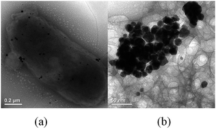
Next, the as-synthesized AuNPs were separated from bacterial cells through heating at 121 °C for 20 min, and characterized by UV-visible, dynamic light scattering (DLS) and transmission electron microscopy (TEM) analyses. The AuNPs5 and AuNPs10 showed the absorption peaks at 520 and 600 nm, respectively. DLS measurements of the AuNPs showed that the size distributions of AuNPs5 and AuNPs10 were 13.67 ± 1.46 nm and 60.46 ± 24.81 nm. The higher concentration of HAuCl4, i.e. 10 mM, contained more reducing moieties, which increased the excess growth of AuNPs through secondary reduction process. Therefore, a larger diameter of AuNPs formed as a result. Given that a smaller size of AuNPs usually had better catalytic and sensing performance, AuNPs5 was further characterized by TEM.8 TEM images showed both microbe associated (Fig. 1a) and intracellular AuNPs (Fig. 1b) released after heating at 121 °C.
![]() | ||
Fig. 1 TEM images of biogenic AuNPs produced by Cupriavidus metallidurans SHE (5 mM HAuCl4). (a) AuNPs formed around the cell wall of strain SHE. (b) Intracellular AuNPs. |
Then, the application of AuNPs in colorimetric detection of various metal ions was assayed. 1 mM of Pb2+, Hg2+, Au3+, Ag+, Cu2+, Co2+, Mn2+ and Ni2+ were added into colloid AuNPs, respectively. The metal ions except Hg2+ showed no significant shift in the SPR band of AuNPs, and the colors of the dispersions remained wine-red for at least 24 h (Fig. 2a). Unlike the L-tyrosine mediated AuNPs, there was no significant response (red-shift) for Pb2+ in this case, which exhibited a better selectivity on Hg2+.9 In the case of Hg2+, a significant shoulder peak of the SPR absorption spectra was observed, and the color of dispersions became slightly orange within 1 h. Meanwhile, gray precipitates could be observed in the bottom of tube after 12 h (Fig. 2b). This phenomenon was different from most of the AuNPs-based Hg2+ determination methods.10 According to the previous report, a blue shift of SPR band usually meant the formation of core–shell Hg–Au amalgam, and the decrease in intensity depended on the thickness of Hg shell.11 As the elemental mercury Hg0 could be easily separated from the liquid, the biogenic AuNPs may act as both sensor and scavenger of Hg2+.
![]() | ||
Fig. 2 (a) UV-visible absorption spectra of the biogenic AuNPs in the presence of 1 mM different metal ions. (b) AuNPs with (right) and without (left) 1 mM Hg2+. |
Due to the specificity on Hg2+, the colorimetric detection of Hg2+ by biogenic AuNPs was investigated in details. In the assays with different Hg2+ concentrations (ranging from 100 nM to 2 mM), it was shown that the λmax of SPR band firstly exhibited a gradually blue shift from 520 nm to 509 nm. Then, a shoulder peak appeared around 500 nm when Hg2+ concentration increased to 200 μM, and the original peak disappeared (Fig. 3a). The change of SPR band caused the color of the system changed from wine-red to orange. Fig. 3b showed the calibration curve for the detection of Hg2+ by AuNPs. When Hg2+ concentrations were in the range of 100 nM to 100 μM, a linear correlation between the absorbance ratio (A509/A520) and the Hg2+ concentration was observed (R2 = 0.9958) with the lowest detection limit of 13.2 nM, which is comparable to the other AuNPs or AgNPs based methods (Table S1†)9,12–14 Therefore, the biogenic AuNPs could be used for the detection of Hg2+ in relatively wide concentrations (100 nM to 100 μM). When the concentration of Hg2+ was higher than 100 μM, it exhibited a non-linear change. The colorimetric sensor was also tested in a real drinking water sample. There was no Hg2+ could be detected using both AuNPs and ICP-MS analyses, thus standard solutions containing Hg2+ were spiked into the drinking water samples. The results showed that no matter Hg2+ existed alone, or coexisted with other metal ions, the concentrations detected by AuNPs was in agreement with the result of ICP analysis (Table S2†). It indicated this colorimetric sensor could be a simple and expedient method for Hg2+ determination in drinking water.
![]() | ||
Fig. 3 (a) UV-visible spectra of biogenic AuNPs with different concentrations (100 nM to 200 μM) of Hg2+. (b) The calibration curve for the detection of Hg2+ by AuNPs. |
Besides the colorimetric performance of biogenic AuNPs, the capacity of biogenic AuNPs for removing mercury was also assayed by ICP-MS. Five different initial concentrations of Hg2+ were added into the colloid AuNPs and incubated for 48 h. As shown in Fig. 4, at all tested concentrations, the Hg2+ in solution could be eliminated in different degree. The elimination process of mercury could be divided into two regimes: a quick drop of Hg0 elimination rates from 60% to 23% (25 μM to 100 μM), and a much slower decrease from 23% to 13% (100 μM to 1000 μM). It could be explained by chemical adsorption of Hg0 (quick stage) and formation of amalgam (slow stage). In other reports, the amalgamation of Au–Hg at higher Hg2+ concentration and long exposure would always lead to the aggregation of particles, thus resulting in a significant red-shift in UV-visible spectrum.1 However, in our case, the red-shift was not observed neither with even 5 mM Hg2+, nor incubating the mixture for 144 h. The possible explanation should be that the coated biological molecules were charged, which could avoid the aggregation of larger amalgam particles by coulomb repulsion. This result showed that the biogenic AuNPs could remove Hg2+ efficiently.
As we thought the blue shift of SPR band was caused by the formation of Au–Hg amalgam, TEM and DLS were further employed to compare the morphology and size distribution of AuNPs with different Hg2+ concentrations. This microscopic morphology change of AuNPs was shown in Fig. 5. When the Hg2+ content was 20 μM, the size of AuNPs did not have significant difference with the sole AuNPs (Fig. 5a and b). However, after adding 1 mM Hg2+, the particles were still spheres but partly aggregated. In addition, there were some biological molecules adsorbed on the surface of nanoparticles. EDX analysis showed the local molar ratios of Hg–Au increased from 58.6/41.4 to 93.3/6.7 (atom%), which indicated AuNPs were embedded by thicker Hg shell at higher Hg concentrations (Fig. S3†). The size distributions of amalgam particles were analysed by DLS (Fig. 5c). The original AuNPs and AuNPs with 20 μM Hg2+ had similar polydispersity, and the particle size of the latter had a slightly increase, which corresponded to the coated Hg0 atoms. When 1 mM Hg2+ was incubated with AuNPs, the size distribution became narrower, and the average hydrodynamic diameter further increased to 203 ± 27 nm. The larger particles should be a fusion of AuNPs embedded into the mercury-rich matrix. Thick Hg shell dramatically affected the SPR band of AuNPs, leading to the appearance of shoulder peak in UV-visible spectrum.
![]() | ||
Fig. 5 TEM images and DLS analysis of AuNPs and Au–Hg amalgams. (a) AuNPs with 20 μM Hg2+, (b) AuNPs with 1 mM Hg2+, (c) Z-average diameter of AuNPs and Au–Hg amalgams. |
Since no extra reduction agents, such as NaBH4 and sodium citrate, were added into the colloid AuNPs, it is interesting to know how Hg2+ was reduced. After AuNPs released from bacterial cells after sterilization, there would be many biological molecules, such as peptides, DNA and oligosaccharides, adsorbed on the surface of AuNPs due to their highly biological affinity.15 Comparative experiment was performed using the sterilization product of the cells without AuNPs, and gray precipitates also appeared. Since the previous report showed free thiols in amino acid sidechain (such as cysteine) could convert Hg2+ to Hg0, We determined whether the colloid AuNPs contained proteins or peptides by Bradford method.16 This result showed protein/peptide existed in the system with a concentration about 11.4 μg mL−1, which might mediate the Hg2+ reduction. The existence of proteins/peptides capping in the surface of AuNPs was also confirmed by FTIR studies (Fig. S4†). The peaks centered at 1653 cm−1 and 1517 cm−1 corresponded to the amide I and amide II regions of proteins/peptides. In addition, the peaks centered at 1046 cm−1 in the crude extracts without AuNPs became two peaks centered at 1063 cm−1 and 1037 cm−1 when interacted with AuNPs, indicating the AuNPs may interacted with C–O group.17 Meanwhile, although mercury reductases existed in many Cupriavidus spp. strains, they should not be the reducers of Hg2+ in this case due to the high temperature treatment.18 Overall, the biogenic AuNPs generation from strain SHE and working mechanism was proposed in Fig. 6. The membrane associated Au3+ related reductase firstly reduced Au3+ to intracellular AuNPs, then AuNPs coated with biological molecule released into the solution after sterilization. Those peptides or amino acids with free thiols could reduce Hg2+ to Hg0, and make Hg0 easily adsorb on the surface of AuNPs. When the concentration of Hg2+ was relatively low (<100 μM), AuNPs could be used as a colorimetric sensor for Hg2+ due to the chemical adsorption of Hg0. Meanwhile, the biogenic AuNPs could remove biological reduced Hg through forming amalgam.
In conclusion, we demonstrated the biogenic AuNPs from Cupriavidus sp. SHE had two roles when meeting Hg2+: (1) an unusual blue-shift response for determining the concentrations of Hg2+ in a wide range, and (2) an effective mercury remover based on formation of Au–Hg amalgam. As many metabolic products of bacteria contain free thiols, this difunctional biogenic AuNPs should be easily synthesized by various other microbes. To the best of our knowledge, this is the first report about AuNPs without any extra modification could be used as an effective colorimetric sensor and scavenger of Hg2+.
Footnote |
† Electronic supplementary information (ESI) available: Experimental details, digital images of as-synthesized AuNPs and EDX spectra of AuNPs with different concentrations of Hg2+. See DOI: 10.1039/c5ra03174e |
This journal is © The Royal Society of Chemistry 2015 |