Discovery of 3-O-β-chacotriosyl oleanane-type triterpenes as H5N1 entry inhibitors†
Gaopeng Song‡
*a,
Xintian Shen‡b,
Sumei Lic,
Hongzong Sid,
Yibin Lia,
Haiye Luane,
Jihong Fana,
Qianqian Lianga and
Shuwen Liu*b
aCollege of Resources and Environment, South China Agricultural University, 510642, Guangzhou, China. E-mail: vinsin1021@126.com; Fax: +86-20-85280292; Tel: +86-20-85280293
bSchool of Pharmaceutical Sciences, Southern Medical University, 510515, Guangzhou, China. E-mail: liusw@smu.edu.cn; Fax: +86-20-6164-8655; Tel: +86-20-6164-8538
cDepartment of Human Anatomy, School of Medicine, Jinan University, 510632, Guangzhou, China
dInstitute for Computational Science and Engineering, Qingdao University, 266071, Qingdao, China
eInstitute of Agricultural Science in Coastal Area of Jiangsu Province, 224002, Yancheng, China
First published on 9th April 2015
Abstract
A series of 3-O-β-chacotriosyl oleanane-type triterpenes have been designed, synthesized and evaluated as H5N1 entry inhibitors, based on a small molecule inhibitor, saponin 1, previously discovered by us. Detailed structure–activity relationship (SAR) studies on the aglycone of compound 1 indicated that oleanane-type triterpenes with conserved structural features as the aglycone favored the antiviral activity. The results suggested that the introduction of bulky groups onto the 28-COOH of OA was helpful for significantly improving the selectivity index while keeping the antiviral activities, which was opposite to what was found for the GA analogs. Compound 5 was selected for further mechanistic studies because of its distinguished inhibition activity and good selectivity index. Molecular simulation analysis confirmed that compound 5 stabilized the HA2 subunit of hemagglutinin (HA) by binding with amino acid residues THR-189, LYS-156, and ARG-133A, and therefore may prevent HA from undergoing conformational rearrangement, which is a critical step for viral entry.
1. Introduction
The H5N1 avian influenza A virus is a serious health threat and a future pandemic risk, and has caused acute upper respiratory tract infections with high morbidity and mortality.1 Currently, the two classes of anti-influenza drugs developed for the interruption of specific processes in influenza infection are neuraminidase inhibitors, like zanamivir and oseltamivir,2 or inhibitors of the viral M2 protein, such as amantadine and rimantadine.3 However, the emergence of drug-resistant influenza viruses has limited the use of those drugs,4,5 making the identification of novel anti-influenza drugs an urgent task.Pentacyclic triterpenes are secondary plant metabolites found in different plant organs which display inhibitory activity against various viruses in vitro and in vivo.6–8 It is suggested that the protective activities of pentacyclic triterpenes stem from their ability to prevent various pathogen and herbivore attacks on the host.9 For example, oleanolic acid (OA), an oleanane-type triterpene, and betulinic acid (BA), a lupane-type triterpene, have been confirmed to display inhibitory activity against HIV entry.10–12 Notably, certain triterpenoic glycosides exhibit good anti-influenza virus activity, of which the biological effects are attributed to the presence of the aglycone as well as the sugar moiety.13–15 Glycyrrhizic acid (GL), the most intensively investigated bioactive compound of licorice roots, is a glycosyl triterpenoic acid whose aglycone, glycyrrhetinic acid (GA), can be obtained easily from the extract of liquorice roots.16 GL is well known for its broad activity against several viruses in vitro and in vivo, including influenza A viruses (IAV).13 The inhibition of virus penetration was proposed as a mechanism of action of GL against various virus infections, which can lead to an inhibition of fusion pore formation and hence reduce infection by various viruses.13 In addition, compound Y3, an OA–acetyl galactose conjugate, is a member of the triterpenoic glycosides that has displayed strong anti-influenza virus activity in vitro.15
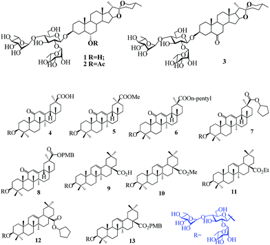
Influenza virus infection starts with the attachment of viral particles to the host cell. This process is mediated by hemagglutinin (HA), which is a type of viral envelope glycoprotein that binds to a sialic acid receptor on the host cell, leading to viral endocytosis.17 Influenza virus entry is a potential target for discovering novel anti-influenza drugs because blocking the first step of influenza virus infection could result in an efficient inhibition of virus propagation. In our previous study, we used an efficient HIV-based pseudotyping system to screen a saponin library generated from semisynthesis, and discovered three small molecule H5N1 viral entry inhibitors 1–3 (Fig. 1A), which show good inhibitory activity against H5N1 entry with IC50 values of 7.2–12.0 μM.18 The trisaccharide moiety of these inhibitory molecules, namely chacotrioside, has been the subject of several SAR studies dealing with alkylations,19 removal or positional change of some of its α-L-rhamnosyl residues,18,19 and conjugation to triterpene aglycones,18 which indicate that the chacotriosyl residue is essential for activity and subtle modifications of the aglycone may be tolerated without losing antiviral activity.
Based on the above results, given that GL shows good antiviral activity against IAV entry, we replaced the chlorogenin moiety of active compound 1 with GA to attempt the synthesis of several 3-O-β-chacotriosyl GA analogs 4–8. Structurally, OA belongs to the oleanane-type triterpenes, as does GA, with the C-29 COOH group shifted to C-28. Compound 9 was synthesized to investigate the effect of subtle differences in oleanane-type triterpenes as the aglycone residue on the inhibitory activity. In addition, different hydrocarbyl oleanolates were selected as the aglycone substitutes to derive saponins 10–13 (Fig. 1B), which provides a means to judge the effects of esterification of the 28-COOH group of OA on the inhibitory activity.
2. Results and discussion
2.1 Chemistry
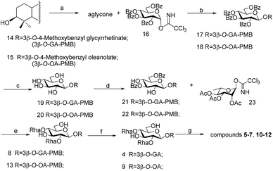
As depicted in Scheme 1, treatment of glycyrrhetinic acid or oleanolic acid with 4-methoxybenzyl chloride in the presence of K2CO3 in DMF provided compound 14 or 15, respectively, in good yield. The activation of the anomeric center is mandatory for the glycoside synthesis. Ample examples show that the trichloroacetimidate group activated by the Lewis acid trifluoromethanesulfonate (TMSOTf) or boron fluoride ethyl ether (BF3·Et2O) becomes a suitable leaving group for the nucleophilic attack of an alcohol. Hence, 2,3,4,6-tetra-O-benzoyl-D-glucopyranosyl trichloroacetimidate 16 was prepared via a three-step protocol.18 Glycosylation of the intermediates 14,15 with trichloroacetimidate 16 under the action of TMSOTf afforded the 3-O-β-glucopyranosides 17,18, respectively. Removal of the benzoyl group was achieved using NaOMe in MeOH to yield compounds 19,20, which were reacted with 1-(benzoyloxy)-benzotriazole (1-BBTZ), effecting selective protection of the 3,6-OHs of the β-glucopyranosyl residues to obtain compounds 21,22. Subsequent glycosylation of the 2,4-OHs in 21,22 with 2,3,4-tri-O-acetyl-L-rhamnopyranosyl trichloroacetimidate 23 (ref. 18) under the “inverse addition conditions” with TMSOTf as the promoter, followed by deprotection of the acyl groups with MeONa in MeOH, afforded saponins 8 and 13, respectively. Debenzylation of compounds 8 and 13 was carried out via hydrogenolysis using Pd/C to provide compounds 4 and 9. Finally, treatment of 4 or 9 with different halohydrocarbon in the presence of K2CO3 in DMF yielded compounds 5–7 or 10–12, respectively.2.2 Bioactivity
Due to the safety concerns in studying viral H5N1 pathogens, the single-cycle pseudovirus was used instead of live H5N1 avian influenza virus to evaluate the inhibitory activities of compounds 4–13 against H5N1 entry. In this report, compounds 1–13 were evaluated for the inhibitory activity against the entry of H5N1 influenza virus based on an efficient HIV-based pseudotyping system with a luciferase report element, established by us,20,21 while the VSV-G/HIV pseudovirions were used as specificity controls. The obtained IC50 values are reported in Table 1 and indicate that all the compounds showed inhibitory activity against the H5N1 pseudovirus with potencies ranging from moderate (IC50 > 10 μM) to potent (IC50 < 5 μM, which is comparable to the IC50 of a positive control compound, CL-385319 (ref. 21 and 22)). Notably, the active compounds in series 5–6 and 10–13 display stronger inhibition activity than compound 1, whilst having weaker cytotoxicity against Madin Darby Canine Kidney (MDCK) cells than compound 1.
| Compound | IC50a | CC50b | SIc |
|---|---|---|---|
| a IC50: compound concentration required to achieve 50% inhibition of replication of H5N1, as determined by plaque reduction assays.b CC50: compound concentration required to cause 50% death of uninfected MDCK cells, as determined by the MTT method.c SI: selectivity index as CC50/IC50. | |||
| 1 | 7.72 ± 0.11 | 12.02 ± 0.32 | 1.7 |
| 2 | 9.16 ± 0.23 | 16.85 ± 0.24 | 1.8 |
| 3 | 11.62 ± 0.55 | 17.22 ± 0.30 | 1.5 |
| 4 | 22.08 ± 0.25 | 35.80 ± 0.53 | 1.6 |
| 5 | 3.35 ± 0.44 | 33.83 ± 0.58 | 10.1 |
| 6 | 6.86 ± 0.12 | 15.25 ± 0.18 | 2.2 |
| 7 | 10.35 ± 0.26 | 14.42 ± 0.15 | 1.4 |
| 8 | 11.72 ± 0.58 | 15.20 ± 0.85 | 1.3 |
| 9 | 9.58 ± 0.22 | 20.65 ± 0.88 | 2.2 |
| 10 | 4.13 ± 0.97 | 18.64 ± 2.26 | 4.5 |
| 11 | 6.19 ± 0.53 | 37.36 ± 0.35 | 6.0 |
| 12 | 4.80 ± 0.62 | 56.25 ± 0.85 | 11.7 |
| 13 | 7.62 ± 1.53 | 55.41 ± 1.81 | 7.3 |
| GA | >50 | 21.56 ± 0.65 | |
| Zanamivir | 0.92 ± 0.08 | >100 | >108.6 |
| CL-385319 | 4.45 ± 1.25 | 1480 ± 10 | 332.3 |
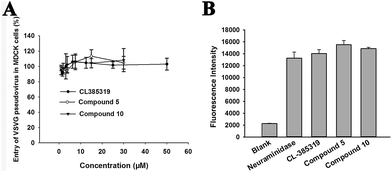
The representative compounds 5 and 10 had no effect on the VSV-G enveloped pseudovirus (Fig. 2A), similarly to the HA targeted compound CL-385319.21,22 These results demonstrate that these molecules did not inhibit the HIV-backbone and luciferase reporting activity. In addition, compounds 5 and 10 at 20 μM did not inhibit neuraminidase (NA) (Fig. 2B), a glycoprotein enveloped in the pseudovirus system. Thus, these results revealed that compounds 4–13 might interfere with the entry of influenza virus by targeting hemagglutinin, the only other glycoprotein enveloped in the pseudovirus system.
The SAR exploration first focused on the oleanane-type triterpenes GA and OA as aglycone residues and their inhibition activities. Interestingly, compounds 5–6 and 10–12 showed stronger inhibitory activity than the reported H5N1 entry inhibitor 1, suggesting that replacement of the aglycone moiety of compound 1 with oleanane-type triterpenes with conserved structural features can enhance inhibitory activity. Then we turned our attention to the influence of esterification of the COOH group, with different carbon chain lengths and sizes, on the inhibition activity and cytotoxicity against MDCK cells. It was found that esterification of the COOH group of either GA or OA enhanced the inhibitory activity (5 > 4, 10 > 9). We supposed that the hydroxyl group of COOH did not contribute to the interaction of the aglycone residue with the receptor. The extent of the inhibitory activities of the compounds, depending on the substitution of the COOH group in the side chain, can be ordered as follows: GA derivatives were in the order of 5 > 6 > 7 ≈ 8, and OA derivatives were in the order of 10 ≈ 12 > 11 > 13. Replacement of the hydroxy group at the 29-COOH position of GA with a methyl group resulted in the most significant increases in potency, while a dramatic loss of inhibition activity was observed when 29-COOH of GA was substituted with an n-pentyl group or cyclopentyl group, indicating that the introduction of a bulky group may increase the steric hindrance and decrease binding with the receptor. However, when the 29-COOH of the GA derivatives was substituted with a fatty alkyl group, the cytotoxicity against MDCK cells was significantly enhanced as the length of the carbon chain increased (8 ≈ 7 > 6 > 5). Taken together, these results suggested that when the 29-COOH group of GA was esterified, introduction of short, straight alkyl groups was helpful for enhancing the inhibitory activity and decreasing cytotoxicity, whereas the introduction of bulky groups was not tolerated.
In contrast to the GA derivatives, 28-COOH of OA was substituted with an Et or PMB group (to give compound 11 or 12) to lead to slightly reduced inhibition activity compared with compound 10, suggesting that the inhibition activities of the OA derivatives were not affected significantly by the carbon length and size in the side chains of the COOH group. This finding was different to that from the modification of the 29-COOH group in GA, which was presumably due to the obvious steric hindrance of the 28-COOH group in OA. Compound 12, an analog of compound 10, had an improved selective index (4.5 to 11.7), indicating that the introduction of a bulky group at the 28-COOH position of OA could significantly improve the safety of these compounds while keeping their antiviral activity.
![[thin space (1/6-em)]](https://www.rsc.org/images/entities/char_2009.gif) :
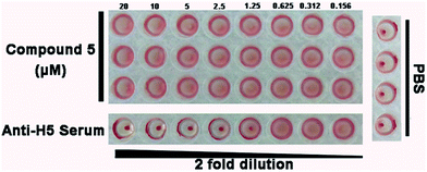
:![[thin space (1/6-em)]](https://www.rsc.org/images/entities/char_2009.gif) 10 (Fig. 3). This result suggested that compound 5 may not target the receptor binding domain of HA1.
10 (Fig. 3). This result suggested that compound 5 may not target the receptor binding domain of HA1.
2.3 Molecular modeling of compound 5 binding to H5 hemagglutinin
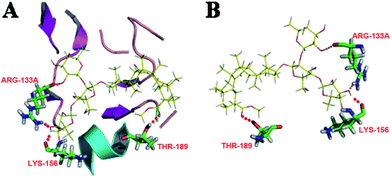
We found that the HA2 sequence of A/Thailand/Kan353/2004 was 100% identical to that of A/Vietnam/1194/2004, of which the neutral-pH crystal structure of HA has been recently well-defined. On the basis of the results, we performed a docking study using the Surflex-Dock program of SYBYL 7.3 in order to determine the mechanism and mode of interaction between compound 5 and the HA structure of A/Vietnam/1194/2004. The crystal structures of HA (1RVT) were extracted from the Protein Data Bank. Compound 5 was docked within the active site of HA (1RVT) and the results showed that compound 5 exhibited high binding affinity with a score of 4.267. Compound 5 could fit inside the same cavity in the stem region of HA2 near the fusion peptide through hydrophobic contacts, van der Waals interactions, and hydrogen bonding networks. As shown in Fig. 4, at the top of the cavity, compound 5 occupied the binding site with C2–OH of the L-rhamnose moiety linked to C2–OH of D-glucose forming stable hydrogen bonds with ARG-133A. The hydroxyl group of the L-rhamnose moiety linked to C4–OH of D-glucose and the carbonyl group at the C-29 position of GA could also form stable hydrogen bonds with LYS-156 and THR-189 at the bottom of the cavity, respectively. As expected, the analysis of the binding models also gave our preceding study support, suggesting that the chacotriosyl residue might play a very important role in the anti-H5N1 activity, since changing the β-chacotriosyl moiety into an α-L-rhamnopyranosyl-(1 → 4)-β-D-glucopyranosyl moiety or α-L-rhamnopyranosyl-(1 → 2)-β-D-glucopyranosyl moiety resulted in the loss of activity.18 The results also show that compound 5 could be a potential anti-H5N1 entry inhibitor, likely targeting the HA2 protein, which is different from the other known entry inhibitors.21,222.4 Pharmacokinetic properties and bioavailability of compound 5
In this study, we have employed the ACD lab program to predict the pharmacokinetic properties of compound 5. As shown in Table 2, compound 5 showed no obvious carcinogenic toxicity and mutagenic toxicity. However, compound 5 exhibited poor gastrointestinal absorption and subsequent hepatic first-pass metabolism, which might lead to low oral bioavailability. The logarithm of the partition coefficient (log![[thin space (1/6-em)]](https://www.rsc.org/images/entities/char_2009.gif) P) is a parameter which reflects the drug equilibrium partition ratio between polar (water) and non-polar (octanol) phases, and it has been proven to determine the absorption, distribution, biological availability, and pharmacological activity of drugs. Compound 5 exhibited high anhydrous solubility with a log
P) is a parameter which reflects the drug equilibrium partition ratio between polar (water) and non-polar (octanol) phases, and it has been proven to determine the absorption, distribution, biological availability, and pharmacological activity of drugs. Compound 5 exhibited high anhydrous solubility with a log![[thin space (1/6-em)]](https://www.rsc.org/images/entities/char_2009.gif) P value of 3.32, suggesting that the water solubility of compound 5 should be improved. In a recent study, Cao et al.23 showed that propylene glycol-linked amino acid/dipeptide diester prodrugs of OA showed better stability, permeability, affinity, and bioavailability. In order to further increase the bioavailability and optimize the ADME properties of compound 5, we next plan to design and synthesize a series of compound 5 analogs as prodrugs of 3-O-β-chacotriosyl GA, of which 29-COOH is modified with polar groups such as amino acids and sugars.
P value of 3.32, suggesting that the water solubility of compound 5 should be improved. In a recent study, Cao et al.23 showed that propylene glycol-linked amino acid/dipeptide diester prodrugs of OA showed better stability, permeability, affinity, and bioavailability. In order to further increase the bioavailability and optimize the ADME properties of compound 5, we next plan to design and synthesize a series of compound 5 analogs as prodrugs of 3-O-β-chacotriosyl GA, of which 29-COOH is modified with polar groups such as amino acids and sugars.
![[thin space (1/6-em)]](https://www.rsc.org/images/entities/char_2009.gif) P and toxicity of the lead compound 5a
P and toxicity of the lead compound 5a
| Compd | log![[thin space (1/6-em)]](https://www.rsc.org/images/entities/char_2009.gif) P P |
Carcinogenic toxicity | Mutagenic toxicity | ||||
|---|---|---|---|---|---|---|---|
| CP | CIP | Pred. | MP | MIP | Pred. | ||
a log![[thin space (1/6-em)]](https://www.rsc.org/images/entities/char_2009.gif) P means octanol–water partition coefficient, which is equal to the logarithm of the ratio of concentrations of an unionized compound between octanol and water. For the carcinogenic toxicity, when the predicted value is more than 70%, and the value of CIP (Carcinogenic Impossibility) is more than 0.65, meanwhile its CIP value is greater than CP (Carcinogenic Possibility), the compound is considered as non-carcinogenic. Otherwise, they are considered to be carcinogenic. For the mutagenic toxicity, if the predictability value is more than 70% and its MIP (Mutagenic Impossibility) value is greater than the MP (Mutagenic Possibility), the compound is considered as nonmutagenic. Otherwise, they are considered to be mutagenic. P means octanol–water partition coefficient, which is equal to the logarithm of the ratio of concentrations of an unionized compound between octanol and water. For the carcinogenic toxicity, when the predicted value is more than 70%, and the value of CIP (Carcinogenic Impossibility) is more than 0.65, meanwhile its CIP value is greater than CP (Carcinogenic Possibility), the compound is considered as non-carcinogenic. Otherwise, they are considered to be carcinogenic. For the mutagenic toxicity, if the predictability value is more than 70% and its MIP (Mutagenic Impossibility) value is greater than the MP (Mutagenic Possibility), the compound is considered as nonmutagenic. Otherwise, they are considered to be mutagenic. |
|||||||
| 5 | 3.32 | 0.001 | 0.345 | 98% | 0.01 | 0.39 | 98% |
3. Conclusion
Based on our previously discovered small molecule inhibitor 1, a series of 3-O-β-chacotriosyl oleanane-type triterpenes were designed and synthesized as H5N1 entry inhibitors, of which intensive SAR studies on the aglycone were conducted. Our results showed that oleanane-type triterpenes with conserved structural features as the aglycone can improve inhibitory activity. When the 29-COOH of GA was esterified, introduction of short, straight saturated alkyl groups was helpful in enhancing inhibitory activity, but the introduction of bulky groups should be avoided. Conversely, the introduction of bulky groups onto 28-COOH of OA maintained the antiviral activity and greatly decreased cytotoxicity against MDCK cells.Molecular simulation analysis confirmed that compound 5 stabilized the HA2 subunit of hemagglutinin (HA) by binding with amino acid residues THR-189, LYS-156, and ARG-133A; therefore compound 5 may prevent HA from undergoing conformational rearrangement induced by acidification in the cell endosome, which is a critical step for viral entry. Compound 5, which can block the entry of H5N1 avian influenza by interfering with viral entry, can serve as a lead for optimization in order to design new compounds with improved potency and reduced toxicity.
4. Experimental protocols
4.1 General methods
Solvents were purified in a conventional manner. Thin layer chromatography (TLC) was performed on precoated E. Merck silica gel 60 F254 plates. Flash column chromatography was performed on silica gel (200–300 mesh, Qingdao, China). 1H NMR and 13C NMR spectra were obtained using a JEOL JNM-ECP 600 spectrometer with tetramethylsilane as an internal standard, and chemical shifts are recorded in ppm values. Mass spectra were recorded on a Q-TOF Global mass spectrometer.4.2 Synthesis
![[thin space (1/6-em)]](https://www.rsc.org/images/entities/char_2009.gif) :
:![[thin space (1/6-em)]](https://www.rsc.org/images/entities/char_2009.gif) 1
1![[thin space (1/6-em)]](https://www.rsc.org/images/entities/char_2009.gif) :
:![[thin space (1/6-em)]](https://www.rsc.org/images/entities/char_2009.gif) 1) to afford compound 17 (2.12 g, 89%) as a white solid. 1H NMR (CDCl3): δ 6.91–8.03 (m, 24H, Ar-H), 5.92 (t, 1H, J = 9.6 Hz, H-3′), 5.60 (t, 1H, J = 9.5 Hz, H-4′), 5.58 (s, 1H, H-12), 5.57 (t-like, 1H, J = 9.5, 8.1 Hz, H-2′), 5.14 (d, 1H, J = 11.9 Hz, Ar-CH2-1), 5.07 (d, 1H, J = 11.9 Hz, Ar-CH2-2), 4.88 (d, 1H, J = 8.0 Hz, H-1′), 4.66 (dd, 1H, J = 11.9, 3.2 Hz, H-6′-1), 4.49 (dd, 1H, J = 11.9, 6.9 Hz, H-6′-2), 4.14–4.17 (m, 1H, H-5′), 3.83 (s, 3H, OCH3), 3.15 (dd, 1H, J = 11.6, 4.7 Hz, H-3), 2.75 (dt, 1H, J = 13.4, 4.1 Hz, H-1), 2.22 (s, 1H, H-9), 1.31, 1.15, 1.10, 1.08, 0.87, 0.73, 0.71 (each s, each 3H, CH3); 13C NMR (CDCl3): δ 199.8 (C-11), 176.2 (C-30), 166.6, 165.9, 165.3, 165.0, 159.7, 133.4, 133.3, 133.2, 133.0, 130.1, 129.9, 129.8, 129.7, 129.6, 129.5, 128.9, 128.8, 128.5, 128.4 (two), 128.3 (two), 114.0, 103.1 (C-1′), 90.5, 73.0, 72.2, 72.0, 70.3, 66.0, 63.4, 61.7, 55.3, 55.2, 48.2, 45.3, 43.9, 43.1, 39.1, 39.0, 37.6, 36.8, 32.7, 31.7, 31.2, 28.4, 28.3, 27.6, 26.4 (two), 25.8, 23.3, 18.6, 17.3, 16.3, 16.2; ESIMS calcd for C72H80O14Na 1191.5; found 1191.5.
1) to afford compound 17 (2.12 g, 89%) as a white solid. 1H NMR (CDCl3): δ 6.91–8.03 (m, 24H, Ar-H), 5.92 (t, 1H, J = 9.6 Hz, H-3′), 5.60 (t, 1H, J = 9.5 Hz, H-4′), 5.58 (s, 1H, H-12), 5.57 (t-like, 1H, J = 9.5, 8.1 Hz, H-2′), 5.14 (d, 1H, J = 11.9 Hz, Ar-CH2-1), 5.07 (d, 1H, J = 11.9 Hz, Ar-CH2-2), 4.88 (d, 1H, J = 8.0 Hz, H-1′), 4.66 (dd, 1H, J = 11.9, 3.2 Hz, H-6′-1), 4.49 (dd, 1H, J = 11.9, 6.9 Hz, H-6′-2), 4.14–4.17 (m, 1H, H-5′), 3.83 (s, 3H, OCH3), 3.15 (dd, 1H, J = 11.6, 4.7 Hz, H-3), 2.75 (dt, 1H, J = 13.4, 4.1 Hz, H-1), 2.22 (s, 1H, H-9), 1.31, 1.15, 1.10, 1.08, 0.87, 0.73, 0.71 (each s, each 3H, CH3); 13C NMR (CDCl3): δ 199.8 (C-11), 176.2 (C-30), 166.6, 165.9, 165.3, 165.0, 159.7, 133.4, 133.3, 133.2, 133.0, 130.1, 129.9, 129.8, 129.7, 129.6, 129.5, 128.9, 128.8, 128.5, 128.4 (two), 128.3 (two), 114.0, 103.1 (C-1′), 90.5, 73.0, 72.2, 72.0, 70.3, 66.0, 63.4, 61.7, 55.3, 55.2, 48.2, 45.3, 43.9, 43.1, 39.1, 39.0, 37.6, 36.8, 32.7, 31.7, 31.2, 28.4, 28.3, 27.6, 26.4 (two), 25.8, 23.3, 18.6, 17.3, 16.3, 16.2; ESIMS calcd for C72H80O14Na 1191.5; found 1191.5.![[thin space (1/6-em)]](https://www.rsc.org/images/entities/char_2009.gif) :
:![[thin space (1/6-em)]](https://www.rsc.org/images/entities/char_2009.gif) 1) and then NaOMe was added until pH = 10. After stirring at r.t. for 6 h, the solution was neutralized with Dowex 50 × 8 (H+) resin until pH = 7, filtered and concentrated. Then the residue was purified by silica gel column chromatography (CH2Cl2–MeOH, 10
1) and then NaOMe was added until pH = 10. After stirring at r.t. for 6 h, the solution was neutralized with Dowex 50 × 8 (H+) resin until pH = 7, filtered and concentrated. Then the residue was purified by silica gel column chromatography (CH2Cl2–MeOH, 10![[thin space (1/6-em)]](https://www.rsc.org/images/entities/char_2009.gif) :
:![[thin space (1/6-em)]](https://www.rsc.org/images/entities/char_2009.gif) 1) to provide compound 19 (1.11 g, 93%); 1H NMR (CDCl3): δ 7.31 (d, 2H, J = 8.7 Hz, Ar-H), 6.90 (d, 2H, J = 8.7 Hz, Ar-H), 5.56 (s, 1H, H-12), 5.13 (d, 1H, J = 11.8 Hz, Ar-CH2-1), 5.03 (d, 1H, J = 11.8 Hz, Ar-CH2-2), 4.38 (d, 1H, J = 7.1 Hz, H-1′), 3.82–3.85 (m, 2H, H-6′), 3.81 (s, 3H, OCH3), 3.63 (t, 1H, J = 9.3 Hz, H-4′), 3.55 (t, 1H, J = 9.0 Hz, H-3′), 3.43–3.45 (m, 1H, H-5′), 3.31 (d, 1H, J = 10.5 Hz, H-3), 3.19 (t-like, 1H, J = 8.8, 7.8 Hz, H-2′), 2.77–2.80 (m, 1H, H-1), 2.31 (s, 1H, H-9), 1.34, 1.15, 1.14, 1.05, 0.86, 0.73 (each s, each 3H, CH3); 13C NMR (CDCl3): δ 200.0 (C-11), 176.2 (C-30), 169.0, 159.6, 130.0 (two), 128.5, 128.4, 114.1 (two), 105.1 (C-1′), 89.8, 76.4, 75.3, 73.9, 69.7, 66.0 (two), 61.8, 60.4, 55.3, 48.1, 45.4, 44.0, 43.1, 41.1, 39.5, 39.2, 37.6, 36.8, 32.8, 31.7, 31.1, 28.4, 28.2, 28.0, 26.5, 26.1, 23.4, 18.7, 17.4, 16.7, 16.7, 16.4; ESIMS calcd for C44H64O10Na 775.4; found 775.4.
1) to provide compound 19 (1.11 g, 93%); 1H NMR (CDCl3): δ 7.31 (d, 2H, J = 8.7 Hz, Ar-H), 6.90 (d, 2H, J = 8.7 Hz, Ar-H), 5.56 (s, 1H, H-12), 5.13 (d, 1H, J = 11.8 Hz, Ar-CH2-1), 5.03 (d, 1H, J = 11.8 Hz, Ar-CH2-2), 4.38 (d, 1H, J = 7.1 Hz, H-1′), 3.82–3.85 (m, 2H, H-6′), 3.81 (s, 3H, OCH3), 3.63 (t, 1H, J = 9.3 Hz, H-4′), 3.55 (t, 1H, J = 9.0 Hz, H-3′), 3.43–3.45 (m, 1H, H-5′), 3.31 (d, 1H, J = 10.5 Hz, H-3), 3.19 (t-like, 1H, J = 8.8, 7.8 Hz, H-2′), 2.77–2.80 (m, 1H, H-1), 2.31 (s, 1H, H-9), 1.34, 1.15, 1.14, 1.05, 0.86, 0.73 (each s, each 3H, CH3); 13C NMR (CDCl3): δ 200.0 (C-11), 176.2 (C-30), 169.0, 159.6, 130.0 (two), 128.5, 128.4, 114.1 (two), 105.1 (C-1′), 89.8, 76.4, 75.3, 73.9, 69.7, 66.0 (two), 61.8, 60.4, 55.3, 48.1, 45.4, 44.0, 43.1, 41.1, 39.5, 39.2, 37.6, 36.8, 32.8, 31.7, 31.1, 28.4, 28.2, 28.0, 26.5, 26.1, 23.4, 18.7, 17.4, 16.7, 16.7, 16.4; ESIMS calcd for C44H64O10Na 775.4; found 775.4.![[thin space (1/6-em)]](https://www.rsc.org/images/entities/char_2009.gif) :
:![[thin space (1/6-em)]](https://www.rsc.org/images/entities/char_2009.gif) 8
8![[thin space (1/6-em)]](https://www.rsc.org/images/entities/char_2009.gif) :
:![[thin space (1/6-em)]](https://www.rsc.org/images/entities/char_2009.gif) 2) to provide 21 (1.01 g, 70%) as a white solid; 1H NMR (CDCl3): δ 8.07–8.11 (m, 4H, Ar-H), 7.60 (t, 2H, J = 8.2 Hz, Ar-H), 7.45–7.50 (m, 4H, Ar-H), 7.33 (d, 2H, J = 8.6 Hz, Ar-H), 6.93 (d, 2H, J = 8.6 Hz, Ar-H), 5.60 (s, 1H, H-12), 5.23 (t, 1H, J = 9.1 Hz, H-3′), 5.15 (d, 1H, J = 11.9 Hz, Ar-CH2-1), 5.07 (d, 1H, J = 11.9 Hz, Ar-CH2-2), 4.72 (dd, 1H, J = 11.2, 3.2 Hz, H-6′-1), 4.62 (dd, 1H, J = 11.2, 5.6 Hz, H-6′-2), 4.52 (d, 1H, J = 7.8 Hz, H-1′), 3.83 (s, 3H, OCH3), 3.74–3.76 (m, 3H, H-4′, H-5′, H-2′), 3.22 (d, 1H, J = 11.8, 4.6 Hz, H-3), 2.75 (dt, 1H, J = 13.4, 3.1 Hz, H-3), 2.27 (s, 1H, H-9), 1.34, 1.16, 1.14, 1.11, 1.03, 0.86, 0.74 (each s, each 3H, CH3); 13C NMR (CDCl3): δ 199.9 (C-11), 176.3 (C-30), 169.0, 167.7, 166.7, 159.5, 133.5, 133.3, 130.1 (two), 130.0 (two), 129.8 (two), 129.7, 129.4, 128.5 (two), 128.4 (two), 128.3, 114.0 (two), 104.8 (C-1′), 90.1, 78.6, 74.2, 72.8, 70.0, 66.0, 64.0, 61.7, 55.3, 55.2, 48.2, 45.3, 44.0, 43.1, 41.1, 39.3, 39.0, 37.6, 36.8, 32.7, 31.8, 31.2, 28.4, 28.3, 28.2, 26.5, 26.4, 25.9, 23.3, 18.7, 17.3, 16.6, 16.3; ESIMS calcd for C58H72O12Na 983.5; found 983.5.
2) to provide 21 (1.01 g, 70%) as a white solid; 1H NMR (CDCl3): δ 8.07–8.11 (m, 4H, Ar-H), 7.60 (t, 2H, J = 8.2 Hz, Ar-H), 7.45–7.50 (m, 4H, Ar-H), 7.33 (d, 2H, J = 8.6 Hz, Ar-H), 6.93 (d, 2H, J = 8.6 Hz, Ar-H), 5.60 (s, 1H, H-12), 5.23 (t, 1H, J = 9.1 Hz, H-3′), 5.15 (d, 1H, J = 11.9 Hz, Ar-CH2-1), 5.07 (d, 1H, J = 11.9 Hz, Ar-CH2-2), 4.72 (dd, 1H, J = 11.2, 3.2 Hz, H-6′-1), 4.62 (dd, 1H, J = 11.2, 5.6 Hz, H-6′-2), 4.52 (d, 1H, J = 7.8 Hz, H-1′), 3.83 (s, 3H, OCH3), 3.74–3.76 (m, 3H, H-4′, H-5′, H-2′), 3.22 (d, 1H, J = 11.8, 4.6 Hz, H-3), 2.75 (dt, 1H, J = 13.4, 3.1 Hz, H-3), 2.27 (s, 1H, H-9), 1.34, 1.16, 1.14, 1.11, 1.03, 0.86, 0.74 (each s, each 3H, CH3); 13C NMR (CDCl3): δ 199.9 (C-11), 176.3 (C-30), 169.0, 167.7, 166.7, 159.5, 133.5, 133.3, 130.1 (two), 130.0 (two), 129.8 (two), 129.7, 129.4, 128.5 (two), 128.4 (two), 128.3, 114.0 (two), 104.8 (C-1′), 90.1, 78.6, 74.2, 72.8, 70.0, 66.0, 64.0, 61.7, 55.3, 55.2, 48.2, 45.3, 44.0, 43.1, 41.1, 39.3, 39.0, 37.6, 36.8, 32.7, 31.8, 31.2, 28.4, 28.3, 28.2, 26.5, 26.4, 25.9, 23.3, 18.7, 17.3, 16.6, 16.3; ESIMS calcd for C58H72O12Na 983.5; found 983.5.![[thin space (1/6-em)]](https://www.rsc.org/images/entities/char_2009.gif) :
:![[thin space (1/6-em)]](https://www.rsc.org/images/entities/char_2009.gif) 3) to give the desired crude trisaccharide. The trisaccharide mixture was dissolved in CH2Cl2 and CH3OH (v/v = 1
3) to give the desired crude trisaccharide. The trisaccharide mixture was dissolved in CH2Cl2 and CH3OH (v/v = 1![[thin space (1/6-em)]](https://www.rsc.org/images/entities/char_2009.gif) :
:![[thin space (1/6-em)]](https://www.rsc.org/images/entities/char_2009.gif) 1) and then NaOMe was added until pH = 10. After stirring at r.t. for 12 h, the solution was neutralized with Dowex 50 × 8 (H+) resin until pH = 7, filtered and concentrated. Then the residue was purified by silica gel column chromatography (CH2Cl2–MeOH, 6
1) and then NaOMe was added until pH = 10. After stirring at r.t. for 12 h, the solution was neutralized with Dowex 50 × 8 (H+) resin until pH = 7, filtered and concentrated. Then the residue was purified by silica gel column chromatography (CH2Cl2–MeOH, 6![[thin space (1/6-em)]](https://www.rsc.org/images/entities/char_2009.gif) :
:![[thin space (1/6-em)]](https://www.rsc.org/images/entities/char_2009.gif) 1) to provide compound 8 (0.36 g, 68%) as a white solid; 1H NMR (CD3OD): δ 7.60 (d, 2H, J = 8.6 Hz, Ar-H), 6.94 (d, 2H, J = 8.6 Hz, Ar-H), 5.44 (s, 1H, H-12), 5.38 (s, 1H, Rha-1-H), 5.20 (d, 1H, J = 11.8 Hz, Ar-CH2-1), 5.03 (d, 1H, J = 11.8 Hz, Ar-CH2-2), 4.59 (s, 1H, Rha-1-H), 4.44 (d, 1H, J = 7.7 Hz, H-1′), 3.98–4.00 (m, 2H), 3.91–3.92 (m, 1H), 3.85 (dd, 1H, J = 3.0, 1.8 Hz, Rha-H-2), 3.81 (dd, 1H, J = 12.0, 1.8 Hz, H-6′-1), 3.81 (s, 3H, OCH3), 3.75 (dd, 1H, J = 9.5, 3.2 Hz, Rha-H-3), 3.68 (dd, 1H, J = 12.0, 4.0 Hz, H-6′-2), 3.63 (dd, 1H, J = 9.4, 3.3 Hz, Rha-H-3), 3.60 (t, 1H, J = 8.8 Hz), 3.55 (t, 1H, J = 9.2 Hz), 3.46 (t, 1H, J = 8.3 Hz), 3.36–3.45 (m, 3H), 3.20 (dd, 1H, J = 11.8, 4.3 Hz, H-3), 2.71 (td, 1H, J = 13.3, 4.1 Hz, H-1), 2.42 (s, 1H, H-9), 2.11 (td, 1H, J = 13.6, 4.1 Hz, H-18), 1.39, 1.15, 1.14, 1.13, 1.08, 0.89, 0.71 (each s, each 3H, CH3), 1.28 (d, 3H, J = 6.2 Hz, Rha-H-6), 1.23 (d, 3H, J = 6.2 Hz, Rha-H-6); 13C NMR (CD3OD): δ 201.1 (C-11), 176.5 (C-30), 171.0, 159.9, 130.1 (two), 128.4, 127.5, 113.6 (two), 104.1 (C-1′), 101.7 (Rha-C-1), 100.6 (Rha-C-1), 88.7, 79.1, 78.1, 77.9, 76.8, 75.1, 72.6, 72.3, 71.0, 70.7, 70.6, 69.4, 68.6, 65.7, 61.8, 60.6, 55.3, 54.4, 48.2, 45.3, 43.7, 43.2, 41.0, 39.2 (two), 37.4, 36.6, 32.4, 31.4, 30.7, 27.6, 27.0 (two), 26.1, 25.9, 25.8, 22.4, 17.9, 17.1, 17.0, 16.6, 16.5, 15.8, 15.7; HRESIMS calcd for C56H84O18Na 1067.5555; found 1067.5551.
1) to provide compound 8 (0.36 g, 68%) as a white solid; 1H NMR (CD3OD): δ 7.60 (d, 2H, J = 8.6 Hz, Ar-H), 6.94 (d, 2H, J = 8.6 Hz, Ar-H), 5.44 (s, 1H, H-12), 5.38 (s, 1H, Rha-1-H), 5.20 (d, 1H, J = 11.8 Hz, Ar-CH2-1), 5.03 (d, 1H, J = 11.8 Hz, Ar-CH2-2), 4.59 (s, 1H, Rha-1-H), 4.44 (d, 1H, J = 7.7 Hz, H-1′), 3.98–4.00 (m, 2H), 3.91–3.92 (m, 1H), 3.85 (dd, 1H, J = 3.0, 1.8 Hz, Rha-H-2), 3.81 (dd, 1H, J = 12.0, 1.8 Hz, H-6′-1), 3.81 (s, 3H, OCH3), 3.75 (dd, 1H, J = 9.5, 3.2 Hz, Rha-H-3), 3.68 (dd, 1H, J = 12.0, 4.0 Hz, H-6′-2), 3.63 (dd, 1H, J = 9.4, 3.3 Hz, Rha-H-3), 3.60 (t, 1H, J = 8.8 Hz), 3.55 (t, 1H, J = 9.2 Hz), 3.46 (t, 1H, J = 8.3 Hz), 3.36–3.45 (m, 3H), 3.20 (dd, 1H, J = 11.8, 4.3 Hz, H-3), 2.71 (td, 1H, J = 13.3, 4.1 Hz, H-1), 2.42 (s, 1H, H-9), 2.11 (td, 1H, J = 13.6, 4.1 Hz, H-18), 1.39, 1.15, 1.14, 1.13, 1.08, 0.89, 0.71 (each s, each 3H, CH3), 1.28 (d, 3H, J = 6.2 Hz, Rha-H-6), 1.23 (d, 3H, J = 6.2 Hz, Rha-H-6); 13C NMR (CD3OD): δ 201.1 (C-11), 176.5 (C-30), 171.0, 159.9, 130.1 (two), 128.4, 127.5, 113.6 (two), 104.1 (C-1′), 101.7 (Rha-C-1), 100.6 (Rha-C-1), 88.7, 79.1, 78.1, 77.9, 76.8, 75.1, 72.6, 72.3, 71.0, 70.7, 70.6, 69.4, 68.6, 65.7, 61.8, 60.6, 55.3, 54.4, 48.2, 45.3, 43.7, 43.2, 41.0, 39.2 (two), 37.4, 36.6, 32.4, 31.4, 30.7, 27.6, 27.0 (two), 26.1, 25.9, 25.8, 22.4, 17.9, 17.1, 17.0, 16.6, 16.5, 15.8, 15.7; HRESIMS calcd for C56H84O18Na 1067.5555; found 1067.5551.![[thin space (1/6-em)]](https://www.rsc.org/images/entities/char_2009.gif) :
:![[thin space (1/6-em)]](https://www.rsc.org/images/entities/char_2009.gif) 1) to give 4 (168.5 mg, 95%) as a white solid; 1H NMR (DMSO-d6): δ 5.39 (s, 1H, H-12), 5.16 (s, 1H, Rha-1-H), 4.65 (s, 1H, Rha-1-H), 4.26 (d, 1H, J = 7.6 Hz, H-1′), 3.75–3.79 (m, 2H), 3.74 (dd, 1H, J = 3.1, 1.8 Hz, Rha-H-2), 3.62 (dd, 1H, J = 3.2, 1.8 Hz, Rha-H-2), 3.56 (dd, 1H, J = 11.1, 1.7 Hz, H-6′-1), 3.48 (dd, 1H, J = 9.4, 3.3 Hz, Rha-H-3), 3.42 (dd, 1H, J = 9.2, 3.2 Hz, Rha-H-3), 3.33–3.40 (m, 4H), 3.26 (t-like, J = 8.1, 7.8 Hz), 3.20 (t, 1H, J = 9.3 Hz), 3.19 (t, 1H, J = 9.3 Hz), 3.14–3.15 (m, 1H, H-3), 3.02–3.05 (m, 1H, H-1), 2.29 (s, 1H, H-9), 1.39, 1.05, 0.98, 0.97, 0.92, 0.71, 0.70 (each s, each 3H, CH3), 1.09 (d, 3H, J = 6.2 Hz, Rha-H-6), 1.04 (d, 3H, J = 6.2 Hz, Rha-H-6); 13C NMR (DMSO-d6): δ 177.1 (C-30), 143.4 (C-13), 122.5 (C-12), 104.1 (C-1′), 100.9 (Rha-C-1), 100.5 (Rha-C-1), 88.5, 77.6, 77.5, 76.6, 75.5, 72.3, 72.2, 70.9, 70.7, 70.6, 70.5, 69.2, 68.6, 65.7, 61.8, 60.6, 55.7, 47.5, 46.5, 41.6, 41.4, 36.6, 33.1, 30.7, 27.8, 26.0, 23.7, 23.3, 23.0, 18.2 (two), 17.0, 16.6, 15.6, 15.5; HRESIMS calcd for C48H76O17Na 947.4975; found 947.4978.
1) to give 4 (168.5 mg, 95%) as a white solid; 1H NMR (DMSO-d6): δ 5.39 (s, 1H, H-12), 5.16 (s, 1H, Rha-1-H), 4.65 (s, 1H, Rha-1-H), 4.26 (d, 1H, J = 7.6 Hz, H-1′), 3.75–3.79 (m, 2H), 3.74 (dd, 1H, J = 3.1, 1.8 Hz, Rha-H-2), 3.62 (dd, 1H, J = 3.2, 1.8 Hz, Rha-H-2), 3.56 (dd, 1H, J = 11.1, 1.7 Hz, H-6′-1), 3.48 (dd, 1H, J = 9.4, 3.3 Hz, Rha-H-3), 3.42 (dd, 1H, J = 9.2, 3.2 Hz, Rha-H-3), 3.33–3.40 (m, 4H), 3.26 (t-like, J = 8.1, 7.8 Hz), 3.20 (t, 1H, J = 9.3 Hz), 3.19 (t, 1H, J = 9.3 Hz), 3.14–3.15 (m, 1H, H-3), 3.02–3.05 (m, 1H, H-1), 2.29 (s, 1H, H-9), 1.39, 1.05, 0.98, 0.97, 0.92, 0.71, 0.70 (each s, each 3H, CH3), 1.09 (d, 3H, J = 6.2 Hz, Rha-H-6), 1.04 (d, 3H, J = 6.2 Hz, Rha-H-6); 13C NMR (DMSO-d6): δ 177.1 (C-30), 143.4 (C-13), 122.5 (C-12), 104.1 (C-1′), 100.9 (Rha-C-1), 100.5 (Rha-C-1), 88.5, 77.6, 77.5, 76.6, 75.5, 72.3, 72.2, 70.9, 70.7, 70.6, 70.5, 69.2, 68.6, 65.7, 61.8, 60.6, 55.7, 47.5, 46.5, 41.6, 41.4, 36.6, 33.1, 30.7, 27.8, 26.0, 23.7, 23.3, 23.0, 18.2 (two), 17.0, 16.6, 15.6, 15.5; HRESIMS calcd for C48H76O17Na 947.4975; found 947.4978.4.3 General procedure for the preparation of 5–7 and 10–12
A mixture of compound 4 or 9 (1 mmol) and K2CO3 (2 mmol) in DMF (10 mL) was stirred at room temperature for 4 h. Iodomethane (6 mmol) or bromoalkane (3 mmol) was then dripped slowly into the mixture. After being stirred for another 10 h, the reaction mixture was poured into 40 mL distilled water and partitioned with butyl alcohol (3 × 80 mL). The organic layer was washed with saturated sodium chloride, dried over Na2SO4, and purified via silica gel column chromatography (MeOH–CH2Cl2, 1![[thin space (1/6-em)]](https://www.rsc.org/images/entities/char_2009.gif) :
:![[thin space (1/6-em)]](https://www.rsc.org/images/entities/char_2009.gif) 5) to provide 5–7 and 10–12, respectively, as white solids.
5) to provide 5–7 and 10–12, respectively, as white solids.
4.4 Measurement of the inhibitory activity against H5N1pseudovirus
MDCK cells and 293T cells were obtained from the American Type Culture Collection (ATCC). Cells were grown in Dulbecco’s Modified Eagle Medium (DMEM, Gibco) containing glutamine, supplemented with 10% fetal calf serum (FCS). The H5N1 pseudoviruses were prepared by transfecting HA plasmid from the H5 subtype strain A/Thailand/Kan353/2004 (H5N1) and the NA plasmid from the N1 subtype strain A/Thailand/Kan353/2004. Briefly, 293T cells (70–80% confluent) were co-transfected with 1 μg HA plasmid, 1 μg NA plasmid and 3 μg HIV backbone plasmid (pNL4-3.luc.R_E_) into a six-well plate with polyethylenimine (PEI).20 Forty-eight hours after transfection, the culture supernatants were harvested and centrifuged at 2000 g for 10 min. Aliquots were stored at −80 °C. For measuring the inhibitory activities of test compounds, MDCK cells (104 per well) were seeded in 96-well plates and grown overnight. Tested compounds at indicated concentrations were incubated with pseudotyped particles for 30 min at 37 °C. Subsequently, the virus-compound mixture was transferred to the cells and incubated for an additional 48 h. Cells were washed with phosphate buffered saline (PBS) and lysed with luciferase cell culture lysis reagent (Promega, Madison, WI). Aliquots of cell lysates were transferred to 96-well flat bottom luminometer plates (Costar), followed by the addition of luciferase assay substrate (Promega). The luciferase activity was measured in a microplate luminometer (Genios Pro, Tecan, US). As a negative control, VSV-G pseudotyped particles were incubated with the tested compound instead of the H5N1 pseudovirus.4.5 Neuraminidase activity assay
The neuraminidase activity was measured using a fluorescence based assay with a Neuraminidase Inhibitors Screen Kit by following the manufacturer’s instructions (Beyotime Institute of Biotechnology, China). Briefly, 10 μL of purified N1-typed neuraminidase was added to 70 μL of detection buffer, followed by adding 10 μL of a test compound and 10 μL of neuraminidase substrate sequentially. After incubation at 37 °C for 30 min, the fluorescence intensity was measured at an excitation wavelength of 340 nm and an emission wavelength of 535 nm using a microplate reader (Genios Pro, Tecan, US).4.6 Hemagglutination inhibition assay
The inhibitory activity of the compound towards HA-mediated hemagglutination of avian RBCs was tested using a hemagglutination inhibition assay. Briefly, 25 μL of H5 standard antigen (4 hemagglutination units) was incubated with 25 μL H5 standard antiserum (both H5 standard antigen and antiserum were provided by the Harbin Veterinary Research Institute, China) or a test compound at indicated concentration for 1 h at room temperature. Then, 50 μL chicken RBCs (0.5%) in saline solution were added to each well and incubated at room temperature for 45 min. The hemagglutination was recorded.4.7 Molecular modeling assay
The recently published X-ray structure of H5 hemagglutinin trimeric protein (A/Vietnam/1194/2004 PDB: 2IBX) was used for the molecular docking of compound 5.21 The structure of compound 5 was drawn using the SYBYL 7.3 sketch molecule program. The molecular structure optimization and conformation energy minimization were performed using the Concord clean-up command. The H5N1 hemagglutinin protein was prepared using the SYBYL7.3 protein preparation tool, removing water, adding polar hydrogen atoms and fixing incomplete side chains. GRID was used to identify the potential binding site since HA is in an unligated state. The HA2 stem region was selected as a probable binding site for docking. The docking calculation of compound 5 was performed using the Surflex-Dock module of SYBYL 7.3 with default docking parameters.Acknowledgements
This project was supported by the National Science Foundation for Young Scientists of China (no. 21202047), National Nature Science Foundation of China (U1301224), Foundation for Distinguished Young Talents in Higher Education of Guangdong, China (no. LYM10037), The PhD Start-up Fund of Natural Science Foundation of Guangdong Province, China (S2012040007711) and the Jiangsu Agriculture Science and Technology Innovation Fund (no. SCX(14)5076).Notes and references
- D. Schade, J. Kotthaus, L. Riebling, J. Kotthaus, H. Müller-Fielitz, W. Raasch, O. Koch, N. Seidel, M. Schmidtke and B. Clement, J. Med. Chem., 2014, 57, 759 CrossRef CAS PubMed.
- M. von Itzstein, W. Y. Wu, G. B. Kok, M. S. Pegg, J. C. Dyason, B. Jin, T. Van Phan, M. L. Smythe, H. F. White, S. W. Oliver and A. Et, Nature, 1993, 363, 418 CrossRef CAS PubMed.
- H. P. Hsieh and J. T. Hsu, Curr. Pharm. Des., 2007, 13, 3531–3542 CrossRef CAS.
- A. Moscona, N. Engl. J. Med., 2009, 360, 953 CrossRef CAS PubMed.
- Y. C. Xie, D. Q. Xu, B. Huang, X. L. Ma, W. B. Qi, F. Y. Shi, X. Y. Liu, Y. J. Zhang and W. F. Xu, J. Med. Chem., 2014, 57, 8445 CrossRef CAS PubMed.
- V. V. Grishko, N. V. Galaiko, I. A. Tolmacheva, I. I. Kucherov, V. F. Eremin, E. I. Boreko, O. V. Savinova and P. A. Slepukhin, Eur. J. Med. Chem., 2014, 83, 601 CrossRef CAS PubMed.
- F. Yu, Q. Wang, Z. Zhang, Y. Peng, Y. Qiu, Y. Shi, Y. Zheng, S. Xiao, H. Wang, X. Huang, L. Zhu, K. Chen, C. Zhao, C. Zhang, M. Yu, D. Sun, L. Zhang and D. Zhou, J. Med. Chem., 2013, 56, 4300 CrossRef CAS PubMed.
- T. Ikeda, K. Yokomizo, M. Okawa, R. Tsuchihashi, J. Kinjo, T. Nohara and M. Uyeda, Biol. Pharm. Bull., 2005, 28, 1779 CAS.
- H. Wang, Q. Wang, S. L. Xiao, F. Yu, M. Ye, Y. X. Zheng, C. K. Zhao, D. A. Sun, L. H. Zhang and D. M. Zhou, Eur. J. Med. Chem., 2013, 64, 160 CrossRef CAS PubMed.
- K. Qian, R. Y. Kuo, C. H. Chen, L. Huang, S. L. Morris-Natschke and K. H. Lee, J. Med. Chem., 2010, 53, 3133 CrossRef CAS PubMed.
- P. Wang, J. Wang, T. T. Guo and Y. X. Li, Carbohydr. Res., 2010, 345, 607 CrossRef CAS PubMed.
- B. K. Cassels and M. Asencio, Phytochem. Rev., 2011, 10, 545–564 CrossRef CAS PubMed.
- A. Wolkerstorfer, H. Kurz, N. Bachhofner and O. H. J. Szolar, Antiviral Res., 2009, 83, 171 CrossRef CAS PubMed.
- M. Takei, M. Kobayashi, X. D. Li, R. B. Pollard and F. Suzuki, Pathobiology, 2005, 72, 117–123 CrossRef CAS PubMed.
- M. R. Yu, L. L. Si, Y. F. Wang, Y. M. Wu, F. Yu, P. X. Jiao, Y. Y. Shi, H. Wang, S. L. Xiao, G. Fu, K. Tian, Y. T. Wang, Z. H. Guo, X. S. Ye, L. H. Zhang and D. M. Zhou, J. Med. Chem., 2014, 57, 10058 CrossRef CAS PubMed.
- L. A. Baltina, Curr. Med. Chem., 2003, 10, 155 CrossRef CAS.
- E. Vanderlinden and L. Naesens, Med. Res. Rev., 2014, 34, 301 CrossRef CAS PubMed.
- G. P. Song, S. Yang, W. Zhang, Y. L. Cao, P. Wang, N. Ding, Z. H. Zhang, Y. Guo and Y. X. Li, J. Med. Chem., 2009, 52, 7368–7371 CrossRef CAS PubMed.
- N. Ding, Q. Chen, W. Zhang, S. M. Ren, Y. Guo and Y. X. Li, J. Med. Chem., 2012, 53, 316 CrossRef CAS PubMed.
- Z. B. Zhu, R. M. Li, G. K. Xiao, Z. P. Chen, J. Yang, Q. H. Zhu and S. W. Liu, Eur. J. Med. Chem., 2012, 57, 211 CrossRef CAS PubMed.
- S. W. Liu, R. M. Li, R. T. Zhang, C. C. S. Chan, B. M. Xi, Z. B. Zhu, J. Yang, V. K. M. Poon, J. Zhou, M. Chen, J. Münch, F. Kirchhoff, S. Pleschka, T. Haarmann, U. Dietrich, C. G. Pan, L. Y. Du, S. B. Jiang and B. J. Zheng, Eur. J. Pharmacol., 2011, 660, 460 CrossRef CAS PubMed.
- S. J. Plotch, B. O’Hara, J. Morin, O. Palant, J. LaRocque, J. D. Bloom Jr, S. A. Lang, M. J. DiGrandi, M. Bradley, R. Nilakantan and Y. Gluzman, J. Virol., 1999, 73, 140 CAS.
- F. Cao, Y. Gao, M. Wang, L. Fang and Q. Ping, Mol. Pharm., 2013, 10, 1378 CrossRef CAS PubMed.
Footnotes |
| † Electronic supplementary information (ESI) available: Scans of spectral data of the new compounds. See DOI: 10.1039/c5ra02677f |
| ‡ These authors contributed equally to this work. |
| This journal is © The Royal Society of Chemistry 2015 |

