Core–shell structured MgO@mesoporous silica spheres for enhanced adsorption of methylene blue and lead ions

Chen-Xi Gui, Qian-Jie Li, Ling-Ling Lv, Jin Qu*, Qian-Qian Wang, Shu-Meng Hao and Zhong-Zhen Yu*
State Key Laboratory of Organic-Inorganic Composites, College of Materials Science and Engineering, Beijing University of Chemical Technology, Beijing 100029, China. E-mail: qujin@mail.buct.edu.cn; yuzz@mail.buct.edu.cn; Fax: +86-10-64428582; Tel: +86-10-64428582

Received 10th February 2015 , Accepted 13th February 2015

First published on 13th February 2015


Abstract

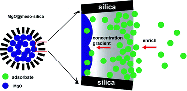
Core–shell structured MgO@mesoporous silica spheres are synthesized by a two-step programmed method. MgO@mesoporous silica exhibits a high BET specific surface area of 567 m2 g−1 and a pore volume of 1.08 cm3 g−1. The stable mesoporous silica coating not only serves as a strong shell to improve the mechanical stability of MgO, but also enriches the adsorbates in the mesopores to reach a higher adsorption rate. The core–shell MgO@mesoporous silica spheres exhibit excellent removal capabilities of 3297 mg g−1 for Pb2+ and 420 mg g−1 for methylene blue, which are much higher than those of MgO itself.


Introduction

Pollutants, such as heavy metal ions and toxic organics from industry, agriculture and other natural processes could cause various diseases including cancer, malformation, mutation, nausea, coma and mental retardation.1–6 Various nanomaterials present a bright future for the efficient removal of pollutants from natural ground water and drinking water. Among the promising technologies, such as membrane filtration,7,8 co-precipitation9–11 and adsorption,12–14 adsorption is a very effective method to water remediation due to its low cost, easy operation, and performance stability, especially in developing countries.15 Active carbon based adsorbents have been sold as water filter for purifying drinking water for years. Besides carbon based nanomaterials, metal oxides and their composites are also important adsorbents because of their advantages such as high adsorption capacity, selective adsorption, low cost and simple preparation.16–21 Recently, Song et al. reported that flowerlike magnesium oxide (MgO) could adsorb lead ions (Pb2+) or cadmium ions by a solid–liquid interfacial cation-exchange adsorption mechanism.22 Such a chemical process results in superb adsorption properties of MgO for Pb2+ and cadmium ions, as high as 1980 and 1500 mg g−1, respectively. High specific surface area and effective mass transfer are two important factors for adsorption efficiency.23,24 Size reduction and ordered mesopore construction are the main approaches to achieve this result. However, such nano-sized materials are difficult to be separated from the solution, which may cause second pollution.

Coating with a mesoporous silica shell is an effective method. It has been reported that nanocomposite could effectively improve the separability and adsorption efficiency.25,26 For example, the presence of a magnetic Fe3O4 core made the hierarchical core–shell Fe3O4@magnesium silicate easier to be separated and recycled upon the application of an external magnetic field.26 Recently, we reported that a sandwich-like magnesium silicate/reduced graphene oxide with all magnesium silicate nanopetals exposed on the surface exhibits a much higher adsorption efficiency than other silicate nanomaterials, and it could be separated from solution under gravity owing to the micrometer scale reduced graphene oxide.27 Different from these, the mesoporous structure affords a better mass transfer and concentration gradient in some specific regions, which are the driving force of diffusion in solution. Core–shell structured Pd@meso-silica, CuO@meso-silica, Fe2O3@meso-silica and Au@TiO2 show improved catalytic properties in organic reaction or dye degradation process.28–31 The improved mass transfer would also be beneficial for adsorption.

In this study, a programmed method is used to fabricate core–shell structured MgO@meso-silica spheres. The mesoporous silica shell not only improves the mechanical stability of MgO spheres to avoid structure destruction during stirring, but also enriches adsorbates from the solution to the adsorbent due to the better mass transfer and concentration gradient. Thus, the as-prepared core–shell MgO@meso-silica spheres should exhibit better removal performance than MgO alone.

Experimental section

Materials

Ethanol, ethylene glycol, ammonium hydroxide (NH3·H2O, 28%) were bought from Beijing Chemical Factory (China). Magnesium acetate tetrahydrate (Mg(CH3COO)2·4H2O), polyvinylpyrrolidone (PVP, K30), cetyltrimethyl ammonium bromide (CTAB), tetraethyl orthosilicate (TEOS), lead nitrate, and methylene blue (MB) were supplied by Sinopharm Chemical Reagent Co. Ltd. (China). All chemicals are analytical grade and were used without further purifications.

Synthesis of spherical MgO

Spherical MgO was synthesized by a simple method. Mg(CH3COO)2·4H2O (0.428 g) and PVP (0.99 g) were dissolved in 40 mL ethylene glycol, and the mixture was magnetically stirred in an oil bath at 180 °C for 2 h. The resultant was centrifuged, washed with ethanol for 3 times, dried in an oven at 80 °C for 12 h, and finally calcinated in a muffle furnace at 500 °C for 5 h.

Synthesis of MgO@meso-silica spheres

MgO precursor (200 mg) was dispersed in the solution of 80 mL deionized water and 60 mL ethanol by ultrasonication for 20 min. After adding 1140 μL NH3·H2O and 0.28 g CTAB and stirring for 30 min, 400 μL TEOS was added dropwise. The mixture was stirred for 6 h using an IKA disperser at 500 rpm, washed with ethanol and deionized water for 3 times, successively. After drying in an oven at 80 °C for 12 h, the resultant was calcinated in the furnace at 500 °C for 5 h to remove the cationic surfactant. For comparison, mesoporous silica was also fabricated as reported.32

Characterization

MgO and MgO@meso-silica were characterized with a Thermo VG RSCAKAB 250× high resolution X-ray photoelectron spectroscopy (XPS) device. X-ray diffraction (XRD) measurement was carried out using a Rigaku D/Max 2500 diffractometer with CuKα radiation (λ = 1.54 Å) at a generator voltage of 40 kV and a generator current of 40 mA. The morphology and microstructure of MgO and MgO@meso-silica were observed with a Hitachi S4700 field emission scanning electron microscope (SEM) and a JEOL JEM-3010 transmission electron microscope (TEM), and their specific surface areas were measured by the Brunauer–Emmet–Teller (BET) method using N2 adsorption and desorption isotherms on an Autosorb-1 analyzer at 77.3 K.

Adsorption experiment

Pb2+ or MB adsorption experiments were carried out as follows: Pb2+ solutions were prepared using Pb(NO3)2 as the cation source. The adsorption isotherms of Pb2+ and MB were obtained using 20 mg adsorbents mixed with 20 mL solutions at different concentrations and stirred overnight at constant temperature of 25 °C. The adsorption kinetics data resulted from measuring the adsorption capacity at different time intervals. MB concentration was measured with a Thermal Scientific Evolution 200 UV-visible (UV-vis) Spectrophotometer (USA). The concentration of Pb2+ was analyzed using a Shimadzu ICPS-7500 inductively coupled plasma atomic emission spectroscopy (ICP-AES).

Results and discussion

For practical usage, excellent structural and mechanical stability of nanostructured adsorbents has always been important for being operated without second pollution. Hierarchical nanomaterials were constructed with various nano-sized building blocks, which were kept together through weak interactions, such as van der Waals force, hydrogen bonding and electrostatic attraction. Although crystalline nanoparticles are resistant to structural damage, physical impact from stirring and frequent collision in water causes the damage of hierarchical nanostructures, leading to the breakdown of overall structure. Recently, many studies have shown that coating with a hard and porous shell is an effective approach to improve the mechanical stability and chemical property of the absorbents.28–30 Herein, we constructed the mesoporous silica shell by a programmed method. The fabrication process for MgO@meso-silica spheres is illustrated in Fig. 1. First, spherical MgO precursor was prepared by a ethylene glycol mediated process reported for fabrication of V2O5 and ZnS.33,34 Second, mesoporous silica shell was constructed outside of the MgO precursor through the TEOS hydrolysis with the assistance of CTAB in alkaline media. Finally, the core–shell MgO@meso-silica spheres were obtained after calcinating at 500 °C for 5 h. As shown in Fig. 2, MgO spheres composed of thousands of ca. 15 nm nanoparticles endow MgO with a high BET surface area and more active sites for high adsorption performance. However, the spheres tend to break into nanoparticles during stirring in the adsorption process (Fig. S1). It is speculated that a hard mesoporous silica shell would serve as an exoskeleton to protect the MgO core from structural damage and improve the adsorption property of MgO.
image file: c5ra02596f-f1.tif
Fig. 1 Schematic of the preparation of MgO@meso-silica spheres.

image file: c5ra02596f-f2.tif
Fig. 2 (a) XRD patterns of MgO and MgO@meso-silica; SEM images of (b) MgO and (c) MgO@meso-silica; (d) TEM image of MgO@meso-silica.

The XRD patterns of MgO and MgO@meso-silica are shown in Fig. 2a. MgO and MgO@meso-silica nearly have same peaks, and all peaks could be indexed to MgO (JCPDS: 45-0946). However, there is a broad peak in the range of 20–40° for MgO@meso-silica, which is attributed to the amorphous mesoporous silica shell. The peak widths of MgO@meso-silica are broader than those of MgO, indicating that the size of nanoparticles in MgO@meso-silica is much smaller than that in MgO spheres. The size decrease would result in a high BET specific surface area and more adsorption sites, which is beneficial for adsorption. Besides, no other impurities are observed. The morphologies of MgO and MgO@meso-silica were observed with SEM and TEM. As shown in Fig. 2b and S2, MgO spheres have an average diameter of about 400 nm. The entire structure is built from nanoparticles with a diameter of about 15 nm, which are connected with each other to form the three-dimensional spherical hierarchical structure by self-assembly. After the hydrolysis of TEOS, the rough surface becomes smooth, confirming the successful formation of silica shells, and the diameter increases to ca. 500 nm (Fig. 2c). TEM image also shows the core–shell structure. The light gray core is MgO, while the 50 nm-thick silica shell shows a dark gray circled shadow owing to the different electron scattering abilities with different chemical components (Fig. 2d). The high magnification TEM image shows that the silica shell has a worm-like mesoporous structure (inset of Fig. 2d). Furthermore, Si 2s and Si 2p peaks are observed in the XPS curve of MgO@meso-silica (Fig. S3). All these results confirm that the mesoporous silica has been successfully coated on the surface of MgO. Such a hard and mesoporous shell not only improves the mechanical stability of the composite, but also enhances the mass transfer to obtain a high activity that has been reported in catalyst and drug-delivery fields.28,29,35,36 It is envisioned that coating with the mesoporous silica shell would improve the structure stability and adsorption performance of the adsorbent.

Fig. 3a shows the nitrogen adsorption–desorption isotherms for MgO and MgO@meso-silica. MgO@meso-silica exhibits a typical type IV adsorption isotherm with a remarkably sharp capillary condensation step between 0.2 and 0.4 P/P0, indicating that the specimen exhibits a narrow size distribution. In addition, MgO@meso-silica exhibits a high BET specific surface area of 567 m2 g−1 and a pore volume of 1.08 cm3 g−1. Both values are much higher than those of MgO with a BET specific surface area of 103 m2 g−1 and a pore volume of 0.70 cm3 g−1. The pore size distributions estimated by Density Functional Theory (DFT) method are shown in Fig. 3b. MgO has a fairly broad peak from 4–20 nm, which is attributed to the void space between MgO nanoparticles. Interestingly, MgO@meso-silica exhibits two evident pore radius peaks at 1.8 nm and a range of over 3.0 nm. The pores around 1.8 nm relate to the mesoporous shell, while the void space between MgO nanoparticles decreases due to the coating of mesoporous silica shell. The pore size distributions also show that there are almost no micropores in both materials. Much smaller pores are responsible for the higher BET specific surface area. As the surface of an adsorbent is where adsorption occurs, and pore structure determines the efficiency of mass transfer. They are two main important factors affecting adsorbent's capacity and efficiency. Such high BET specific surface area and pore volume benefit the improvement of the adsorption performance of MgO@meso-silica.


image file: c5ra02596f-f3.tif
Fig. 3 (a) Nitrogen adsorption–desorption isotherms, and (b) pore size distribution curves of MgO and MgO@meso-silica spheres.

To confirm the advantage of the mesoporous silica shell, Fig. 4 shows the adsorption capacities and adsorption isotherms of MgO and MgO@meso-silica for MB and Pb2+. Langmuir model is employed for adsorption analysis as follows:37,38

Qe = QmbCe/(1 + bCe)
where Ce is the equilibrium concentration of an adsorbate (mg L−1), Qe is the amount of adsorbate adsorbed per unit weight of the adsorbent at equilibrium (mg g−1), Qm is the maximum adsorption capacity (mg g−1), and b is a constant in Langmuir equation. The adsorption data of MB fit the Langmuir adsorption isotherm well (Fig. 4a, Table 1). The values of determination coefficients for the fitting Langmuir model are satisfactory. To further confirm the adsorption model, the values of 1/Qe are fitted against 1/Ce based on the Langmuir equation below:
1/Qe = 1/Qm + 1/(KLQmCe)


image file: c5ra02596f-f4.tif
Fig. 4 Adsorption isotherms of (a) MB and (c) Pb2+ on MgO and MgO@meso-silica at 25 °C. Ce (mg L−1): the equilibrium concentration of the MB or Pb2+ solution; Qe (mg g−1): the amount of MB or Pb2+ adsorbed at equilibrium. Time-dependent concentration of (b) MB and (d) Pb2+ with MgO, MgO@meso-silica, and meso-silica as adsorbents at 25 °C (initial concentrations of MB and Pb2+ are 100 and 1750 mg L−1, respectively).
Table 1 Langmuir parameters associated with adsorption isotherms of MB and Pb2+ on MgO and MgO@meso-silicaa
Adsorbates Adsorbents Qm (mg g−1) b (L mg−1) R2
a Qm: the maximum adsorption capacity of MB or Pb2+.
MB MgO@meso-silica 420 ± 5 0.153 ± 0.006 0.999
MB MgO 61 ± 8 0.025 ± 0.007 0.969
Pb2+ MgO@meso-silica 3297 ± 134 0.189 ± 0.049 0.976
Pb2+ MgO 2454 ± 82 0.132 ± 0.032 0.985


KL is the Langmuir adsorption constant (L mg−1) associated with the free energy of adsorption. The linear fitting data are given in Fig. S5a and b. All correlation coefficient values for MB or Pb2+ adsorption are above 0.945. However, if the adsorption data are fitted according to the Freundlich equation:

ln[thin space (1/6-em)]Qe = ln[thin space (1/6-em)]KF + bF[thin space (1/6-em)]ln[thin space (1/6-em)]Ce
where bF and KF (mg g−1(L mg−1)1/n) are the Freundlich constants relating to adsorption intensity and adsorption capacity, respectively. All correlation coefficient values for MB or Pb2+ adsorption are below 0.886 (Fig. S5c and d). So Langmuir model is more suitable for the adsorption process than Freundlich model.

The maximum adsorption capacity of MgO@meso-silica for MB is 420 mg g−1, which is nearly 6 times higher than that of MgO (61 mg g−1, Table 1) and much higher than reported value for bare mesoporous silica (Table S1), so the higher capacity should be contributed by the MgO component. Furthermore, the fitting isotherm curve of MgO@meso-silica rises more steeply than that of MgO, indicating that the b value (a constant, related to the adsorption energy and the binding strength) of MgO@meso-silica is much higher than that of MgO (Table 1). It was reported that a porous shell could enrich molecules or ions from the bulk solution, resulting in a higher concentration in the void space of pores.26,30,39 That is, the concentration of MB in the shell layer is very high while that in the core is very low. So a concentration gradient from the shell to the core is formed, enriching MB from bulk solution to be adsorbed steadily by MgO. Such an enriching effect makes the core–shell structured MgO@meso-silica adsorb MB more easily, leading to a higher b value. Time-dependent concentration experiments also support the conclusion. In the first hour, 75% MB is adsorbed by MgO@meso-silica, while the uptake of MB is only 20% by MgO and 5% by bare mesoporous silica under the same initial MB concentration (Fig. 4b). Ten hours later, MgO@meso-silica adsorbs nearly 100% MB, but there is little change in the control experiment.

Pb2+ is also investigated as a model ion. As shown in Fig. 4c and Table 1, the maximum adsorption capacity increases from 2454 mg per g of MgO to 3297 mg per g of MgO@meso-silica, which is higher than reported values (Table S1).22,40,41 Surprisingly, almost 100% Pb2+ is removed by MgO@meso-silica during the initial few minutes, while MgO alone needs more than 10 hours (Fig. 4d). MgO@meso-silica also has a higher b value (Table 1). Besides, SEM image of MgO@meso-silica after adsorption shows that the spherical structure retains well after mechanical stirring (Fig. S4). On the contrary, the uncoated MgO is destroyed to much smaller nanoparticles (Fig. S1). It is true that the core–shell structure with a stable mesoporous silica shell exhibits a much higher efficiency for adsorption with enhanced mechanical stability and improved mass transfer ability.

As adsorption occurs at interfaces, large BET surface area is beneficial for high adsorption capacity. However, the high BET surface area of MgO@meso-silica mainly comes from the mesoporous silica shell that is not an ideal adsorbent for adsorption. Thus, the outstanding adsorption performance is mainly attributed to the nanoreactor feature of the core–shell structure. As discussed above and reported, adsorbates could be enriched within the mesopores to form a powerful concentration gradient from the un-active silica shell to active MgO core (Fig. 5). The concentration gradient contributes to the steady adsorption of MB and Pb2+ compared to MgO alone under the same initial concentration.


image file: c5ra02596f-f5.tif
Fig. 5 Schematic of the adsorption mechanism of the core–shell MgO@meso-silica spheres.

Even if the concentration of adsorbates decreases to a relatively low level along with the adsorption process, the enriching effect could still make MgO component of the composite work. To confirm this, the adsorption performances of MgO@meso-silica and MgO are measured using a large amount of dilute solution with low initial concentrations of 5 or 10 mg L−1 MB. As shown in Fig. 6a, at the low initial concentration of 10 mg L−1, MgO@meso-silica quickly adsorbs nearly 84% MB while MgO just reaches to 59% in 10 minutes. Even at 5 mg per L of MB (Fig. 6b), the concentration easily decreases to ca. 0.9 mg L−1 by MgO@meso-silica while MgO just reduces the concentration to ca. 2.0 mg L−1. Furthermore, MgO@meso-silica could continue removing the adsorbates while MgO itself almost has no further adsorption below concentration of 2.0 mg L−1, which confirms the enriching effect of mesoporous silica coating. It is clear that the core–shell structure has enhanced adsorption performance, which shows good potential in water remediation applications.


image file: c5ra02596f-f6.tif
Fig. 6 Time-dependent concentration of MB using MgO@meso-silica and MgO as adsorbents at MB initial concentration of (a) 10 and (b) 5 mg L−1. Dosage of adsorbents: 25 mg; amount of MB solution: 100 mL; temperature: 25 °C.

Conclusions

Core–shell structured MgO@meso-silica spheres are prepared with a programmed method. XRD patterns and XPS spectra index the crystallographic information and chemical composition of the spheres. SEM and TEM observations clearly show the core–shell structure. The stable mesoporous silica shell improves the mechanical stability of MgO spheres, avoiding destruction of the structure during mechanical stirring in solutions. Furthermore, the enriching effect of the shell powers adsorbates to diffuse from the solution to the adsorbent by a better mass transfer and concentration gradient. The core–shell MgO@meso-silica spheres exhibit excellent removal capabilities of 3297 mg g−1 for Pb2+and 420 mg g−1 for MB, which are 34% and 6 times higher than those of MgO alone, respectively. The MgO@meso-silica spheres would have good potential application in water remediation and such a coating treatment could be used to improve the adsorption performance of other inorganic adsorbents.

Acknowledgements

Financial support from the National Natural Science Foundation of China (51125010, 51402012) and the Fundamental Research Funds for the Central Universities, and the State Key Laboratory of Organic-Inorganic Composites (201404) is gratefully acknowledged.

Notes and references

  1. H. Sun, L. Cao and L. Lu, Nano Res., 2011, 4, 550–562 CrossRef CAS PubMed .
  2. Z. H. Huang, X. Y. Zheng, W. Lv, M. Wang, Q. H. Yang and F. Y. Kang, Langmuir, 2011, 27, 7558–7562 CrossRef CAS PubMed .
  3. G. X. Zhao, X. M. Ren, X. Gao, X. L. Tan, J. X. Li, C. L. Chen, Y. Y. Huang and X. K. Wang, Dalton Trans., 2011, 40, 10945–10952 RSC .
  4. T. E. Milja, K. P. Prathish and T. P. Rao, J. Hazard. Mater., 2011, 188, 384–390 CrossRef CAS PubMed .
  5. B. Paul, D. Yang, W. N. Martens and R. L. Frost, J. Colloid Interface Sci., 2011, 356, 240–247 CrossRef CAS PubMed .
  6. I. Tabushi, Y. Kobuke and T. Nishiya, Nature, 1979, 280, 665–666 CrossRef CAS .
  7. I. Hajdu, M. Bodnár, Z. Csikós, S. Wei, L. Daróczi, B. Kovács, Z. Győri, J. Tamás and J. Borbély, J. Membr. Sci., 2012, 409–410, 44–53 CrossRef CAS PubMed .
  8. M. Soylak, Y. E. Unsal, N. Kizil and A. Aydin, Food Chem. Toxicol., 2010, 48, 517–521 CrossRef CAS PubMed .
  9. Z. G. Meng, G. P. Korfiatis, C. Christodoulatos and S. Bang, Water Res., 2001, 35, 2805–2810 CrossRef .
  10. O. D. Uluozlu, M. Tuzen, D. Mendil and M. Soylak, J. Hazard. Mater., 2010, 176, 1032–1037 CrossRef CAS PubMed .
  11. M. M. Matlock, B. S. Howerton and D. A. Atwood, Ind. Eng. Chem. Res., 2002, 41, 1579–1582 CrossRef CAS .
  12. S. Komarneni, N. Kozai and W. J. Paulus, Nature, 2001, 410, 771 CrossRef CAS PubMed .
  13. H. H. Tran, F. A. Roddick and J. A. O'Donnell, Water Res., 1999, 33, 2992–3000 CrossRef CAS .
  14. C. T. Yavuz, J. T. Mayo, W. W. Yu, A. Prakash, J. C. Falkner, S. Cong, L. Yean, H. J. Shipley, A. Kan, M. Tomson, D. Natelson and V. L. Colvin, Science, 2006, 314, 964–967 CrossRef PubMed .
  15. Y. Q. Wang, G. Z. Wang, H. Q. Wang, C. H. Liang, W. P. Cai and L. D. Zhang, Chem.–Eur. J., 2010, 16, 3497–3503 CrossRef CAS PubMed .
  16. J. S. Hu, L. S. Zhong, W. G. Song and L. J. Wan, Adv. Mater., 2008, 20, 2977–2982 CrossRef CAS .
  17. L. S. Zhong, J. S. Hu, H. P. Liang, A. M. Cao, W. G. Song and L. J. Wan, Adv. Mater., 2006, 18, 2426–2431 CrossRef CAS .
  18. Y. Ide, N. Ochi and M. Ogawa, Angew. Chem., 2011, 123, 680–682 CrossRef .
  19. D. Yang, Z. Zheng, H. Liu, H. Zhu, X. Ke, Y. Xu, D. Wu and Y. Sun, J. Phys. Chem. C, 2008, 112, 16275–16280 CAS .
  20. Q. Cao, F. Huang, Z. Y. Zhuang and Z. Lin, Nanoscale, 2012, 4, 2423–2430 RSC .
  21. X. Y. Yu, T. Luo, Y. Jia, R. X. Xu, C. Gao, Y. X. Zhang, J. H. Liu and X. J. Huang, Nanoscale, 2012, 4, 3466–3474 RSC .
  22. C. Y. Cao, J. Qu, F. Wei, H. Liu and W. G. Song, ACS Appl. Mater. Interfaces, 2012, 4, 4283–4287 CAS .
  23. Z. Wu and D. Zhao, Chem. Commun., 2011, 47, 3332–3338 RSC .
  24. B. Kuchta, L. Firlej and G. Maurin, J. Mol. Model., 2005, 11, 293–300 CrossRef CAS PubMed .
  25. V. Chandra, J. Park, Y. Chun, J. W. Lee, I. C. Hwang and K. S. Kim, ACS Nano, 2010, 4, 3979–3986 CrossRef CAS PubMed .
  26. S. Zhang, W. Xu, M. Zeng, J. Li, J. Li, J. Xu and X. Wang, J. Mater. Chem. A, 2013, 1, 11691–11697 CAS .
  27. C. X. Gui, Q. Q. Wang, S. M. Hao, J. Qu, P. P. Huang, C. Y. Cao, W. G. Song and Z. Z. Yu, ACS Appl. Mater. Interfaces, 2014, 6, 14653–14659 CAS .
  28. Z. Chen, Z. M. Cui, F. Niu, L. Jiang and W. G. Song, Chem. Commun., 2010, 46, 6524–6526 RSC .
  29. C. Q. Chen, J. Qu, C. Y. Cao, F. Niu and W. G. Song, J. Mater. Chem., 2011, 21, 5774–5779 RSC .
  30. Z. M. Cui, Z. Chen, C. Y. Cao, L. Jiang and W. G. Song, Chem. Commun., 2013, 49, 2332–2334 RSC .
  31. Y. Yu, C. Y. Cao, Z. Chen, H. Liu, P. Li, Z. F. Dou and W. G. Song, Chem. Commun., 2013, 49, 3116–3118 RSC .
  32. S. W. Bian, Z. Ma, L. S. Zhang, F. Niu and W. G. Song, Chem. Commun., 2009, 1261–1263 RSC .
  33. J. S. Hu, L. L. Ren, Y. G. Guo, H. P. Liang, A. M. Cao, L. J. Wan and C. L. Bai, Angew. Chem., Int. Ed., 2005, 44, 1269–1273 CrossRef CAS PubMed .
  34. A. M. Cao, J. S. Hu, H. P. Liang and L. J. Wan, Angew. Chem., Int. Ed., 2005, 44, 4391–4395 CrossRef CAS PubMed .
  35. Z. Li, J. C. Barnes, A. Bosoy, J. F. Stoddart and J. I. Zink, Chem. Soc. Rev., 2012, 41, 2590–2605 RSC .
  36. Y. Yang, X. Liu, X. Li, J. Zhao, S. Bai, J. Liu and Q. Yang, Angew. Chem., Int. Ed., 2012, 51, 9164–9168 CrossRef CAS PubMed .
  37. I. Langmuir, J. Am. Chem. Soc., 1916, 38, 2221–2295 CrossRef CAS .
  38. I. Langmuir, J. Am. Chem. Soc., 1918, 40, 1361–1403 CrossRef CAS .
  39. B. Li, F. Li, S. Bai, Z. Wang, L. Sun, Q. Yang and C. Li, Energy Environ. Sci., 2012, 5, 8229–8233 CAS .
  40. C. H. Lin, D. S. H. Wong and S. Y. Lu, ACS Appl. Mater. Interfaces, 2014, 6, 16669–16678 CAS .
  41. C. Gao, W. Zhang, H. Li, L. Lang and Z. Xu, Cryst. Growth Des., 2008, 8, 3785–3790 CAS .

Footnote

Electronic supplementary information (ESI) available: SEM images of MgO and MgO@meso-silica after adsorption; TEM image of MgO; XPS spectra and adsorption properties of MgO and MgO@meso-silica. See DOI: 10.1039/c5ra02596f

This journal is © The Royal Society of Chemistry 2015
Click here to see how this site uses Cookies. View our privacy policy here.