Metabolomic study of Chinese medicine Huang Qin decoction as an effective treatment for irinotecan-induced gastrointestinal toxicity

Jing Wangab, Hongyan Fanab, Yong Wangc, Xu Wangab, Pei Zhangab, Jiaqing Chenab, Yuan Tianab, Wei Zhangd, Fengguo Xu*ab and Zunjian Zhang*ab
aMOE Key Laboratory of Drug Quality Control and Pharmacovigilance (Ministry of Education), China Pharmaceutical University, Nanjing, 210009, China. E-mail: fengguoxu@gmail.com; zunjianzhangcpu@hotmail.com; Fax: +86 025 83271021; Fax: +86 025 83271454; Tel: +86 025 83271021 Tel: +86 025 83271454
bState Key Laboratory of Natural Medicine, China Pharmaceutical University, Nanjing, 210009, China
cJiangsu Institute for Food and Drug Control, Nanjing 210008, China
dState Key Laboratory for Quality Research in Chinese Medicines, Macau University of Science and Technology, Taipa, Macau, China

Received 10th February 2015 , Accepted 5th March 2015

First published on 5th March 2015


Abstract

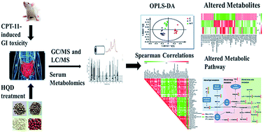
The Huang Qin Decoction (HQD) has been used in China for over 1800 years for the treatment of gastrointestinal ailments. As a formulation derived from HQD, PHY906 shows an attenuation effect on chemotherapeutic-induced gastrointestinal toxicity. Method: to explore the mechanism of irinotecan (CPT-11)-induced gastrointestinal toxicity and the ameliorative effect of HQD, an integrated gas and liquid chromatography-mass spectrometry (GC/MS, LC/MS) approach was applied to detect serum metabolome changes in rats following a treatment of CPT-11 with/without HQD. Results: significant alterations in metabolic profiling were observed in CPT-11-treated groups versus control groups. HQD attenuated the side-effects with reversed glutamine, tryptophan and lipid metabolisms. Conclusion: this study demonstrated that HQD can effectively decrease CPT-11-induced side-effects, and the metabolism pathways involved were speculated to be novel targets for reducing CPT-11-induced gastrointestinal toxicity.
Introduction

Irinotecan (CPT-11) is a water-soluble derivative of camptothecin, and it has been shown to be highly effective in the treatment of colon, lung as well as other types of cancer.1–4 It is accepted as a first-line therapy for the treatment of metastatic colorectal cancer in combination with 5-fluorouracil (5-FU) and leucovonin (LV), and is also approved as a monotherapy in the second-line indication after 5-FU treatment.5–8 However, several adverse effects have been observed in patients treated with CPT-11, such as neutropenia, alopecia, fatigue, nausea/vomiting, diarrhea and other symptoms.9,10 Among which, late diarrhea is recognized as the dose-limiting toxicity that limits the use of more aggressive CPT-11 therapy. The incidence of CPT-11- or combination therapy-induced diarrhea has been reported to be as high as 80% of treated patients with up to one third of patients experiencing severe diarrhea (grade 3 or 4).7,9,11,12 It is of high value to find out effective modulators of chemotherapeutic agent in cancer therapy.

Various agents potentially inhibiting CPT-11-induced diarrhea were applied in clinic as proposed mechanisms behind the late diarrhea caused by CPT-11 are diverse. Notably, high dose of loperamide was used as the preferred method to relieve the diarrhea, but the outcomes were not always satisfactory.12–17 HQD, a traditional Chinese medicine, has been used in China for over 1800 years for the treatment of gastrointestinal ailments, including diarrhea, nausea and vomiting. A formulation derived from HQD, PHY906 was proved to be a modulator of chemotherapeutic agent in cancer therapy, and shown to effectively decrease gastrointestinal toxicity induced by CPT-11 both in preclinical and clinical studies.18,19 These evidences suggest that, as a botanical medicine, HQD may be helpful for the chemotherapeutic treatment of cancer by reducing the severity of the following late diarrhea.

In recent years, some investigations on the mechanism of CPT-11-induced diarrhea as well as the effectiveness of HQD (PHY906) have been reported. Some studies hypothesized that CPT-11 and its active metabolite SN-38 cause DNA damage and cell apoptosis in the intestine, as well as a destruction of the epithelium mucosal architecture resulting in an imbalance between absorption and secretion of fluid, which lead to diarrhea.20,21 Other studies suggested that CPT-11-induced late diarrhea could be a consequence of intestinal bacterial flora disturbance, inflammatory cytokines and cyclooxygenase-2 (COX-2) induction, secondary to colonic mucosal damage.22–26 The effects of PHY906 on the intestinal toxicity caused by CPT-11 were reported by promoting the regeneration of intestinal progenitor or stem cells and several Wnt signaling components, exhibiting anti-inflammatory effects as well as inhibiting nuclear factor κB (NF-κB), COX-2 and inducible nitric oxide synthase (iNOS).19 However, the understanding of the molecular process is incomprehensive and the systematical metabolic response to CPT-11 as well as the effect of HQD in a whole living biosystem is still unclear.

Metabolomics, primarily focus on identification and quantitation of small-molecule metabolites (molecular weight less than 1500 Da) which act as downstream to DNA, RNA and protein. It represents the comprehensive and simultaneous systematic profiles of multiparametric metabolic changes that occur in living systems in response to disease, pharmacological treatment or toxicological insult.27–30 As a complementary to biochemical and histological findings, metabolomics has been proved to be an effective tool for evaluating drug efficacy and toxicity.31–34 Till now, metabolomics have not been used to study CPT-11-induced diarrhea and thus may offer new insight on the molecular mechanism.

In the present study, we applied an integrated gas and liquid chromatography mass spectrometry-based metabolomics approach to identify CPT-11-altered metabolite changes as well as the ameliorative effect after HQD exposure. In complement, diarrhea score and histopathology examination provided evidence to support the therapeutic value of HQD for late diarrhea induced by CPT-11. Our result, for the first time, displayed the HQD-mediated metabolic changes associated with CPT-11-induced gastrointestinal toxicity.

Materials and methods

Materials and chemicals

Irinotecan hydrochloride for injection (CPT-11) was obtained from Hengrui (Jiangsu, China). The four component herbs of HQD, Huangqin (Shanxi province), Baishao (Zhejiang province), Gancao (Gansu province) and Dazao (Henan province) were identified by Assistant Professor Zhang Wei (State Key Laboratory for Quality Research in Chinese Medicines, Macau University of Science and Technology). Authentic compounds, methoxyamine hydrochloride, N-methyl-N-trifluoroacetamide (MSTFA) and pyridine were purchased from Sigma-Aldrich (St. Louis, MO, USA). Methanol, acetonitrile and ethyl acetate for high-performance LC-grade were obtained from Honeywell (Burdick & Jackson, NJ, USA). Deionized water was purified using a Milli-Q system (Millipore, Bedford, MA, USA).

Decoction preparation, animal study and sample collection

Huang Qin Decoction (HQD) was water-boiling extracts of four herbs, Huangqin (dried roots of Scutellaria baicalensis Georgi), Baishao (dried roots of Paeonia lactiflora Pall), Gancao (dried and honey-fried roots and rhizomes of Glycyrrhiza uralensis Fisch) and Dazao (dried fruits of Ziziphus jujuba Mill) in the weight ratio 3[thin space (1/6-em)]:[thin space (1/6-em)]2[thin space (1/6-em)]:[thin space (1/6-em)]2[thin space (1/6-em)]:[thin space (1/6-em)]2. The extracts were concentrated to dryness using a freeze dry system, and then suspended in Milli-Q water to form a concentration of 1 g crude drug per milliliter before intragastric administration.

Thirty-six male Sprague-Dawley rats (weighing 200 ± 20 g) were purchased from the Sino-British SIPPR/BK Lab Animal Ltd. (Shanghai, China) and housed in a temperature-controlled environment under 12/12 h – dark/light cycle. All experiments were performed according to the Guide for the Care and Use of Laboratory Animals and were authorized by the Animal Ethics Committee of China Pharmaceutical University. Rats were acclimatized for 7 days with a standard rodent diet and water available ad libitum. Then they were randomly divided into three groups with twelve rats in each group. CPT-11 diluted with saline was administered intravenously (i.v.) to the animals (group T and H/T) via the tail vein with 150 mg kg−1 once a day for two consecutive days. Animals in control group (group C) received a corresponding administration of saline. HQD (10 g kg−1) was given to rats in H/T group twice per day for 5 days, starting from day 0, 0.5 h prior to CPT-11 administration. Animals in group C and T received equivalent Milli-Q water. Body weight and diarrhea score were monitored twice per day (before intragastric administration). Serum samples were collected on day 5 after CPT-11 administration in the first two days. The samples were clotted, centrifuged at 5000 rpm for 15 min, then stored at −80 °C until metabolomic analysis. All rats were euthanized after the serum samples had been collected. The stomach, jejunum, ileum, cecum and colon were removed and used for histological examination.

Sample preparation

Serum samples were thawed at room temperature. 100 μL acetonitrile and 40 μL glibenclamide (internal standard (IS), 5 μg mL−1) were added to 20 μL serum and the mixture was vortexed for 5 min to extract metabolites. After centrifuged twice at 16[thin space (1/6-em)]000 rpm (4 °C) for 10 min, the supernatant was transferred and analyzed by LC/MS.

100 μL of cold methanol (containing 5 μg mL−1 heptadecanoic acid, working as IS) was added to 10 μL of thawed serum and vortex-mixed for 15 min to extract metabolites. After a second centrifugation (16[thin space (1/6-em)]000 rpm, 10 min, 4 °C), a 80 μL supernatant was obtained and transferred to a screw vial (1 mL) followed by the addition of methyoxyamine hydrochloride (25 μL, 10 mg mL−1 in dry pyridine) and incubation at 37 °C for 90 min. The mixture was evaporated to dryness and then silylated with 120 μL MSTFA/ethyl acetate (v/v, 1/1). After incubation for 2 hours at a temperature of 37 °C, the mixture was prepared for GC/MS analysis.

Histopathology examination

All animals were inspected daily for any clinically abnormal signs and mortalities. At the end of the treatment period, the animals were sacrificed for histopathology examination. The stomach, jejunum, ileum, cecum, colon slices of control and treatment rats were fixed in 10% formalin, stained with hematoxylin–eosin, then examined by light microscopy for histopathology assessment.

LC/MS analysis

LC/MS analysis was performed on Shimadzu ultrafast LC-ion trap time-of flight MS system equipped with a electrospray ionization (ESI) source (Shimadzu, Kyoto, Japan). Chromatographic separation was achieved on a Phenomenex Kinelex C18 column (100 × 2.1 mm, 2.6 μm, Phenomenex, Torrance, CA, USA) using a gradient elution involved 5–95% acetonitrile (0.1% formic acid)-aqueous formic acid (0.1% formic acid). The column oven was maintained at 40 °C and the flow rate of 0.4 mL min−1. The ESI-MS were acquired in both positive and negative ion mode with an interface voltage of 4.5 kV and −3.5 kV respectively. The scan range was 100–1000 m/z. The flow rate of nebulizing gas was 1.5 L min−1 and pressure of drying gas was 100 kPa. The temperature of heat block and curved desorption line were both 200 °C. LC/MS solution version 3.0 (Shimadzu, Kyoto, Japan) was used for mass spectra acquisition and chromatograms procession.

GC/MS analysis

Analysis was performed on Shimadzu GCMS-QP2010 Ultra (Shimadzu, Kyoto, Japan) equipped with a 30.0 m × 0.25 mm i.d. fused-silica capillary column with 0.25 μm Rtx-5MS stationary phase (Agilent, Shanghai, China). Helium was used as carrier gas and set at 1 mL min−1. An injection volume of 1 μL was used with the split ratio of 50[thin space (1/6-em)]:[thin space (1/6-em)]1. The column temperature was initially kept at 70 °C for 3 min and then increased to 320 °C at 10 °C min−1, where it was held for 2 min. The injector temperature, interface temperature and ion source temperature were set at 250 °C, 200 °C, 250 °C, respectively. Masses were acquired from m/z 45 to 600 in scan mode. The acceleration voltage was turned on after a solvent delay of 5 min. Mass spectra and chromatograms were acquired and processed with GC/MS solution version 2.7 (Shimadzu, Kyoto, Japan).

Data preprocessing and analysis

Each chromatogram obtained was processed by profiling solution version 1.1 (Shimadzu, Kyoto, Japan) for peak deconvolution and alignment. The primary parameters were set as follows: ion m/z tolerance (500 mDa) for GC/MS and (25 mDa) for LC/MS analysis, ion retention time tolerance (0.05 min) for GC/MS and (0.3 min) for LC/MS analysis, and ion intensity threshold (5000 counts) and (8000 counts) respectively. The resulting data tables were exported to an excel table, handled according to the “80% rule”: only the variables with values above zero in at least 80% of one or more groups were kept. Then, the individual ion fragment intensity was normalized to the intensity of the sum intensity of all peaks in the relative chromatogram. After normalization, variables with relative standard deviation (RSD) lower than 30% in quality control (QC) samples were kept for correlation analysis and pattern recognition.

The preprocessed data sets were imported into SIMCA-P (version 13.0, Umetrics, Sweden) to perform principle components analysis (PCA) and orthogonal partial least squares discriminant analysis (OPLS-DA), with Pareto scaling (Par) method. A number of variables were identified from OPLS-DA as being responsible for the differences between treatment and control groups with VIP value >1.0. Furthermore, the nonparametric Wilcoxon, Mann–Whitney U test was performed to determine the significance of each metabolite using MultiExperiment Viewer v.4.8. Differences of body weight and diarrhea scores were statistically analyzed by one-way ANOVA test using PASW Statistics 18 (SPSS Inc., Chicago, USA).

Identification of potential biomarkers and pathway analysis

Preliminary compound identification in GC/MS by using the NIST and Wiley EI mass spectral library search was performed with the Shimadzu GC/MS solution software (version 2.7). Peaks with similarity index more than 70% were assigned compound names. Metabolites detected in LC/MS were validated by mass-to-change ratio and MS/MS fragmentation patterns from databases (HMDB, METLIN). Identities of selected metabolites were further confirmed with commercially available authentic standards by comparison of their mass spectra and MS/MS fragmentation and retention times. Moreover, the heat map was used to visualize the change trend between groups using MultiExperiment Viewer v.4.8. Statistical correlations were calculated by Spearman correlation analysis using PASW Statistics 18 (SPSS Inc., Chicago, USA). Finally, the potential metabolites were connected by MetaboAnalyst for pathway analysis (http://www.metaboanalyst.ca/MetaboAnalyst/). The most biologically vital and correlative pathways involved in the metabolic modulations were under investigation.

Method validation

QC samples were prepared by mixing equal aliquots of all serum samples and were analyzed in the same way as other samples. They were analyzed before, during and after the run to monitor the reproducibility and stability of the method. Besides, relative standard deviation (RSD) of IS were calculated from all QC samples. The common metabolites detected by both LC/MS and GC/MS methods were validated by correlation analysis.

Results

Body weight and histopathology examination

The late diarrhea started on day 4, accompanied by lower body weight. It showed that the most serious late diarrhea occurred in the CPT-11 group with the average score of (2.13 ± 0.19) and significantly decreased body weight ratio (88.7 ± 1.4%) compared with day 0. On the contrary, protective effect was already seen in the H/T with significantly reversed score of (0.47 ± 0.21) as well as increased body weight ratio (95.1 ± 1.7%) as revealed in Table 1.
Table 1 The average score of late diarrhea and relative body weight monitored in all groupsa
Treatment Average diarrhea score (D4-5) ± SE Relative body weight (D5/D0%) ± SE
a Note: the average score of late diarrhea was calculated for each animal by counting observations with scores from day 4 to 5. Relative body weight on day 5 was calculated relative to animals' weight at the beginning of the experiment (day 0). *p < 0.05 compared with group C; #p < 0.05 compared with group T.
C (control) 0 ± 0 105.8 ± 0.4
T (CPT-11) 2.13 ± 0.19* 88.7 ± 1.4*
H/T (HQD/CPT-11) 0.47 ± 0.21# 95.1 ± 1.7#


Hematoxylin/eosin (HE) stained tissue sections revealed that after CPT-11-treatment several pathological damages were observed, including mild epithelial degeneration, villus shortening and adhesions as well as part of the gland atrophy compared with normal tissues of C. The damages were more serious in ileum and cecum while fewer damages were showed after HQD treatment (H/T) (ESI Fig. 2).

CPT-11 exposure leads to altered serum metabolome

In order to reveal global metabolic profile, PCA was firstly applied to display the trends of the samples in control and CPT-11 groups. The score plots revealed that the diarrhea and control rats were obviously different. Then, a series of OPLS-DA score plots were applied to identify the metabolites that related with CPT-11 exposure (Fig. 1). Statistical analysis revealed a total of 44 metabolites were significantly altered by CPT-11 (VIP > 1 and p < 0.05) (ESI Table 1). The Spearman correlation analysis was used to identify potential links among altered metabolites, diarrhea scores and body weight (Fig. 2). Seen from the overall trend of most of the metabolites in each class, we got the following results. Significantly positive correlation was observed among amino acids (AAs), sterols and lysophosphatidylethanolamine (lysoPEs). Bile acids, lysophosphatidylcholine (lysoPCs), fatty acids and citric acid cycle (TCA cycle) intermediates were found to be negatively related to amino acids, sterols and lysoPEs levels. According to examination index, diarrhea score was negatively related to body weight (Spearman correlation coefficients −0.852) as well as bile acids, lysoPCs, fatty acids (except arachidonic acid, p > 0.05), sugars, TCA cycle intermediates, and has a positive relationship with creatine, amino acids, sterols and lysoPEs. CPT-11 exposure induced substantial increase in AA and sterol metabolism and losses in energy and lipid metabolism.
image file: c5ra02581h-f1.tif
Fig. 1 Score plots of PCA (A, C and E) and OPLS-DA (B, D and F) models of serum metabolite profiles after CPT-11 (T) exposure detected by (A and B) LC/MS(+) analysis, R2X = 0.706, Q2 = 0.517; R2X = 0.473, R2Y = 0.983, Q2 = 0.872; (C and D) LC/MS(−) analysis, R2X = 0.791, Q2 = 0.456; R2X = 0.509, R2Y = 0.955, Q2 = 0.839; (E and F) GC/MS analysis, R2X = 0.762, Q2 = 0.564; R2X = 0.717, R2Y = 0.989, Q2 = 0.929. q (QC, quality control).

image file: c5ra02581h-f2.tif
Fig. 2 Spearman correlation analysis of serum marker metabolites induced by CPT-11, diarrhea scores as well as body weight ratios. Green squares indicate significant negative correlations (−0.404 to −0.863, p < 0.05), white squares indicate nonapplicable correlations, and red squares indicate significant positive correlations (0.404 to 0.969, p < 0.05).

HQD restores metabolic changes in experimental diarrhea

Metabolic profiles of HQD-exposed rats were observed to distinctively separate from CPT-11 and control groups as detected by GC/MS and LC/MS, with R2Y and Q2 greater than 0.5 (Fig. 3). An overview of 13 metabolites in glutamine, tryptophan and lipid metabolism significantly reversed by HQD was represented in a heatmap (Fig. 4). HQD reversed the CPT-11-induced metabolites changes, causing reduction in glutamine, N-acetylglutamine, tryptophan, cholesterol, deoxycholic acid and lysoPE (20[thin space (1/6-em)]:[thin space (1/6-em)]0) and considerable increase in phosphoric acid, propanoic acid, palmitic acid, oleic acid, glycocholic acid, lysoPC (14[thin space (1/6-em)]:[thin space (1/6-em)]0) and lysoPC (20[thin space (1/6-em)]:[thin space (1/6-em)]1) compared with group T.
image file: c5ra02581h-f3.tif
Fig. 3 OPLS-DA models of serum metabolite profiles after CPT-11 (T) and HQD/CPT-11 (H/T) exposure detected by (A) LC/MS(+) analysis, R2X = 0.462, R2Y = 0.819, Q2 = 0.592; (B) LC/MS(−) analysis, R2X = 0.513, R2Y = 0.894, Q2 = 0.695; (C) GC/MS analysis, R2X = 0.67, R2Y = 0.708, Q2 = 0.523. (n = 12 for T, n = 10 for H/T, n = 12 for C).

image file: c5ra02581h-f4.tif
Fig. 4 Serum metabolites changes are represented as a heatmap depicting significant metabolite changes induced by CPT-11 (T group vs. C, p < 0.05) in C, T and H/T groups detected by either LC/MS or GC/MS. Mann–Whitney U test: *H/T group vs. T, p < 0.05.

Metabolic pathways related to CPT-11 exposure

As shown in ESI Fig. 3, a number of endogenous metabolites belonging to various metabolic pathways can be perturbed by CPT-11 exposure. It was found from pathway analysis that metabolic regulations owing to CPT-11 treatment were chiefly amino acid metabolism and bile acid metabolism. Pathways identified from serum with impact higher than 0.10 and p value below 0.05 were phenylalanine, tyrosine and tryptophan biosynthesis, phenylalanine metabolism, glycine, serine and threonine metabolism, alanine, aspartate and glutamate metabolism and primary bile acid biosynthesis.

Method validation

The analytical performance of GC/MS and LC/MS was found to be satisfactory as the QC samples were clustered closely compared with the samples of dose group and control group in the PCA plots (Fig. 1). Furthermore, RSDs of IS in all QC samples were also calculated and both of them were below 15%. Moreover, tryptophan and phenylalanine were detected by both LC/MS and GC/MS. The Spearman and Pearson correlation analysis showed significantly positive correlation with coefficients (0.740/0.751, 0.859/0.910).

Discussion

In our preliminary experiment, late diarrhea induced by CPT-11 started on day 4 (approximately 48 h after the second injection) and became most serious in the morning, then recovered gradually, accompanied by lower body weight, which decreased gradually and reached its lowest value on day 6 (ESI Fig. 1). In the current study, the serious late diarrhea and decreased body weight as well as gastrointestinal damage suggested the success of the modeling of gastrointestinal toxicity induced by CPT-11. The results also supported that CPT-11 induced changes to the plasma metabolic profiles after two days administration. The biomarkers selected by comparison of samples on day 5 were related with the gastrointestinal side effect in some degree.

Our investigation reveals comprehensive diarrhea-associated metabolism changes relating to amino acid, energy and lipids metabolism (Fig. 5). We found most of the amino acids were up-regulated after CPT-11 exposure, which may be a consequence of increased protein catabolism and decreased protein synthesis. Similarly CPT-11 exposure increased the levels of sterols and creatine, but conversely reduced the levels of sugars and lysoPCs. Fatty acids and metabolites involved in TCA cycle were also found at lower amounts in CPT-11 treatment groups compared with control group.


image file: c5ra02581h-f5.tif
Fig. 5 The perturbed metabolic pathways in response to CPT-11 treatment as well as HQD modification. Elevation (up arrows) and reduction (down arrows) of the levels of metabolites observed in rats are indicated.

Amino acids metabolism

The metabolism of tryptophan, tyrosine and phenylalanine are believed to be associated with gut microbes.35 Tryptophan metabolized into indole-3-acetic acid (IAA) by the action of bacteria in the mammalian gut. Significantly increased tryptophan and decreased IAA were found after CPT-11 exposure. Similarly, the increase in plasma tryptophan is also observed in diarrhea-predominant irritable bowel syndrome patients possibly due to alterations in tryptophan metabolism.36 In addition, tryptophan metabolism has been reported as pivotal pathways in various intestinal diseases.37,38 Konturek et al. reported that tryptophan could protect the gastric mucosa from NSAID-induced damage.39 Significant increases in serum phenylalanine and phenylalanine–tyrosine ratio have potential value for estimating the presence of an inflammatory disease and the catabolic state.40 Increased phenylalanine and decreased tyrosine implied the inflammatory condition and disturbed gut microbial homeostasis induced by CPT-11. HQD reversed the altered level of tryptophan but had no significant influence on IAA, phenylalanine and tyrosine content, which indicated a weak effect on the gut bacteria.

The up-regulated glutamine metabolism in the diarrhea model group has attracted much attention. It has been reported that glutamine stimulates proliferation in intestinal epithelial cells.41 Supplemental use of glutamine can protect intestine and prevent gut and oral toxic side-effects induced by cancer chemotherapy.42 In addition, glutamine could affect intestinal microbiota and prevent CPT-11-induced increase of β-glucuronidase activity which mediates CPT-11 intestinal toxicity in the cecum. Orally administered glutamine protects rats against CPT-11-induced diarrhea.43 The elevated levels of glutamine and N-acetylglutamine were perhaps released from the damaged intestinal tract caused by CPT-11 exposure. In our study, increased serum glutamine and N-acetylglutamine were suppressed by HQD. This indicated that glutamine metabolism is likely mediating the protective effect of HQD in late diarrhea.

Histidine was observed correlated most positively with diarrhea score. It is a precursor for histamine and carnosine biosynthesis, with the properties of anti-oxidant, anti-inflammatory and anti-secretory.44 Additionally, histidine appears to suppress pro-inflammatory cytokine expression in adipocytes possibly via the NF-κB pathway,45 which also mediates the inflammation induced by CPT-11.19

Energy metabolism

One of the significant changes found in the study was the down-regulated succinic acid and citric acid in TCA cycle and a decreased level of sugars, resulting from the energy consumption status under weakened physical conditions, and ultimately resulted in weight loss. The reduced level of TCA cycle intermediates indicated that the respiration chain was inhibited in rats exposed to CPT-11. Myo-inositol, the isomer of glucose, acting as a membrane metabolite, decreased after CPT-11 exposure. The same result was also revealed in the colonic tissue of ulcerative colitis patients.46 Creatine, acting as an emergency energy regulator whose increased levels may suggest a competition for ATP availability in response to the CPT-11-induced dysfunction of TCA cycle.47 The metabolite of creatine, creatinine, was also found to be altered in our study. It was also the biomarker of gastric ulcer models induced by different causes.48 Previous study demonstrated that uric acid is an antioxidant that can react with O2−, H2O2, NO3 and NO. The reactions of uric acid with oxidants result in its stepwise degradation, and cannot be renewed once degraded.49 Down-regulated uric acid level is probably the consequence of the depletion with superoxide radicals released by chemotherapy agent.

Lipids metabolism

A significant decrease of free fatty acids and lysoPCs as well as a suppression of primary bile acid biosynthesis revealed that exposure to CPT-11 caused a down-regulation of lipid metabolism. We observed the total levels of fatty acids decreased markedly in treatment groups compared with the control. As important pathology indicators for various diseases, alteration of free fatty acids is closely related to antioxidant system, reactive oxygen species (ROS) and inflammation.50 Short-chain fatty acids (SCFAs) are important energy substrate of colonic mucosa, providing metabolic energy for the growth and proliferation of colonic epithelial cells and mucous membranes.51–53 HQD reversed the decreased propanoic acid content in CPT-11 group, which is consistent with the phenomenon of less damage observed in epithelial cells in HQD treatment group.

We observed substantial increase of cholesterols and corresponding decreases of lysoPCs and bile acids after CPT-11 exposure, and the changes were reversed after HQD administration. Cholesterol was significantly positively correlated with diarrhea score, while bile acids were negatively related. Modifications of cholesterol content in lipid rafts of intestinal epithelial cells is a well-known phenomenon in the pathophysiology of Crohn disease, leading to increased paracellular permeability.54 The altered serum cholesterol indicates the dysregulated cholesterol metabolism in intestines, which needs further confirmation. It is known that the inflammatory cytokines and substances such as TNF-α, NO could cause an increased paracellular permeability.55 Previous report represented that the gastrointestinal (GI) toxicity of CPT-11 was linked to genetic variants in ABCG1 gene. ABCG1 gene appears to play a critical role in lipid homeostasis and be involved in the transport of cholesterol and phospholipids in macrophages.56 HQD seems to protect the intestinal membrane by decreasing the biosynthesis and release of these inflammatory cytokines19 as well as restoring the cholesterol.

Bile acid biosynthesis was one of the most important metabolite pathways involved in CPT-11 exposure. The diarrhea model serum showed a much lower concentration for most of bile acid species (except deoxycholic acid), which is consistent with the level in colitis.57 As bile acids modulate lipid, glucose and energy metabolism, disturbance of bile acid metabolism is a vital risk in several disease, including fatty liver diseases and GI cancer.58–62 A recent study showed that intestinal peroxisome proliferator-activated receptor α (PPARα)-UDP-glucuronosyltransferases (UGTs) signaling plays an important role in bile acid homostasis.57 As the active metabolite of CPT-11, SN-38 is metabolized to inactive SN-38G via UGT. We may hypothesis that CPT-11 metabolism as well as its toxicity is correlated with bile acid metabolism in some way. In addition, perturbing gut microbiome leads to altered bile acid profiles, and bile acids are also likely to affect intestinal flora as well as the absorption of water and electrolytes by the colonic mucosa.63 HQD reversed deoxycholic acid and glycocholic acid may be partly owing to the protection of bacterial homostasis. However, the connection between effect of HQD on CPT-11-induced gastrointestinal toxicity, bile acid metabolism and gut flora remains to be answered. From the above, we could draw the conclusion that HQD may protect the gastrointestinal tract by altering the lipids metabolism as well as glutamine and tryptophan metabolism.

Conclusion

This was the first-hand experimental understanding of HQD-mediated metabolic alterations in CPT-11-induced gastrointestinal toxicity. We employed an integrated LC/MS and GC/MS metabolomics analysis to identify potential relevant metabolite changes in the CPT-11-induced diarrhea model with/without HQD treatment. Significant metabolic changes revealed that CPT-11 treatment induced decreases in the concentrations of the fatty acids, bile acids, lysoPCs as well as TCA cycle intermediates and increases in production of the amino acids and sterols. The altered biomarkers were helpful for clinically monitoring of CPT-11 treatment. In addition, HQD was demonstrated to be an effective treatment for CPT-11-induced gastrointestinal toxicity by reversing glutamine, tryptophan and lipids metabolism. The study provided mechanistic insights into the therapeutic strategy against CPT-11-induced gastrointestinal toxicity.

Ethical conduct of research

The authors state that they have obtained appropriate institutional review board approval or have followed the principles outlined in the Declaration of Helsinki for all human or animal experimental investigations. In addition, for investigations involving human subjects, informed consent has been obtained from the participants involved.

Acknowledgements

This study was financially supported by the National Science Foundation of China (no. 81302733), the research project of Chinese Ministry of education (no. 113036A), the Program for Jiangsu province Innovative Research Team (no. CXZZ13-0329), the Program for New Century Excellent Talents in University (no. NCET-13-1036), the Fundamental Research Funds for the Central Universities (no. JKZD2013004) and the Open Project Program of State Key Laboratory of Natural Medicines, China Pharmaceutical University (no. SKLNMZZYQ201303, SKLNMKF201220). The authors have no relevant affiliations or financial involvement with any organization or entity with a financial interest in or financial conflict with the subject matter or materials discussed in the manuscript. This includes employment, consultancies, honoraria, stock ownership or options, expert testimony, grants or patents received or pending, or royalties. No writing assistance was utilized in the production of this manuscript.

References

  1. S. Negoro, M. Fukuoka, N. Masuda, M. Takada, Y. Kusunoki, K. Matsui, N. Takifuji, S. Kudoh, H. Niitani and T. Taguchi, J. Natl. Cancer Inst., 1991, 83, 1164–1168 CrossRef CAS PubMed.
  2. Y. Shimada, M. Yoshino, A. Wakui, I. Nakao, K. Futatsuki, Y. Sakata, M. Kambe, T. Taguchi and N. Ogawa, J. Clin. Oncol., 1993, 11, 909–913 CAS.
  3. K. Futatsuki, A. Wakui, I. Nakao, Y. Sakata, M. Kambe, Y. Shimada, M. Yoshino, T. Taguchi and N. Ogawa, Gan to Kagaku Ryoho, 1994, 21, 1033–1038 CAS.
  4. S. Y. Yi, Y. S. Park, H. S. Kim, H. J. Jun, K. H. Kim, M. H. Chang, M. J. Park, J. E. Uhm, J. Lee, S. H. Park, J. O. Park, J. K. Lee, K. T. Lee, H. Y. Lim and W. K. Kang, Cancer Chemother. Pharmacol., 2009, 63, 1141–1145 CrossRef CAS PubMed.
  5. C. D. Blanke, D. G. Haller, A. B. Benson, M. L. Rothenberg, J. Berlin, M. Mori, Y. C. Hsieh and L. L. Miller, Ann. Oncol., 2001, 12, 1575–1580 CrossRef CAS PubMed.
  6. J. Y. Douillard, D. Cunningham, A. D. Roth, M. Navarro, R. D. James, P. Karasek, P. Jandik, T. Iveson, J. Carmichael, M. Alakl, G. Gruia, L. Awad and P. Rougier, Lancet, 2000, 355, 1041–1047 CrossRef CAS.
  7. L. B. Saltz, J. V. Cox, C. Blanke, L. S. Rosen, L. Fehrenbacher, M. J. Moore, J. A. Maroun, S. P. Ackland, P. K. Locker, N. Pirotta, G. L. Elfring, L. L. Miller and Irinotecan Study Group, N. Engl. J. Med., 2000, 343, 905–914 CrossRef CAS PubMed.
  8. C. S. Fuchs, J. Marshall, E. Mitchell, R. Wierzbicki, V. Ganju, M. Jeffery, J. Schulz, D. Richards, R. Soufi-Mahjoubi, B. Wang and J. Barrueco, J. Clin. Oncol., 2007, 25, 4779–4786 CrossRef CAS PubMed.
  9. B. Glimelius, Drug Saf., 2005, 28, 417–433 CrossRef CAS PubMed.
  10. L. R. Wiseman and A. Markham, Drugs, 1996, 52, 606–623 CrossRef CAS PubMed.
  11. C. S. Fuchs, M. R. Moore, G. Harker, L. Villa, D. Rinaldi and J. R. Hecht, J. Clin. Oncol., 2003, 21, 807–814 CrossRef CAS PubMed.
  12. J. A. Maroun, L. B. Anthony, N. Blais, R. Burkes, S. D. Dowden, G. Dranitsaris, B. Samson, A. Shah, M. P. Thirlwell, M. D. Vincent and R. Wong, Curr. Oncol., 2007, 14, 13–20 CrossRef CAS PubMed.
  13. F. Saliba, R. Hagipantelli, J. L. Misset, G. Bastian, G. Vassal, M. Bonnay, P. Herait, C. Cote, M. Mahjoubi, D. Mignard and E. Cvitkovic, J. Clin. Oncol., 1998, 16, 2745–2751 CAS.
  14. X. X. Yang, Z.P. Hu, S. Y. Chan, E. Chan, B. C. Goh, W. Duan and S. F. Zhou, Curr. Med. Chem., 2005, 12, 1343–1358 CrossRef CAS PubMed.
  15. A. Alimonti, A. Gelibter, I. Pavese, F. Satta, F. Cognetti, G. Ferretti, D. Rasio, A. Vecchione and M. Di Palma, Cancer Treat. Rev., 2004, 30, 555–562 CrossRef CAS PubMed.
  16. S.J. Chen, M. F. Yueh, C. Bigo, O. Barbier, K. P. Wang, M. Karin, N. Nguyen and R. H. Tukey, Proc. Natl. Acad. Sci. U. S. A., 2013, 110, 19143–19148 CrossRef CAS PubMed.
  17. D. Abigerges, J. P. Armand, G. G. Chabot, L. Dacosta, E. Fadel, C. Cote, P. Herait and D. Gandia, J. Natl. Cancer Inst., 1994, 86, 446–449 CrossRef CAS PubMed.
  18. S. H. Liu and Y. C. Cheng, J. Ethnopharmacol., 2012, 140, 614–623 CrossRef CAS PubMed.
  19. W. Lam, S. Bussom, F. L. Guan, Z. L. Jiang, W. Zhang, E. A. Gullen, S. H. Liu and Y. C. Cheng, Sci. Transl. Med., 2010, 2, 45ra59 Search PubMed.
  20. N. Ikuno, H. Soda, M. Watanabe and M. Oka, J. Natl. Cancer Inst., 1995, 87, 1876–1883 CrossRef CAS PubMed.
  21. R. J. Gibson, J. M. Bowen, M. R. B. Inglis, A. G. Cummins and D. M. K. Keefe, J. Gastroenterol. Hepatol., 2003, 18, 1095–1100 CrossRef CAS PubMed.
  22. A. M. Stringer, R. J. Gibson, J. M. Bowen, R. M. Logan, K. Ashton, A. S. J. Yeoh, N. Al-Dasooqi and D. M. K. Keefe, Int. J. Exp. Pathol., 2009, 90, 489–499 CrossRef CAS PubMed.
  23. M. L. P. Melo, G. A. C. Brito, R. C. Soares, S. Carvalho, J. V. Silva, P. M. G. Soares, M. L. Vale, M. Souza, F. Q. Cunha and R. A. Ribeiro, Cancer Chemother. Pharmacol., 2008, 61, 775–784 CrossRef CAS PubMed.
  24. Y. Kase, T. Hayakawa, Y. Togashi and T. Kamataki, Jpn. J. Pharmacol., 1997, 75, 399–405 CrossRef CAS PubMed.
  25. H. Sakai, T. Sato, N. Hamada, M. Yasue, A. Ikari, B. Kakinoki and N. Takeguchi, J. Physiol., 1997, 505, 133–144 CrossRef CAS.
  26. O. C. Trifan, W. F. Durham, V. S. Salazar, J. Horton, B. D. Levine, B. S. Zweifel, T. W. Davis and J. L. Masferrer, Cancer Res., 2002, 62, 5778–5784 CAS.
  27. D. G. Robertson, Toxicol. Sci., 2005, 85, 809–822 CrossRef CAS PubMed.
  28. R. H. Weiss and K. Kim, Nat. Rev. Nephrol., 2011, 8, 22–33 CrossRef PubMed.
  29. Y. Y. Yan, Q. H. Wang, W. W. Li, Z. J. Zhao, X. Yuan, Y. P. Huang and Y. X. Duan, RSC Adv., 2014, 4, 25430–25439 RSC.
  30. X. J. Zheng, A. H. Zhao, G. X. Xie, Y. Chi, L. J. Zhao, H. K. Li, C. R. Wang, Y. Q. Bao, W. P. Jia, M. Luther, M. M. Su, J. K. Nicholson and W. Jia, Sci. Transl. Med., 2013, 5, 172ra22 Search PubMed.
  31. Q. J. Yang, J. Bian, L. L. Wan, Y. L. Han, B. Li, Q. Yu, Y. Li, C. Guo and G. J. Yang, RSC Adv., 2015, 5, 11766–11775 RSC.
  32. D. Morvan and A. Demidem, Cancer Res., 2007, 67, 2150–2159 CrossRef CAS PubMed.
  33. H. W. Zhang, P. Fu, B. L. Ke, S. P. Wang, M. Li, L. Han, C. C. Peng, W. D. Zhang and R. H. Liu, J. Ethnopharmacol., 2014, 154, 55–64 CrossRef PubMed.
  34. Q. Shi, Y. H. Kong, B. He, X. X. Chen, Y. Yan and Y. L. Li, RSC Adv., 2015, 5, 13768–13776 RSC.
  35. X. J. Zheng, G. X. Xie, A. H. Zhao, L. J. Zhao, C. Yao, N. H. L. Chiu, Z. X. Zhou, Y. Q. Bao, W. P. Jia, J. K. Nicholson and W. Jia, J. Proteome Res., 2011, 10, 5512–5522 CrossRef CAS PubMed.
  36. D. M. Christmas, A. A. B. Badawy, D. Hince, S. J. C. Davies, C. Probert, T. Creed, J. Smithson, M. Afzal, D. J. Nutt and J. P. Potokar, Nutr. Res., 2010, 30, 678–688 CrossRef CAS PubMed.
  37. M. D. Coates, C. R. Mahoney, D. R. Linden, J. E. Sampson, J. Chen, H. Blaszyk, M. D. Crowell, K. A. Sharkey, M. D. Gershon, G. M. Mawe and P. L. Moses, Gastroenterology, 2004, 126, 1657–1664 CrossRef CAS.
  38. M. A. Ciorba, Curr. Opin. Gastroenterol., 2013, 29, 146–152 CrossRef CAS PubMed.
  39. P. C. Konturek, S. J. Konturek, K. Celinski, M. Slomka, H. Cichoz-Lach, W. Bielanski and R. J. Reiter, J. Pineal Res., 2010, 48, 318–323 CrossRef CAS PubMed.
  40. R. W. Wannemacher, A. S. Klainer, R. E. Dinterman and W. R. Beisel, Am. J. Clin. Nutr., 1976, 29, 997–1006 CAS.
  41. T. C. Ko, R. D. Beauchamp, C. M. Townsend, J. C. Thompson and W. W. Souba, Surgery, 1993, 114, 147–154 CAS.
  42. A. L. Miller, Altern. Med. Rev., 1999, 4, 239–248 CAS.
  43. H. Xue, M. B. Sawyer, C. J. Field, L. A. Dieleman, D. Murray and V. E. Baracos, J. Nutr., 2008, 138, 740–746 CAS.
  44. J. W. Peterson, I. Boldogh, V. L. Popov, S. S. Saini and A. K. Chopra, Lab. Invest., 1998, 78, 523–534 CAS.
  45. R. N. Feng, Y. C. Niu, X. W. Sun, Q. Li, C. Zhao, C. Wang, F. C. Guo, C. H. Sun and Y. Li, Diabetologia, 2013, 56, 985–994 CrossRef CAS PubMed.
  46. J. T. Bjerrum, O. H. Nielsen, F. H. Hao, H. R. Tang, J. K. Nicholson, Y. L. Wang and J. Olsen, J. Proteome Res., 2010, 9, 954–962 CrossRef CAS PubMed.
  47. J. Jung, M. Park, H. J. Park, S. B. Shim, Y. H. Cho, J. Kim, H. S. Lee, D. H. Ryu, D. Choi and G. S. Hwang, Toxicol. Lett., 2011, 200, 1–7 CrossRef CAS PubMed.
  48. K. Takeuchi, M. Ohishi, K. Endo, K. Suzumura, H. Naraoka, T. Ohata, J. Seki, Y. Miyamae, M. Honma and T. Soga, Biomarker Insights, 2014, 9, 61–66 CrossRef CAS PubMed.
  49. K. M. Kim, G. N. Henderson, R. F. Frye, C. D. Galloway, N. J. Brown, M. S. Segal, W. Imaram, A. Angerhofer and R. J. Johnson, J. Chromatogr. B: Anal. Technol. Biomed. Life Sci., 2009, 877, 65–70 CrossRef CAS PubMed.
  50. C. Y. Han, A. Y. Kargi, M. Omer, C. K. Chan, M. Wabitsch, K. D. O'Brien and T. N. Wight, Diabetes, 2010, 59, 386–396 CrossRef CAS PubMed.
  51. S. I. Cook and J. H. Sellin, Aliment. Pharmacol. Ther., 1998, 12, 499–507 CrossRef CAS PubMed.
  52. J. M. W. Wong, R. de Souza, C. W. C. Kendall, A. Emam and D. J. A. Jenkins, J. Clin. Gastroenterol., 2006, 40, 235–243 CrossRef CAS PubMed.
  53. H. M. Blottiere, B. Buecher, J. P. Galmiche and C. Cherbut, Proc. Nutr. Soc., 2003, 62, 101–106 CrossRef CAS PubMed.
  54. D. Lambert, C. A. O'Neill and P. J. Padfield, Biochem. J., 2005, 387, 553–560 CrossRef CAS PubMed.
  55. M. A. McGuckin, R. Eri, L. A. Simms, T. H. J. Florin and G. Radford-Smith, Inflammatory Bowel Dis., 2009, 15, 100–113 CrossRef PubMed.
  56. M. T. Di Martino, M. Arbitrio, E. Leone, P. H. Guzzi, M. S. Rotundo, D. Ciliberto, V. Tomaino, F. Fabiani, D. Talarico, P. Sperlongano, P. Doldo, M. Cannataro, M. Caraglia, P. Tassone and P. Tagliaferri, Cancer Biol. Ther., 2011, 12, 780–787 CrossRef CAS PubMed.
  57. X. Y. Zhou, L. J. Cao, C. T. Jiang, Y. Xie, X. F. Cheng, K. W. Krausz, Y. P. Qi, L. Sun, Y. M. Shah, F. J. Gonzalez, G. J. Wang and H. P. Hao, Nat. Commun., 2014, 5 DOI:10.1038/ncomms5573.
  58. P. Lefebvre, B. Cariou, F. Lien, F. Kuipers and B. Staels, Physiol. Rev., 2009, 89, 147–191 CrossRef CAS PubMed.
  59. M. Watanabe, S. M. Houten, C. Mataki, M. A. Christoffolete, B. W. Kim, H. Sato, N. Messaddeq, J. W. Harney, O. Ezaki, T. Kodama, K. Schoonjans, A. C. Bianco and J. Auwerx, Nature, 2006, 439, 484–489 CrossRef CAS PubMed.
  60. L. Oliva, F. Beauge, D. Choquart, A. M. Montet, M. Guitaoui and J. C. Montet, Alcohol.: Clin. Exp. Res., 1998, 22, 1538–1543 CrossRef CAS.
  61. J. Wei, K. Qiu de and X. Ma, J. Dig. Dis., 2009, 10, 85–90 CrossRef CAS PubMed.
  62. K. Yang, N. Kurihara, K. Fan, H. Newmark, B. Rigas, L. Bancroft, G. Corner, E. Livote, M. Lesser, W. Edelmann, A. Velcich, M. Lipkin and L. Augenlicht, Cancer Res., 2008, 68, 7803–7810 CrossRef CAS PubMed.
  63. A. F. Hofmann, Arch. Intern. Med., 1999, 159, 2647–2658 CrossRef CAS PubMed.

Footnote

Electronic supplementary information (ESI) available. See DOI: 10.1039/c5ra02581h

This journal is © The Royal Society of Chemistry 2015
Click here to see how this site uses Cookies. View our privacy policy here.