Nano-caged shikimate as a multi-site cross-linker of collagen for biomedical applications

Kunnavakkam Vinjimur Srivatsana, Natarajan Duraipandyab, Rachita Lakraa, Sandhiya Ka, Usha Ramamurthya, Purna Sai Korrapatiab and Manikantan Syamala Kiran*ab
aBiomaterials Department, CSIR-Central Leather Research Institute, Chennai, India. E-mail: kiranmsk112@gmail.com, kiran@clri.res.in
bAcademy of Scientific & Innovative Research (AcSIR), New Delhi, India

Received 19th November 2014 , Accepted 11th February 2015

First published on 12th February 2015


Abstract

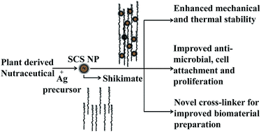
The present study evaluated the application of a nano-biotechnological intervention of nutraceutical shikimate for the development of a potential multi-site cross-linker with enhanced cross linking, anti-microbial and cell proliferative activities. The cross-linking and therapeutic properties of the nutraceutical shikimate were simultaneously utilized by caging them onto silver nanoparticles. The caging of shikimate on silver nanoparticles resulted in the cumulative expression of the physico-chemical properties of both silver nanoparticles and shikimate. We observed that in shikimic acid caged silver nanoparticle (SCS nanoparticle) cross-linked collagen, the viscosity and self-assembly process of collagen, along with its mechanical and thermal properties, were significantly improved when compared with native collagen. The three dimensional conformation of collagen was also retained after cross-linking with SCS nanoparticles. Cell viability with SCS nanoparticle cross-linked collagen was found to be enhanced, in comparison to collagen films cross-linked with shikimic acid and native collagen. SCS nanoparticle-stabilized collagen possessed both cell proliferative and anti-microbial properties, which would make SCS nanoparticles a superior cross-linker of collagen. The results suggested a new strategy for cross-linking collagen and provide scope for alternative biocompatible interventions in the development of biomaterials.
Introduction

Molecules that have more than one functional group are conventionally found to be suitable for cross-linking peptides. The presence of multiple functional groups enables multiple site interactions for enhanced cross-linking of collagenous peptides. Glutaraldehyde,1 dimethyl subrimidate, dimethyl-3,3′-dithio-bis(propionimidate)2,3 and dialdehyde cellulose/starch4 are some of the common multifunctional cross-linkers currently used. Most of these compounds react with side chain amino (NH2) groups of amino acids to cross-link them. On the other hand, carbodiimides provide the same function by cross-linking carboxyl groups.5 Unfortunately, these cross-linking agents have several drawbacks. One such major drawback results from cross-linking agents leaching out of the biomaterial, which causes undesired cross-linking of biomolecules, leading to several side effects. Further, unreacted cross-linking agents present in the scaffold may elicit several side reactions that are highly detrimental for the surrounding cells found near the implant site. Current research thus focuses on identifying suitable cross-linking agents with good biocompatibility and therapeutic properties. Many plant-derived compounds are reported to have multiple functional groups and could be explored as cross-linking molecules. The advantage of plant-derived compounds and nutraceuticals relies on their biocompatibility. Further, most of these compounds possess various therapeutic effects which may become incorporated into the biomaterial when used as a cross-linker of collagenous materials. Several potent plant-derived nutraceuticals are now in focus for their ability to cross-link peptides. Shikimic acid (3,4,5-trihydroxycyclohex-1-ene-1-carboxylic acid) is one such bifunctional molecule that is less explored as a cross-linking agent. Shikimic acid, having several therapeutic functions and multiple functional groups, could be explored as a potential cross-linking agent of collagen for biomaterial applications. Shikimic acid is a precursor in the biosynthesis of amino acids, phenolic compounds and several other secondary metabolites in plants.6,7 Shikimic acid was first isolated in 1885 by Eijkman from the fruit of the Japanese plant Illicium religiosum Sieb.8 Shikimic acid is an important compound that has extensive uses in the pharmaceutical industries. Shikimic acid is water-soluble and is present in plant species. Illicium anisatum and Illicium verum contain about 7 and 6% shikimic acid of their dry weight.9,10 The pharmacological properties of derivatives of shikimic acid have been studied based on recent advances in the structural studies of medicinal plants and various properties including antitumor and antiviral effects have been revealed. Enzymes of the shikimate pathway are good candidate targets in herbicide,11 antiviral12 and antimicrobial production.13 The neuraminidase inhibitor oseltamivir phosphate (Tamiflu) is one important compound, produced to treat swine flu using shikimic acid as precursor.12,14 Shikimic acid has been shown to increase the tolerance of sea urchin larvae to bacterial pathogens.15

Although shikimic acid has been reported to possess several pharmacological properties, there are no known reports on the use of shikimic acid as a cross-linker for collagenous materials. Several reports suggest that mono- and dicarboxylic acids show potent cross linking efficacies. Doillon et al. and Suh et al. studied the effect of collagen treated with hyaluronic acid on wound healing.16,17 Saito et al. studied cross-links using citric acid18 and maleic acid derivatives and their swelling behaviour in response to variations in pH. Mitra et al. studied the effect of oxalic acid, alginic acid, adipic acid, succinic acid and pimelic acid on the stability and physicochemical properties of collagen.19–23 The carboxylic groups of these acids cross-link by reacting with free side chain amino groups of different strands, thereby cross-linking them.15,18,19 Further, several bi-functional natural and plant-derived compounds have been used for their ability to cross-link collagen. We report here in this manuscript for the first time the effect of shikimic acid on the stabilization of collagen and its effect on the physicochemical properties of collagen for tissue engineering applications. As a strategy to improve the cross-linking efficacy, we further explored nano-biotechnological interventions by caging shikimic acid on silver nanoparticles in order to concentrate multi-functionality at the nanoscale level for effective cross-linking of collagen. We observed significant cross-linking efficacy of shikimic acid caged silver nanoparticles for scaffold development with added cell proliferative and anti-microbial properties.

Results and discussion

Synthesis of shikimic acid caged silver (SCS) nanoparticles

Silver nanoparticles were synthesized using silver nitrate and potassium hydroxide as starting materials.24 Highly unstable silver hydroxide formed from the reaction of silver nitrate and potassium hydroxide disintegrates to give nano-sized silver oxide. With shikimic acid present during the breakdown of silver hydroxide to silver oxide, shikimic acid gets caged on the silver nanoparticles, preventing aggregation of nanoparticles.

FTIR analysis

The caging of shikimic acid on silver nanoparticles was confirmed by FTIR analysis of shikimic acid isolated from the silver nanoparticles in comparison to a standard shikimic acid FTIR spectrum. FTIR spectra were obtained for shikimic acid, SCS nanoparticles and solubilized shikimic acid from the SCS nanoparticles. The results are provided in Fig. 1a. The results show strong and broad peaks at 3442, 3450 and 3465 cm−1 which correspond to the OH–H stretching of the alcohol groups. The intensity of these stretching peaks in shikimic acid caged silver nanoparticles was observed to be reduced on conjugation; this may be due to the fact that one or more OH groups out of the three present in shikimic acid play a key role in bonding to the silver nanoparticles. Strong peaks at 1705, 1709 and 1720 cm−1 correspond to the C[double bond, length as m-dash]O stretching vibrations of the acid groups. The medium-weak peaks at 1462 and 1456 cm−1 correspond to the aromatic C[double bond, length as m-dash]C group stretching vibrations.25,26 The spectral peaks for shikimic acid solubilized from the silver nanoparticles and the shikimic acid caged silver nanoparticles were similar to those observed with standard shikimic acid. The presence of similar peaks in all three spectra confirmed conjugation of shikimic acid on the silver nanoparticles.
image file: c5ra02278a-f1.tif
Fig. 1 (a) FTIR analysis of shikimic acid caged silver nanoparticles – (a) represents shikimic acid, (b) shows shikimic acid caged silver nanoparticles (SCS nanoparticles) and (c) shows shikimic acid isolated from the SCSNPs. (b) X-ray diffraction pattern of SCS nanoparticles. (c) UV-Vis spectrum of SCSNPs.

Caging efficiency of shikimic acid on shikimic acid caged silver nanoparticles

In order to understand the loading efficiency of shikimic acid on the silver nanoparticles, we quantified the amount of shikimic acid caged on silver nanoparticle using the p-hydroxybenzaldehyde method.27 The results indicated that the amount of shikimic acid caged on 1 mg of silver nanoparticles was 360 μg mg−1.

X-ray diffraction analysis

The XRD pattern for the shikimic acid caged silver nanoparticles was analyzed and the results are given in Fig. 1b. The diffraction pattern of the silver nanoparticles showed well-defined diffraction peaks corresponding to the crystallographic plane centered at 38.26 = 2θ, which indicated a relatively high degree of crystallinity. The XRD pattern of powdered silver nanoparticles had two peaks at 2θ values of 33.38 and 38.26 degrees corresponding to the (110) and (111) planes. All the reflections corresponded to pure silver metal with face centered cubic symmetry. The lack of spurious diffraction peaks indicated the purity of the synthesized shikimic acid caged silver nanoparticles.

Further, UV-Vis spectral analysis of shikimic acid caged silver nanoparticles was performed to confirm the formation of shikimic acid caged metallic silver nanoparticles. The results provided in Fig. 1c showed an surface plasmon resonance (SPR) absorption peak at around 416 nm, which indicated the purity of the synthesized shikimic acid caged silver nanoparticles. The results are consistent with the XRD analysis.

Particle size analysis

The particle size, zeta potential and dispersity of the nanoparticles were determined by dynamic light scattering (DLS) in aqueous solution. The results showed that the average size of pure silver oxide (control) nanoparticles and that of shikimic acid caged silver nanoparticles were 450 nm and 220 nm, respectively (Fig. 2a and b). The DLS analysis gives the hydrodynamic diameter of the nanoparticles, since water forms a layer that covers the nanoparticle. The larger size of the pure silver oxide (control) nanoparticles may be attributed to aggregation during the synthesis due to the lack of any capping agents. In the case of the SCS nanoparticles, shikimic acid binds with silver during the breakdown of highly labile silver hydroxide [Ag(OH)2], thereby caging the silver nanoparticles, preventing clumps and reducing the size of the SCS nanoparticles. The results are in accordance with earlier observations indicating the aggregation and sedimentation of pure silver oxide (control) nanoparticles. The polydispersities and zeta potentials of the pure silver oxide (control) nanoparticles and SCS nanoparticles are listed in Table 1. The zeta potentials of the pure silver oxide (control) nanoparticles and SCS nanoparticle were −33.0 and −31.0, respectively. The low zeta potential indicates that the nanoparticle solution was stable, mostly due to inter-particulate repulsion. Caged shikimate covers the entire surface of each nanoparticle, preventing agglomeration, and thereby reducing the size and increasing the number of silver nanoparticles.
image file: c5ra02278a-f2.tif
Fig. 2 Size and morphology of the synthesized nanoparticles – (a) and (b) particle size analysis of pure silver oxide (control) nanoparticles and SCS nanoparticles, respectively, by dynamic light scattering; (c) and (d) SEM images of pure silver oxide (control) nanoparticles and SCS nanoparticles, respectively; (e) and (f) EDAX images of pure silver oxide (control) nanoparticles and SCS nanoparticles, respectively.
Table 1 Comparative dynamic light scattering analysis for size dispersity and zeta potential of pure silver oxide (control) nanoparticles and SCS nanoparticles
  Pure silver oxide (control) nanoparticles Shikimic acid caged silver nanoparticles (SCSNPs)
Z-Average (d nm) 450 220
Polydispersity index (Pdi) 0.548 0.621
Zeta potential (mV) −33.0 −31.0


Shape and morphology analysis using electron microscopy

Scanning electron microscopy (SEM) analysis was performed to analyze the shape and morphology of the synthesized pure silver oxide (control) nanoparticles and shikimic acid caged silver nanoparticles. The results (Fig. 2c and d) showed that all the particles were fairly uniform in shape and size with a negligible amount of aggregation of the SCS nanoparticles. In order to confirm the presence of shikimic acid in the synthesized nanoparticles, elemental analysis was also performed. The reduction in oxygen content in the SCS nanoparticles when compared to the pure silver oxide (control) nanoparticles (from 48.01% to 35.18%) showed that shikimic acid has been incorporated on the silver nanoparticles (Fig. 2e and f). SEM and EDAX have shown that shikimic acid has been conjugated with the silver nanoparticles. Transmission electron microscopy (TEM) analysis was also performed to confirm the caging of shikimic acid on the silver nanoparticles. The results are provided in ESI Fig. 1. The TEM microphotograph of the SCSNPs showed a translucent ring around the opaque silver nanoparticles, indicating a uniform caging of shikimic acid over the silver nanoparticles, whereas in the control pure silver nanoparticles, the absence of such a translucent ring confirms the caging of shikimic acid in the case of the SCS nanoparticles. The representative high resolution TEM image of a SCSNP is provided as an inset image, showing the clear translucent ring around the opaque silver nanoparticle.

Physicochemical properties of collagen

Tensile strength. The mechanical stability of collagen cross-linked with SCS nanoparticles was analyzed by estimating their tensile strength and comparing it with collagen scaffolds cross-linked with glutaraldehyde. The results are given in Table 2. Table 2 shows an increase in both the tensile strength and the elongation at break for collagen scaffolds cross-linked with SCS nanoparticles. The tensile strength of collagen cross-linked with SCS nanoparticles was found to be higher than that of native collagen, and shikimic acid and glutaraldehyde cross-linked collagen. The relative mechanical stability was found to be in the following order: SCS nanoparticle cross-linked collagen > glutaraldehyde cross-linked collagen > shikimic acid cross-linked collagen > native collagen scaffolds. These results implied that the SCS nanoparticles cross-linked the collagen better and provided a superior strength to the collagen sheets. Tensile strength is determined at the breakage point of the collagen sheets. When stress is applied, the sheets start to rupture locally and then rupture completely at the breakage point.28
Table 2 Mechanical properties of shikimic acid caged silver nanoparticles (tensile strength and elongation at break). CC denotes the native collagen, Col SCSNP denotes collagen cross-linked with SCS nanoparticles, Col Shi denotes collagen cross-linked with shikimic acid and Col Glu denotes collagen cross-linked with glutaraldehyde
  Maximum load (N) Tensile strength (N mm−2) Elongation at break (%)
a Statistically significant compared to native collagen (p < 0.05).
CC 2.92 ± 0.03 1.33 ± 0.01 6.75 ± 0.03
Col Glu 6.89 ± 0.02a 2.75 ± 0.03a 10.5 ± 0.03a
Col Shi 4.29 ± 0.04a 1.86 ± 0.01 10.17 ± 0.02a
Col SCSNP 6.92 ± 0.02a 3.84 ± 0.01a 15.67 ± 0.01a


Collagen cross-linked with SCS nanoparticles showed a better strength when compared with collagen cross-linked with glutaraldehyde and native collagen. This enhancement in tensile strength can be attributed to the multiple sites for interaction present in the SCS nanoparticles. Multiple site cross-links bring multiple collagen triple helices closer, thereby increasing the combined strength of the resultant collagen matrix.

Rheology. The viscosity of the collagen samples was determined based on the flow rate of collagen with different concentrations of SCS nanoparticles and glutaraldehyde into the capillary of a viscometer probe. Cross-linking collagen greatly influences the stress–relaxation mechanism of collagen based materials.29,30 The flow rate of collagen cross-linked with different concentrations of SCS nanoparticles and glutaraldehyde into the capillary of the viscometer probe tends to vary, based on the viscosity of the collagen samples determined. The rheograms of the native collagen, collagen cross-linked with glutaraldehyde and shikimic acid caged silver nanoparticles at room temperature are given in Fig. 3a and b, representing the viscoelastic nature of the samples being tested. The results indicated that the viscosity was found to increase in SCS nanoparticle cross-linked collagen, which could be due to the facilitation of enhanced cross-linking of collagen by the SCS nanoparticles. A concentration-dependent effect on viscosity was observed with SCS nanoparticles. Nanoparticles conjugated with shikimic acid led to a significant increase in the viscosity of collagen when compared to those without conjugation. The interaction of small molecules and many metal nanoparticles with collagen has already been studied and documented. Nanoparticles have significant adsorption capacities due to their relatively large surface area, which can alter the viscosity by cross-linking collagen. The reported increase in collagen fibre formation kinetics with increasing temperature has been attributed to an increase in intermolecular attractive forces.31 The increase in intermolecular attractive forces contributed by the shikimic acid and silver nanoparticles on conjugation results in giving multiple sites for interaction with collagen molecules, which would plausibly be expected to translate into enhanced fibre rigidity.
image file: c5ra02278a-f3.tif
Fig. 3 Rheology and fibrillation: (a) shows the plot between viscosity vs. shear rate, (b) shows the plot between shear stress vs. shear rate and (c) is the plot showing the fibrillation kinetics of collagen. COL represents native collagen, COLGLU represents collagen cross-linked with glutaraldehyde and COLSNP represents collagen cross-linked with SCS nanoparticles.
Fibrillation assay. Fibrillation is an important assay for studying collagen cross-linking. It shows us the effect of the SCS nanoparticles on fibril-formation kinetics. The change in turbidity at 313 nm gives us an indirect measure of the cross-linking efficacy. Collagen cross-linked glutaraldehyde served as the positive control. Fig. 3c shows the turbidity curves of collagen, collagen glutaraldehyde and SCS nanoparticle cross-linked collagen treated at 32 °C. The results showed that the kinetics of fibril formation is sigmoidal with a lag, growth and plateau phase. The temperature-dependant nucleation process occurred in the lag phase where no visible change in turbidity was detected. Hydrophobic interactions play a pivotal role along with hydrogen and covalent bonding in the microfibril formation and during nucleation process.32,33 The formation of hydrophobic bonds is temperature dependant and after nucleation, the temperature independent fibril growth occurs. The fibril now grows both in diameter and in length.34–37 Fig. 3c shows that the general shape of the turbidity curves was not affected by the differences between glutaraldehyde, SCS nanoparticles and native collagen.38,39 The time taken to reach half the value of final turbidity (t1/2) and the total change in turbidity (Δh) is calculated. The t1/2 and Δh of SCS nanoparticle cross-linked collagen were 16 min and 0.9273, respectively. The results suggested that the SCS nanoparticles stimulated fibril formation and promoted the stabilization of fibrillar forms of collagen by providing multiple sites for nucleation and fibril rearrangements. The relative efficacy for cross-linking collagen was observed to be in the order glutaraldehyde > SCS nanoparticle > control.
Thermal stability. The thermal stabilities of the different collagen sheets were analyzed by Differential Scanning Calorimetry (DSC) and Thermo Gravimetric Analysis (TGA). DSC analysis was performed to understand the hydrothermal stability of the collagen. DSC analysis was carried out to study the stability of materials undergoing physical and chemical changes upon heating. The denaturation temperature (Td) is considered as a key measure to analyze the thermal stability as it gives the temperature at which collagen uncoils or unwinds. The level of cross-linking and stability of collagen is reflected in the Td values. The higher the Td value, the greater the stability and cross-linking.1,40 Typical thermograms of collagen, glutaraldehyde cross-linked collagen and SCS nanoparticle cross-linked collagen are given in Fig. 4a and b. The DSC thermograms (Fig. 4a) show two major endothermic peaks and multiple small peaks. The peaks in the regions of 51 °C to 55 °C correspond to denaturation of collagen and the other major peaks correspond to loss of bound water.
image file: c5ra02278a-f4.tif
Fig. 4 Thermal analysis of collagen: (a) DSC thermograms and (b) TGA thermograms.

The occurrence of small peaks may be due to inconsistent cross-linking leading to differences in the amount of cross-links in different regions of the sample. Broad peaks appear due to the overlap of several peaks that are very close to each other. The endothermic peak centered at 51.27 °C in the DSC thermogram of native collagen is associated with the helix coil transitional denaturation.41,42 It is at this temperature that collagen loses its ordered triple helical structure to form a random coil. The figure also shows that the presence of SCS nanoparticles is responsible for a slight increase in the denaturation temperature of the films, with broader peaks centered at 52.33 °C, compared with that of native collagen films.

The denaturation temperature for glutaraldehyde cross-linked collagen sheets was 54 °C. The results indicated that SCS nanoparticles cross-linked collagen had more hydrogen bonds and fewer hydrophobic bonds, leading to a higher denaturation temperature, whereas the presence of fewer hydrogen bonds and more hydrophobic bonds is responsible for the lower denaturation temperature of native collagen when compared with glutaraldehyde and SCS nanoparticle cross-linked collagens. The difference in denaturation temperature in native collagen and SCS nanoparticle cross-linked collagen is attributed to the water holding capacity of collagen. The water content in collagen fibers is modified by cross-linking and the cross-linkers fill in the water binding sites in the triple helix, so that the heat required for breaking these bonds increases, eventually increasing the denaturation temperature of these sheets. The water loss peak for SCS nanoparticle cross-linked collagen observed at 120.62 °C is almost same as that of glutaraldehyde cross-linked sheets, but is far higher than the peak for native collagen (113.74 °C). The results indicated an increased complexity of cross-links in collagen scaffolds cross-linked with SCSNPs. The change in thermal properties due to cross-linking was investigated using thermogravimetric analysis. Fig. 4b shows the plot of temperature vs. weight loss % of the collagen sheets. The thermal transition of collagen is a four step process. The first step is the removal of absorbed water molecules. The second step is the removal of structural water. The third transition is the partial break down of intermolecular bonding and the fourth is the complete thermal decomposition of the collagen molecules.43–46

The degradation of the polypeptide chain is initiated at 93.7 °C with weight percentage of 87, which is the first transition step of native collagen. In comparison, for the shikimic acid caged silver nanoparticle cross-linked sample there was a shift in the transition temperature and weight percentage to 103.42 °C and 89.89%, respectively. The second transition of collagen cross-linked with SCS nanoparticles starts at 257.19 °C, with an increase in the transition temperature compared to the native collagen. The weights of the collagen films were 83.56 and 86.92 for the native collagen and SCSNP cross-linked collagen, respectively. The partial denaturation of collagen begins at the third transition, where the weight percentage of native collagen was 62.13 and was 70.19 for the SCSNP cross-linked collagen film. Complete degradation of collagen occurs at the fourth temperature transition; the results showed an increased degradation temperature for collagen cross-linked with SCSNP compared with the native collagen. The final weight percentages were 51.35 and 62.77, respectively, for the native collagen and SCS nanoparticle cross-linked collagen films. The data indicated that SCSNPs covalently cross-link collagen, resulting in a relatively higher thermal stability brought about by cross-links induced by SCS nanoparticles.

Structural analysis. The influence of nanoparticle induced cross-links on the conformational change of collagen was studied using circular dichroism. It is important to note that natural collagen possesses a unique CD spectrum in the far UV region, with a small positive peak around 220 nm, a crossover at around 213 nm, and a large negative peak around 197 nm.47,48 The results obtained for collagen scaffolds cross-linked with SCS nanoparticles showed a minimum negative peak at 197 nm and maximum positive peak at 221 nm with a cross over at 214 nm in the CD spectrum (Fig. 5), which are characteristic of the triple helical structure of collagen. The above results indicate that the inter- and intra-helical cross-links created by SCS nanoparticles have very minimal effect on the structural conformation of collagen and do not affect the triple helical nature of collagen.
image file: c5ra02278a-f5.tif
Fig. 5 CD analysis of collagen (Col), glutaraldehyde cross-linked collagen (ColG) and SCS nanoparticle cross-linked collagen (ColShSNP).
Biological activity. The biocompatibility and cell proliferation capacity of the SCS nanoparticle cross-linked collagen scaffolds were analyzed using the MTT assay. The results are given in Fig. 6. The results indicated that for cells seeded on SCS nanoparticle cross-linked collagen scaffolds, the scaffolds were nontoxic and had a concentration dependent effect on the proliferation. Cell proliferation appeared to be significantly higher on collagen matrices cross-linked with SCS nanoparticles when compared to shikimic acid cross-linked collagen matrices. The cell spreading and viability was observed to be 100, 97 and 87% in collagen scaffolds cross-linked with SCS nanoparticles at 100, 200 and 300 μM (corresponding to shikimate), respectively. However the cell viability was observed to be only 70, 20 and 15%, respectively, for collagen scaffolds cross-linked with shikimic acid at 100, 200 and 300 μM (S100, S200 and S300), respectively. The results are in agreement with the photomicrographs, which show that the cell spreading and proliferation was greater for SCS nanoparticle cross-linked collagen scaffolds. The reduction in cytotoxic effect of the SCSNPs when compared to shikimic acid may be due to the fact that shikimic acid may easily leach out from the scaffolds, leading to its toxic effects, whereas conjugation of shikimate may prevent easy leaching of shikimic acid from SCSNPs. Further, it has been reported that the physico-chemical properties of molecules drastically change when conjugated with nanoparticles.49–51 The number of free functional groups available in shikimic acid on conjugation with silver nanoparticles would be much less, decreasing the likelihood of causing undesired side effects compared to unconjugated shikimic acid. Apart from having biocompatibility and a cell proliferative function, it is very important that the scaffolds should prevent any microbial growth while it is implanted or used as a dressing material; otherwise, general rejection of the biomaterial would occur along with other complications. Hence, we analyzed the anti-microbial efficacy of the collagen scaffold cross-linked with SCS nanoparticles. The anti-microbial ability of SCSNP cross-linked collagen sheets was investigated by the agar diffusion method. Assays were carried out using Escherichia coli (gram negative bacteria) and Bacillus subtilis (gram positive bacteria) in LB broth.
image file: c5ra02278a-f6.tif
Fig. 6 Cell proliferation assay of SCS nanoparticles with collagen: (a–h) photomicrographs of cells attached on various collagen scaffolds cross-linked with (a) native collagen (control), (b, c, and d) collagen scaffolds cross-linked with 100, 200 and 300 μM shikimic acid, respectively, (e) collagen scaffold cross-linked with glutaraldehyde (ColGlu) and (f, g, and h) collagen scaffolds cross-linked with 100, 200 and 300 μM SCS nanoparticles, respectively; (i) represents the percentage viability of cells on various collagen matrices cross-linked with glutaraldehyde at 100, 200 and 300 μM, shikimic acid (S100, S200, S300) and 100, 200 and 300 μM SCS nanoparticles (SCSNP 100, SCSNP 200 and SCSNP 300).

Fig. 7 shows the bactericidal kinetics of collagen sheets cross-linked with shikimic acid caged silver nanoparticles. Collagen sheets (300 μM SCSNPs) showed higher inhibition in both organisms which was reflected in the larger zone of inhibition when compared to the collagen scaffold cross-linked with 100 μM SCSNPs. Fig. 7b and c show the anti-microbial activity of collagen sheets against E. coli and B. subtilis, respectively, and Fig. 7a shows the control. The results indicated that SCSNP cross-linked collagen scaffolds showed good anti-microbial property against both gram positive and gram negative bacteria. However, for any material to find application as implants or as wound dressings it may essentially need to have a bactericidal activity of ≥99.9% (3[thin space (1/6-em)]log reduction) and at this MBC concentration it should not have any cytotoxic effect. In order to determine the MBC value of collagen scaffolds cross-linked with various concentrations of SCSNPs (9–180 μg of SCSNPs corresponding to shikimate caged on silver nanoparticles), they were tested for their bactericidal activity using a broth dilution assay. The initial concentrations of microbial cells were taken as 1 × 10⁶ cells per ml and the number of Colony Forming Units (CFUs) formed after treatment with collagen cross-linked with various concentrations of SCSNPs was evaluated.52 The results are presented in the Fig. 7d. The results indicated a concentration dependent bactericidal effect of SCSNP cross-linked collagen on both E. coli and B. subtilis. We observed a 3[thin space (1/6-em)]log reduction of microbial growth at 27 and 36 μg levels of SCSNPs for E. coli and B. subtilis, respectively. E. coli was found to be more susceptible to the SCSNPs when compared to B. subtilis, where the MBC was attained at 27 μg of SCSNPs. The bactericidal activity on E. coli and B. subtilis was found to be similar with 36 μg of SCSNPs. A 4 log reduction in growth was observed with 72 μg of SCSNPs for both E. coli and B. subtilis, and no microbial colonies were observed at higher concentrations tested. The MIC values of the SCSNPs for E. coli and B subtilis were observed to be 6 μg and 10 μg, respectively.


image file: c5ra02278a-f7.tif
Fig. 7 Anti-microbial assay of collagen scaffolds cross-linked with 100 and 300 μM SCSNPs, using the agar diffusion method: (a) blank (b) E. coli [(i) 100 μM and (ii) 300 μM], (c) B. subtilis [(i) 100 μM and (ii) 300 μM]. (d) Minimum bactericidal concentration (MBC) assay for collagen scaffolds crosslinked with various concentrations of SCSNPs showing log reduction in growth of E. coli and B. subtilis. (e) Effect of MBC concentrations of SCSNPs on human normal keratinocyte cell (HaCaT) viability.

The cytotoxic effect of SCSNPs at MBC concentration was evaluated to further confirm the biocompatibility of the SCSNPs for their application as cross-linkers for biomaterial development (Fig. 7e). The cytotoxicity at MBC concentrations was tested using the MTT assay. The results showed that the cell viability was not affected even at concentrations much higher than the MBC concentration ranges.

We observed that although shikimic acid is engineered and expressed in E. coli to meet commercial requirements, its conjugation with silver nanoparticles greatly enhances its anti-microbial properties. The conjugation of shikimic acid to nanoparticles is believed to block the aromatic amino acid synthesis pathway, thereby inhibiting microbial growth. For biomedical applications, the use of biomaterials with anti-microbial properties will be an added advantage. Collagen sheets incorporated with SCS nanoparticles having such antimicrobial activities will prevent infection and sepsis, and could be a good material for preparing wound dressings or implant materials. The results further confirmed the biocompatibility of SCSNPs for their use as cross-linkers of collagen scaffolds with anti-microbial and cell compatible properties for biomaterial applications.

Materials

All the chemicals were procured from Sigma Aldrich, unless otherwise mentioned. HaCaT cells (immortalized human keratinocytes) were purchased from NCCS Pune, India. All the tissue culture wares were from Nunc, Denmark.

Isolation of collagen from rat tail tendon (RTT)

Rat tail tendon collagen was isolated as described previously.53,54 Briefly, tendons from rat tails were washed 4 times with 1[thin space (1/6-em)]:[thin space (1/6-em)]1 diethyl ether and chloroform, followed by two methanol washes. The tendons were then washed with 1% sodium chloride (4 times) and ground in 0.05 M acetic acid. Pellets were removed after centrifugation and the supernatant was precipitated with 5% sodium chloride. The collagen thus obtained was then dialyzed against 0.05 M acetic acid and lyophilized for further use.

Synthesis of shikimic acid caged silver (SCS) nanoparticles

Shikimate caged silver nanoparticles were synthesized by treating 1 ml of 100 mM silver nitrate and 200 μl of 0.5 M KOH with different concentrations of shikimic acid (20 μM to 100 μM), and the solution was vortexed for 10 minutes. The nanoparticles were collected by centrifuging at 7000 rpm for 15 minutes. The particles were freeze dried to obtain a fine powder.

Caging efficiency of shikimic acid

The amount of shikimic acid caged on silver nanoparticles was studied using colorimetric determination of shikimic acid by extracting shikimic acid from a known amount of shikimic acid caged silver nanoparticles. The concentration of shikimic acid caged onto the silver nanoparticle was determined using the p-hydroxybenzaldehyde method.27 Briefly, 1 mg of freeze dried nanoparticle was accurately weighed and dispersed in 1 ml of water. 1 ml of shikimic acid standard (25 μM to 1 mM) was prepared from 100 mM stock solution. 3 ml of concentrated hydrochloric acid was added to 1 ml test solutions kept in ice. Then 1 ml of 0.2% p-hydroxybenzaldehyde was added and the mixture placed in a boiling water bath for 60 min. After cooling to room temperature, the purple colour formed was measured at 570 nm in a BioRad ELISA plate reader.

Fourier transform infrared spectroscopy (FTIR)

SCSNPs, solubilized shikimic acid isolated from SCSNPs and standard shikimic acid were subjected to FTIR analysis using a Spectrum Two model spectrophotometer from Perkin-Elmer Co., USA. Samples were made into a pellet using potassium bromide (KBr) and a scan from 450 to 4000 cm−1 was run with 8 scans and a resolution of 1 cm −1.

Characterization of nanoparticles

Powder XRD. X-ray diffraction patterns were analyzed using a Bruker D8 Advance diffractometer with Cu κα 1.54 Å radiation and detected using a Bruker Lyrix eye detector. Measurement temperature and slit size were set at 25 °C and 0.6, respectively, as the default for all measurements. The X-ray diffraction spectra were recorded in the range 2θ from 10.0 to 60.0 with a step increment of 0.02 and count time of 5 s.
Particle size analysis. The particle sizes and the zeta potentials of the synthesized nanoparticles were determined using Photon Correlation Spectroscopy (PCS) with a Malvern Instruments Zetasizer 3000 HSA equipped with a digital auto-correlator.
SEM with EDAX. The shape and morphology of the synthesized nanoparticles were determined using a Quanta 200 FEG scanning electron microscope equipped with an energy dispersive X-ray (EDAX) spectrometer. Approximately 0.5 mg of nanoparticles was spread on the stub, sputtered for 2–3 minutes with gold for conductivity and analyzed in high vacuum mode.

Physicochemical properties of collagen

Tensile strength. The tensile strength of the films was measured using an Instron Universal Testing machine until the sample ruptured. Samples with uniform thickness and length of 5 cm and width of 1 cm were cut from the film. A load of 10 N was applied and the measuring speed was 5 mm min−1 with a relative humidity of 60% at 25 °C.
Viscosity. The viscosity of the collagen samples was measured using a Brookefield R/S + Rheometer. 0.3% collagen solution was used throughout the study. The solutions were kept overnight with continuous stirring at 4 °C with and without SCS nanoparticles, allowing cross-linking.55
Fibrillation assay. Fibrillation assay was carried out by initializing fibril formation by mixing a 300 μg mL−1 final concentration of collagen with 0.2 M phosphate buffer, 2 M sodium chloride, 0.02 M phosphate buffer and 0.15 M sodium chloride. The pH was adjusted to 7.2 with 1.25 N sodium hydroxide. The turbidity was measured at 313 nm using a Perkin Elmer UV-Vis spectrophotometer. The rate at which fibrils formed was calculated from the time taken to reach half the value of final turbidity (t1/2).
Thermal properties of collagen. Thermal stabilities of hermetically sealed collagen films were determined by DSC analysis using a DSC Q200 (V23.10 Build 79) differential scanning calorimeter from 25 °C to 250 °C under a nitrogen atmosphere at a flow rate of 50 ± 5 ml min−1. The temperature was standardized using iridium as the standard. The heating rate was set at 5 °C min−1. Thermo gravimetric analysis (TGA) measures the amount and rate of change in the mass of a sample as a function of temperature in a controlled atmosphere. Collagen films were prepared by cross-linking with glutaraldehyde and shikimic acid caged silver nanoparticles. Native collagen sheets served as the control. TGAs of the collagen films were carried out using a TGA Q50 (V20.6 Build 31), from 20 °C to 800 °C at a constant heating rate of 20 °C min−1 under a nitrogen atmosphere.
Conformational studies. Circular dichroism (CD) spectra are used extensively for studying the three dimensional conformations of proteins and polypeptides. The influence of the SCS nanoparticles on the conformation of collagen was studied at 20 °C using a circular dichroism spectropolarimeter under a nitrogen atmosphere. 0.5 ml of collagen solution containing 0.3 mg collagen per ml was used for recording the spectra. Two scans per sample and a scan speed of 50 nm per min were set for recording the spectra. A reference spectrum was also recorded with collagen and 0.05 M acetic acid as control and blank, respectively.
Biological activity. The influence of stabilized collagen on cell attachment and proliferation is studied. Collagen films stabilized with shikimic acid, glutaraldehyde and SCS nanoparticles were coated on culture plates to perform the assay. HaCaT cells were used to carry out the assay. Approximately 12[thin space (1/6-em)]000 cells were treated with three different concentrations of shikimic acid caged silver nanoparticles (from 100 μM to 300 μM) and collagen scaffolds cross-linked with SCSNPs (concentrations higher than the minimum bactericidal concentration) were incubated for 24 hours in a carbon dioxide incubator. The cells were then incubated with 3-(4,5 dimethylthiazolyl-2-yl)-2,5-diphenyltetrazolium bromide (MTT) salt at 0.5 mg ml−1 at 37 °C. After 4 hours of incubation, the blue/purple formazan crystals formed were solubilized using DMSO (dimethyl sulphoxide) and the absorbance was read at 570 nm in a Bio-Rad ELISA plate reader.56

Anti-microbial activities of the shikimic acid caged silver (SCS) nanoparticle cross-linked collagen sheets were tested on gram negative Escherichia coli and gram positive Bacillus subtilis by the broth dilution method using Luria Bertani broth57 and the agar diffusion method58 using Mueller Hington agar. The shikimic acid caged silver (SCS) nanoparticle cross-linked collagen sheets (approximately 1 cm2) were placed on agar plates inoculated with each of these microorganisms and incubated overnight. The antimicrobial activity was determined by measuring zones of inhibition formed around the samples. In the broth dilution assay, shikimic acid caged silver (SCS) nanoparticle cross-linked collagen sheets (approximately 1 cm2) were incorporated in a total volume of 2 ml culture media along with culture inoculums and incubated overnight.59 After overnight incubation 10 μl of bacterial culture was diluted 25 times and the optical density was analyzed spectrophotometrically at 630 nm to determine the minimum inhibitory concentration (MIC). The cultures treated with various concentrations of SCSNPs from MIC values of SCSNPs were spread on agar plates and colonies were counted. Minimum bactericidal concentration (MBC) was determined by calculating the log reduction in bacterial colonies.

Log reduction = log[thin space (1/6-em)]A − log[thin space (1/6-em)]B.
where A and B are the number of viable microorganisms before and after SCSNP treatment, respectively.

Conclusions

In this study we analysed the effect of SCS nanoparticles on the stabilization of collagen for biomaterial applications. SCS nanoparticles enhanced the viscosity and self-assembly process of collagen and improved its mechanical and thermal properties. The three dimensional conformation of collagen was also retained after cross-linking with SCS nanoparticles. Cell viability in SCS nanoparticle cross-linked collagen was found to be enhanced, in comparison to a collagen film cross-linked with shikimic acid and native collagen. SCS nanoparticle stabilized collagen, with both cell proliferative and anti-microbial properties, would find applications as an ideal biomaterial scaffold for tissue engineering applications. In conclusion, SCS nanoparticles can be considered as a cross-linking agent for collagen and provide a scope for alternate biocompatible interventions in the development of wound dressings.

Acknowledgements

We thank The Director, CSIR–CLRI, Chennai, India for providing the facilities to carry out the experiments. The financial assistance from CSIR, Govt. of India for the supra-institutional project-STARIT (CSC0201) under XII five year plan and the Department of Biotechnology, Government of India, for the project RGYI (GAP 1062), is greatly acknowledged.

References

  1. N. N. Fathima, B. Madhan, J. R. Rao, B. U. Nair and T. Ramasami, Int. J. Biol. Macromol., 2004, 34, 241–247 CrossRef CAS PubMed.
  2. V. Charulatha and A. Rajaram, Biomaterials, 2003, 24, 759–767 CrossRef CAS.
  3. V. Charulatha and A. Rajaram, J. Biomed. Mater. Res., Part B, 1997, 36, 478–486 CrossRef CAS.
  4. S. V. Kanth, A. Ramaraj, J. R. Rao and B. U. Nair, Process Biochem., 2009, 44, 869–874 CrossRef CAS PubMed.
  5. R. Usha, K. J. Sreeram and A. Rajaram, Colloids Surf., B, 2012, 90, 83–90 CrossRef CAS PubMed.
  6. K. M. Herrmann and L. M. Weaver, Annu. Rev. Plant Physiol. Plant Mol. Biol., 1999, 50, 473–503 CrossRef CAS PubMed.
  7. A. R. Knaggs, Nat. Prod. Rep., 2003, 20, 119–136 RSC.
  8. J. F. Eijkman, Recl. Trav. Chim. Pays-Bas, 2010, 4, 32–54 CrossRef.
  9. D. V. Bochkov, S. V. Sysolyatin, A. I. Kalashnikov and I. a. Surmacheva, J. Chem. Biol., 2012, 5, 5–17 CrossRef PubMed.
  10. H. Adams, N. A. Bailey, R. Brettle, R. Cross, M. Frederickson, E. Haslam, F. S. MaeBeath and G. M. Davies, Tetrahedron, 1996, 52, 8565–8580 CrossRef CAS.
  11. G. M. Kishore and D. M. Shah, Annu. Rev. Biochem., 1988, 57, 627–663 CrossRef CAS PubMed.
  12. G.-W. Wang, W.-T. Hu, B.-K. Huang and L.-P. Qin, J. Ethnopharmacol., 2011, 136, 10–20 CrossRef CAS PubMed.
  13. E. Pinder, J. L. Thain, W. J. Watkins, R. G. Wilson, G. M. Davies, K. J. Barrett-bee, D. A. Jude, M. Lehan, W. W. Nichols, P. E. Pinder, J. L. Thain, W. J. Watkins and R. G. Wilson, Antimicrob. Agents Chemother., 1994, 38, 403–406 CrossRef.
  14. H.-K. Kim and K.-J. J. Park, Tetrahedron Lett., 2012, 53, 1561–1563 CrossRef CAS PubMed.
  15. K. V. Kiselev, N. V. Ageenko and V. V. Kurilenko, Dis. Aquat. Org., 2013, 103, 121–132 CrossRef CAS PubMed.
  16. C. J. Doillon, D. Mantovani, N. Rajan, J. Habermehl and M. Cote, Nat. Protoc., 2007, 1, 2753–2758 CrossRef PubMed.
  17. S.-N. Park, J.-C. Park, H. O. Kim, M. J. Song and H. Suh, Biomaterials, 2002, 23, 1205–1212 CrossRef CAS.
  18. H. Saito, S. Murabayashi, Y. Mitamura and T. Taguchi, Biomacromolecules, 2007, 8, 1992–1998 CrossRef CAS PubMed.
  19. T. Mitra, G. Sailakshmi, A. Gnanamani and A. B. Mandal, J. Therm. Anal. Calorim., 2011, 105, 325–330 CrossRef CAS PubMed.
  20. T. Mitra, G. Sailakshmi, A. Gnanamani, S. T. K. Raja, T. Thiruselvi, V. M. Gowri, N. V. Selvaraj, G. Ramesh and A. B. Mandal, Int. J. Biol. Macromol., 2011, 48, 276–285 CrossRef CAS PubMed.
  21. T. Mitra, G. Sailakshmi, A. Gnanamani and A. B. Mandal, J. Appl. Polym. Sci., 2012, 125, E490–E500 CrossRef CAS.
  22. T. Mitra, G. Sailakshmi, A. Gnanamani and A. B. Mandal, Mater. Res., 2013, 16, 755–765 CrossRef CAS PubMed.
  23. T. Mitra, G. Sailakshmi, A. Gnanamani and A. B. Mandal, Int. J. Polym. Mater., 2013, 62, 572–582 CrossRef CAS.
  24. N. Duraipandy, R. Lakra, K. V. Srivatsan, U. Ramamurthy, P. S. Korrapati and M. S. Kiran, Metallomics, 2014, 6, 2025–2033 RSC.
  25. R. Silverstein, F. Webster and D. J. Kiemle, Spectrometric identification of organic compounds, John Wiley and Sons, New York, 7th edn, 2005 Search PubMed.
  26. P. Griffiths and J. De Haseth, Fourier transform infrared spectrometry, Wiley-Interscience, Hoboken, NJ, 2nd edn, 2007 Search PubMed.
  27. T. Mossor and R. W. Schramm, Anal. Biochem., 1972, 47, 39–45 CrossRef CAS.
  28. N. Cao, Y. Fua and J. Hea, Food Hydrocolloids, 2007, 21, 575–584 CrossRef CAS PubMed.
  29. R. Usha, K. J. Sreeram and A. B. Mandal, Colloids Surf., B, 2013, 109, 121–128 CrossRef CAS PubMed.
  30. D. M. Knapp, J. Rheol., 1997, 41, 971 CrossRef CAS.
  31. J. Rosenblatt, B. Devereux and D. G. Wallace, J. Appl. Polym. Sci., 1993, 50, 953–963 CrossRef CAS.
  32. W. D. Comper and A. Veis, Biopolymers, 1977, 16, 2113–2131 CrossRef CAS PubMed.
  33. M. C. Goh, M. F. Paige, M. A. Gale, I. Yadegari and M. Edirisinghe, Phys. A, 1997, 239, 95–102 CrossRef CAS.
  34. R. A. Gelman, B. R. Williams and K. A. Piez, J. Biol. Chem., 1979, 254, 180–186 CAS.
  35. G. Suarez, M. Veliz and R. L. Nagel, Arch. Biochem. Biophys., 1980, 205, 422–427 CrossRef CAS.
  36. J. Disalvo and M. Schubert, Biopolymers, 1966, 4, 247–258 CrossRef CAS.
  37. H. B. Bensusan and B. L. Hoyt, J. Am. Chem. Soc., 1958, 80, 719–724 CrossRef CAS.
  38. R. Williams, A. Gelman and C. Poppke, J. Biol. Chem., 1978, 253, 6578–6585 Search PubMed.
  39. A. Steplewski and A. Fertala, Fibrog. Tissue Repair, 2012, 5, S29 CrossRef PubMed.
  40. H. A. Scheraga and L. Gierasch, Biopolymers, 2008, 89, 479–485 CrossRef CAS PubMed.
  41. D. G. Wallace, R. A. Condell, J. W. Donovan, A. Paivinen, W. M. Rhee and S. B. Wade, Biopolymers, 1986, 25, 1875–1895 CrossRef CAS PubMed.
  42. H. R. T. Ang and A. D. Covington, J. Agric. Food Chem., 2003, 51, 6652–6656 CrossRef PubMed.
  43. C. Yang, Bull. Mater. Sci., 2012, 35, 913–918 CrossRef CAS PubMed.
  44. Z. Tian, C. Li, L. Duan and G. Li, Connect. Tissue Res., 2014, 55, 239–247 CrossRef CAS PubMed.
  45. Y. Nomura, S. Toki, Y. Ishii and K. Shirai, J. Agric. Food Chem., 2000, 48, 2028–2032 CrossRef CAS PubMed.
  46. K. Yoshimura, M. Terashima, D. Hozan, T. Ebato, Y. Nomura, Y. Ishii and K. Shirai, J. Agric. Food Chem., 2000, 48, 2023–2027 CrossRef CAS PubMed.
  47. D. D. Jenness, C. Sprecher and W. C. Johnson, Biopolymers, 1976, 15, 513–521 CrossRef CAS PubMed.
  48. F. H. Chu and A. Lukton, Biopolymers, 1974, 13, 1427–1434 CrossRef CAS PubMed.
  49. D. L. Leslie-Pelecky, in Biomedical application of nanotechnology, ed. V. Labhasetwar and D. L. Leslie-Pelecky, Wiley-Intescience, New Jersey, 2007, pp. 227–241 Search PubMed.
  50. P. Srinivas, G. Gopinath, A. Banerji, A. Dinakar and G. Srinivas, Mol. Carcinog., 2004, 40, 201–211 CrossRef CAS PubMed.
  51. N. Duraipandy, R. Lakra, K. V. Srivatsan, U. Ramamurthy, P. S. Korrapati and M. S. Kiran, J. Mater. Chem. B, 2015, 3, 1415–1425 RSC.
  52. M. Suarez, M. Haenni, S. Canarelli, F. Fisch, P. Chodanowski, C. Servis, O. Michielin, R. Freitag, P. Moreillon and N. Mermod, Antimicrob. Agents Chemother., 2005, 49, 3847–3857 CrossRef CAS PubMed.
  53. K. P. Sai and M. Babu, Comp. Biochem. Physiol., Part B: Biochem. Mol. Biol., 2001, 128, 81–90 CrossRef CAS.
  54. M. Wohllebe and D. Carmichael, Eur. J. Biochem., 1978, 92, 183–188 CrossRef CAS PubMed.
  55. B. Xu, H. Li and Y. Zhang, Biomatter, 2013, 3, 1–8 CrossRef PubMed.
  56. T. Mosmann, J. Immunol. Methods, 1983, 65, 55–63 CrossRef CAS.
  57. D. Amsterdam, in Antibiotics in laboratory medicine, ed. V. Lorian, Williams and Wilkins, Baltimore, 4th edn, 1996, pp. 52–111 Search PubMed.
  58. A. W. Bauer, W. M. M. Kirby, J. C. Sherris and M. Turck, Am. J. Clin. Pathol., 1966, 45, 493–496 CAS.
  59. I. Wiegand, K. Hilpert and R. E. W. Hancock, Nat. Protoc., 2008, 3, 163–175 CrossRef CAS PubMed.

Footnote

Electronic supplementary information (ESI) available. See DOI: 10.1039/c5ra02278a

This journal is © The Royal Society of Chemistry 2015
Click here to see how this site uses Cookies. View our privacy policy here.