ZnFe2O4 nanoparticles and a clay encapsulated ZnFe2O4 nanocomposite: synthesis strategy, structural characteristics and the adsorption of dye pollutants in water

Azadeh Tadjarodi*, Mina Imani and Mohammad Salehi
Research Laboratory of Inorganic Materials Synthesis, Department of Chemistry, Iran University of Science and Technology, Narmak, Tehran, 16846-13114, Iran. E-mail: tajarodi@iust.ac.ir; Fax: +98 21 77491204; Tel: +98 21 77240516

Received 4th February 2015 , Accepted 5th June 2015

First published on 5th June 2015


Abstract

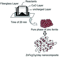
In this work, zinc ferrite nanoparticles were prepared by a one-pot microwave assisted combustion in the solid state with a controllable power up to 900 W and a tunable time of up to 20 min. Then, the ZnFe2O4 nanoparticles were encapsulated in an organoclay using ultrasound assisted impregnation to obtain a zinc ferrite/organoclay nanocomposite as a magnetic heterostructured adsorbent. The combustion reactions were performed in a reaction set-up manufactured from an alumina crucible encircled by a jacket of CuO as a microwave absorbing layer. This workpiece absorbs the distributed microwaves and supplies calcination conditions without any external heat source to prepare a pure phase of zinc ferrite. The effects of the microwave power and the time parameters on the structural and morphological features of the samples are discussed in detail. The adsorption performance of the products for water purification of some dye pollutants was also studied. The UV-Vis results showed that the addition of the as-prepared magnetic nanoparticles to the exfoliated organoclay considerably increased the adsorption of organic dye pollutants from an aqueous solution. Magnetic hysteresis measurements were performed on a vibrating sample magnetometer (VSM) showing the soft paramagnetic properties of the resulting products at room temperature. The structural and morphological analyses were also investigated in detail.


1. Introduction

The discharge of toxic dye wastewaters into the environment from industrial activities is a major environmental problem for bodies of water. They are carcinogenic and toxic to the ecosystem and strongly threaten the survival of organisms. Therefore, the elimination of these hazardous pollutants from bodies of water and the refinement of wastewater by a reasonable route is vital.1–3 Adsorption has been found to be an efficient, low-cost, accessible route for this aim.4 Nano-sized magnetic nanoparticles and their hybrids or composites with clay materials have received increasing attention as adsorbents for the removal of organic pollutants from water sources due to their high surface areas and large adsorption capacities.5 Organoclay is a membrane of clay materials with silica sheets constructed from two basic tetrahedral and octahedral units, which have been modified with organic groups. It is a cheap, readily available material that can be used to adsorb contaminants from the environment. Its structure has a negative charge, which can easily attract and hold cationic pollutants and eliminate them from aqueous solutions.6,7

In order to improve and reinforce the physical and chemical properties of clay materials, nano-sized inorganic particles can be introduced into the body of the clays.8,9 Among various fillers, magnetic nanomaterials have attracted a lot of attention due to their special properties.10,11 Magnetic nanoparticles dispersed in the clay matrix, in addition to expanding the adsorption capacity via electrostatic interactions, increase the simplicity of their recovery from the workplace. In fact, magnetic adsorbents can be employed to remove the pollutants in water and after operation they can be absolutely separated from the environment using their magnetic properties. On the other hand, the tendency of magnetic nanoparticles towards agglomeration leads to an increase in their particle size and decreases their activity as adsorbents or catalysts.12 As a new feasibility, the encapsulation of magnetic metal oxides inside a clay body can prevent the agglomeration and, therefore, produce a new class of nanocomposites with a small particle size and high surface area. Amongst magnetic materials, spinel ferrites, MFe2O4, especially ZnFe2O4 are technologically introduced as an important class of nanomaterials due to their magnetic,13 catalytic,14 photocatalytic,15 optical and electrical properties.16–18 Hence, there is a great potential for their application in the synthesis of magnetic nanohybrid and nanocomposite adsorbents for dealing with environmental problems.19 On the other hand, the discovery of new, low cost and innovative procedures to prepare the single phased magnetic nanomaterials according to the required demands is of great importance to devise technologies for mass production. Most of the described synthesis methods in the literature for the pure phase preparation of zinc ferrites require a high temperature for calcination, severe experimental conditions, complex equipment and several steps or special chemicals.20–22 Among the reported methods, such as co-precipitation,23 hydrothermal/solvothermal,24,25 mechanochemical,26,27 sol–gel,28 thermal decomposition,29 etc., the combustion technique assisted by microwave energy has attracted a lot of attention due to its simplicity, cost effectiveness, and the short reaction times. In contrast to conventional combustion (when the heat is generated from an external heating source, e.g., a hot filament), in the microwave assisted process the heating energy is internal, resulting from the reactant–microwave interactions. In this process, the entire workpiece is exposed to microwave energy, and is open to allow the combustion reaction and yield the prepared product.30–34 However, although this technique is simple and quick, the sintering or calcination post reaction and also the supply of enough energy for this operation appears to be an important subject. Therefore, it is necessary to modify the employed technique to tackle the problems for the synthesis of a pure phase of magnetic ZnFe2O4. Since a single-phased magnetic structure with the fascinating magnetic performance could be a good candidate to be embedded in the matrix of composites to make the applicable materials in water purification, we designed and performed a new method for the synthesis of zinc ferrite nanomaterial in this work. As an innovative idea, we designed a one step reaction of a solid state combustion assisted by microwave heating energy and constructed a reaction set-up encircled by a jacket of a microwave absorber and an insulating fiberglass layer around the bottom. This set-up can convert the absorbed microwaves into heat and create calcinating conditions without the use of any external heat source. It means that the use of the microwave absorber material, CuO, centers the microwave heating energy and engineers the best conditions for the rapid preparation of pure phased zinc ferrite in the nanoscale. It is important to note that the use of a microwave absorbing layer in microwave assisted techniques to supply the sufficient heating energy to form a pure phase of product in a one step reaction has not yet been reported. Because such a technique is operated in the solid state without any organic solvents, it can be introduced as an environmentally friendly method.

Many different approaches have been reported to fabricate metal oxide/clay nanocomposites, such as co-precipitation, molecular self-assembly, sol–gel processing, etc.35–37 A few works have been reported with this aim using an ultrasound assisted procedure, which is an effective, rapid and facile procedure to prepare uniform structures in the range of materials technology.11 This technique is a reliable and simple route, which easily provides a uniformity and reactivity due to the atomic level mixing within the reaction system. In addition, ultrasound waves produce effective droplets and adequate mechanical force leading to the better dispersion of species in aqueous solutions and the better exfoliation of organoclay layers for the encapsulation of magnetic nanoparticles. This technique is not only a simple and rapid method for the formation of a zinc ferrite/organoclay nanocomposite but it will also open a new pathway for the synthesis of various nanocomposites.

Within the objectives of this research, we synthesized a pure phase of zinc ferrite nanoparticles with an acceptable surface area by a simple and available technique without any additional treatment. Then, we successfully prepared a ZnFe2O4/clay nanocomposite by dispersing the magnetic nanoparticles in a commercial exfoliated organoclay using a facile ultrasound assisted impregnation and then promoted their adsorption behavior for water treatment. Malachite Green (MG) and Brilliant Green (BG) were selected as model dye pollutants for the adsorption study at room temperature and neutral pH.

2. Experimental

2.1 Materials

Iron(III) nitrate nonahydrate (Fe(NO3)3·9H2O, 99%), zinc nitrate hexahydrate (Zn(NO3)2·6H2O, 99%), urea (CH4N2O, 99%), glycine (Gly, NH2CH2COOH, 99%) and ammonium nitrate ((NH4)(NO3), 99%) were purchased from Merck Co. and used without further purification. Commercial-grade organoclay with a purity of 95% was purchased from Sahand Petroplastic International Co. (Iran). Brilliant green (C27H33N2·HO4S) and malachite green (C23H25ClN2) dyes were purchased from commercial sources as model dyes. Distilled water was used to prepare all solutions.

2.2 Synthesis of ZnFe2O4 nanoparticles

Fe(NO3)3·9H2O and Zn(NO3)2·6H2O with the molar ratio of 2[thin space (1/6-em)]:[thin space (1/6-em)]1 were mixed with each other in the presence of Gly or urea and ammonium nitrate as fuel and driving agents with different molar ratios to obtain the optimal conditions. The mixture was transferred into the designed reaction set-up and put into a domestic microwave oven with regulated power from 300 to 900 W (33%, 66% and 100% of power at 2450 MHz) for tunable times of 5–20 min. After the treatments, the voluminous sponge-like products were collected, washed with distilled water and ethanol several times to remove the residual initial materials, centrifuged, dried at 80 °C overnight and then analyzed. The effect of the fuel change and also the effect of the variation of the power and time on the formation of the product were discussed in detail. The structural and morphological studies were carried out using FT-IR, XRD, SEM, EDX, VSM and BET analyses.

2.3 Reaction set-up

In order to produce the calcination conditions in a microwave oven without any external thermal source, we designed and manufactured a reaction set-up with the ability to absorb and transform the microwave energy into high temperature heat energy. Fig. 1 illustrates a scheme of this reaction set-up. It was made from an alumina crucible with a jacket layer of CuO around it to absorb the microwave energy and produce calcination heat. The floor of the container was embedded in a thick fiberglass layer to prevent the heat transfer to the bottom and probable damage to the glass plate of the oven.
image file: c5ra02163d-f1.tif
Fig. 1 Schematic diagram of the reaction set-up.

2.4. Synthesis of the ZnFe2O4/organoclay nanohybrid

The as-prepared magnetic zinc ferrite nanoparticles (180 mg) were dispersed and entrapped in an organoclay suspension in water (20 mg, 50 mL) by ultrasound waves with a power of 200 W for 2 h. The sediment was then collected, washed with deionized water several times and dried at 80 °C for 16 h. Finally, a brown colored powder was obtained and the structure and characteristics of this product were studied.

2.5 Characterization techniques

Fourier transform infrared (FT-IR) spectra were recorded on a Shimadzu-8400S spectrometer in the range of 400–4000 cm−1 using KBr pellets. The X-ray diffraction (XRD) patterns were recorded by a STOE powder diffraction system using Cu Kα radiation (wavelength, λ = 1.54060 Å). Scanning electron microscopy (SEM) images were taken on a Hitachi S4160 FESEM with gold coating and energy-dispersive X-ray spectroscopy analysis (EDX) on a VEGA\TESCAN S360 with gold coating. A double-beam UV spectrophotometer (Shimadzu UV-1700) was used for determination of the dye concentrations in the supernatant solutions before and after adsorption. The magnetic properties were recorded using a vibrating sample magnetometer (VSM, MDK6) which was made by the efforts of the Magnetis Daghigh Kavir Company in Iran. The surface area of the final product was obtained using the Brunauer–Emmett–Teller (BET) technique with Micromeritics (Gemini) in the range of relative pressures from 0.0 to 1.0. Before testing, the sample was degassed at 473 K for 2 h. Microwave radiation was supplied using a domestic microwave oven with a controllable power and time (LG model, 2450 MHz).

2.6 The adsorption process

To study the adsorption behavior of the prepared samples, Malachite Green (MG) and Brilliant Green (BG) were selected as model dye pollutants (the molecular structures are in Scheme S1 of ESI). The adsorption experiments were performed by taking 50 mL of the MG and BG dye solutions with an initial concentration of 20 to 200 mg L−1 and introducing 0.02 g of the zinc ferrite nanoparticles and zinc ferrite/organoclay nanohybrid as the adsorbent at room temperature and neutral pH for 4 h in the dark.

The residual concentrations of the dyes were measured using UV-Vis spectrophotometry at the appropriate wavelengths corresponding to the maximum absorptions of BG (624 nm) and MG (617 nm).

3. Results and discussion

3.1. The structural description of the prepared products

Fig. 2a indicates the recorded FT-IR spectrum from the resulting zinc ferrite nanoparticles after reaction using the Gly/ammonium nitrate method in the power of 900 W for 20 min. The peaks at 417 and 550 cm−1 are attributed to the vibration frequencies of Zn2+–O and Fe3+–O bands, respectively.15 Due to the suitable calcination conditions supplied by the reaction set-up having a microwave absorbing jacket made of CuO, all of the organic sections were easily removed and only metal–oxygen bands remained. It was observed that without application of this absorber jacket at the same conditions, peaks at 831, 1381 and 1634 cm−1 appeared (Fig. 2b). They are clearly related to the vibration frequencies of the organic functional groups in the product, which correspond to the C–O bending and stretching vibrations and the C[double bond, length as m-dash]O stretching vibration frequency overlapping with the O–H bending of H2O molecules, respectively. Meanwhile, the observed broad band at the range of 3150–3400 cm−1 can be assigned to the stretching vibration frequencies of the ammonium and hydroxyl groups of the precursors.38 Although the fuel and nitrate groups in the combustion process can produce a high temperature of reaction and oxidation, it is inadequate to synthesize a pure phase zinc ferrite in a short time. Meanwhile, the use of the copper oxide layer as a microwave absorbing material produces the more qualified conditions for the formation of the single phase sample.
image file: c5ra02163d-f2.tif
Fig. 2 FT-IR spectra of the resulting products from the designed technique with the microwave absorber jacket (CuO) (a) and without CuO (b).

Similar X-ray diffraction patterns were obtained for the resulting products in both the designed urea/nitrate (Fig. 3a) and Gly/nitrate (Fig. 3b) models. The XRD patterns confirmed the formation of a single spinel phase of ZnFe2O4 with a lattice parameter a = 8.446 Å. All of the diffraction peaks at 2θ values of 18.17°, 29.89°, 35.21°, 36.83°, 42.78°, 46.84°, 53.07°, 56.57°, 62.11°, 70.44°, 73.45° and 74.44° are in a close agreement with the crystalline planes of 111, 220, 311, 222, 400, 331, 422, 511, 440, 620, 533 and 622 from the cubic system of zinc ferrite (JCPDS-01-079-1150). No other peaks related to impurities were detected.


image file: c5ra02163d-f3.tif
Fig. 3 XRD patterns of the ZnFe2O4 prepared by the urea/nitrate model (a) and the Gly/nitrate model (b).

Fig. 4 indicates the SEM images and histogram graphs of the magnetic samples prepared by utilizing glycine/ammonium nitrate (a and b) and urea/ammonium nitrate (c and d) as the fuel/driving agents in the mentioned optimal conditions. These images revealed a nano-sized particulate morphology of both products with uniformity in the distribution and an average particle size of 32 and 41 nm in the Gly/nitrate and urea/nitrate routes, respectively. Although an agglomeration of nanoparticles is observed in the SEM images, due to the magnetic properties of the synthesized zinc ferrite particles and the tiny sizes of them, the uniform spherical morphology of the products is clear. The histograms (Fig. 4e and f) of the particle size distribution for the resulting products were determined by the microstructure measurement program and Minitab statistical software. It was observed that Gly burns better than urea during the combustion reaction due to the flash point of 176 °C and therefore, it could be a more appropriate fuel for preparing zinc ferrite nanoparticles. Scheme 1 shows a graphical illustration of the production process of the magnetic nanoparticles and escape of gaseous molecules, which can create pores between the particles.


image file: c5ra02163d-f4.tif
Fig. 4 SEM images of the ZnFe2O4 nanoparticles prepared using Gly/NH4NO3 (a and b) and urea/NH4NO3 (c and d), and the statistical graphs of the particle size distribution of the products prepared with Gly/NH4NO3 (e) and urea/NH4NO3 (f).

image file: c5ra02163d-s1.tif
Scheme 1 An illustration of the production process of the magnetic zinc ferrite nanoparticles.

The elemental analysis, measured using EDX, is shown in Fig. 5 and the ICP data demonstrated the presence of the elements Zn and Fe. These data indicate the exact concentration of the iron and zinc elements in the resulting product so that the calculated ratio of iron to zinc is in close accordance with the stoichiometric ratio of 2 that is expected.


image file: c5ra02163d-f5.tif
Fig. 5 EDX analysis of the resulting zinc ferrite nanoparticles.

Nitrogen adsorption–desorption experiments were carried out to determine the surface area of the prepared magnetic product under optimal conditions. The recorded adsorption and desorption isotherm curves (Fig. 6) with a distinct hysteresis loop, similar to the type IV category, represented the mesoporous nature of the resulting product with a Langmuir surface area of 82.13 m2 g−1. This value is considerable for magnetic metal oxides synthesized by a solid state reaction and indicates the presence of a high number of contact sites on the surface of the specimen. The pore size distribution of the resulting nanoparticles was evaluated using the Barrett–Joyner–Halenda (BJH) model from the desorption branch of the nitrogen isotherm (in the inset of Fig. 6). This plot illustrated that the pore size distribution for this product is centered at 4.8 nm.


image file: c5ra02163d-f6.tif
Fig. 6 Nitrogen adsorption (▲) and desorption (■) isotherms for the resulting zinc ferrite nanoparticles. The inset shows the BJH plot for this product.

To study the influence of the reaction time and the power of the irradiation on the resulting nanoparticles, we performed a series of experiments using the urea/nitrate and also the Gly/nitrate models at the different times and powers. Portions of the solid solutions were taken out of the reaction set-up at intervals of 5, 10 and 15 min under identical conditions and analyzed. In addition, the effect of the power parameter on the structure and morphology of the product was investigated by lowering the irradiation power to 600 and 300 W. The observation of some peaks of the organic sections in the recorded FT-IR spectra (ESI Fig. S1a and b, respectively) clearly indicated that the combustion reaction had not completely developed and that the pure phase structure of the final magnetic product failed to appear. The XRD patterns supported the aforementioned FT-IR data (ESI, Fig. S2). An overview of these data indicates the incomplete growth of the crystalline system and particulate morphology, a decrease in crystallinity and so the formation of deficient products. It seems that the decrease of the reaction time or power results in inadequate energy and so the reaction conditions are unsuitable for preparing the single phase zinc ferrite. Although the XRD patterns for the Gly/nitrate model (Fig. S3 of ESI) indicated a small change with the time and power variation, the SEM images indicated the operation time of 20 min with the power of 900 W as an optimal reaction to synthesize the magnetic zinc ferrite nanoparticles (Fig. 7).


image file: c5ra02163d-f7.tif
Fig. 7 SEM images of the product prepared using the Gly/nitrate model at the reaction time and power of 5 min, 900 W (a and b), 10 min, 900 W (c and d), 15 min, 900 W (e and f), 20 min, 300 W (g) and 20 min, 600 W (h).

Therefore, the microwave power of 900 W and an operation time of 20 min in a reaction set-up encircled by a layer of microwave absorbing materials can be introduced as the optimal experimental conditions to produce pure single phase zinc ferrite.

In order to evaluate the reproducibility and repeatability of the designed model for the synthesis of pure phased zinc ferrite nanoparticles, we performed the aforementioned technique three times and analyzed the resulting products by XRD and FT-IR techniques (Fig. 8). The results revealed a pure phase of the ferrite material without any impurities for each of the three runs, which confirmed the suitability of this procedure to synthesize magnetic ferrite nanomaterials.


image file: c5ra02163d-f8.tif
Fig. 8 XRD patterns for the zinc ferrite nanoparticles resulting from three separate reaction runs. The inset shows the FT-IR spectra of these products.

Another objective of this work is the synthesis of a novel zinc ferrite/organoclay nanocomposite by introducing zinc ferrite nanoparticles into organoclay sheets using an ultrasound assisted procedure. The capture of the magnetic nanoparticles using clay layers can be a facile route to prevent the accumulation of magnetic nanoparticles and produce a novel nanocomposite with highly efficient properties. We encapsulated the as-prepared magnetic zinc ferrite nanoparticles with the organoclay layers via an ultrasound assisted impregnation for 2 h and then characterized the products.

The XRD patterns of the commercial organoclay and the processed nanocomposite are shown in Fig. 9a. The reflection peaks of the zinc ferrite nanoparticles are obviously observed in the XRD pattern of the resulting sample, confirming the successful incorporation of the zinc ferrite nanoparticles into the clay sheets. In addition, the reflection peaks of the expanded low angle degree of XRD (LXRD) patterns for unprocessed organoclay (Fig. 9b-i) and the as-prepared composite (Fig. 9b-ii) were compared in detail. An obvious shift is observed from the 2θ values of 6.9° and 3.3° in the raw organoclay pattern to the 2θ values of 4° and 1.4° in the processed product pattern, respectively, which originated from entrapping some zinc ferrite nanoparticles within clay sheets. The interlayer spacings of ca. 12.6 and 22.7 Å were obtained for the employed organoclay and the prepared zinc ferrite/organoclay nanohybrid, respectively. This noticeable expansion is evidence of the successful encapsulation of the zinc ferrite nanoparticles within the organoclay sheets. Scheme 2 indicates a conceptual representation of the intercalation of the zinc ferrite nanoparticles into the interlayers of the organoclay.


image file: c5ra02163d-f9.tif
Fig. 9 XRD patterns of the commercial organoclay (a-i) and the zinc ferrite/clay nanocomposite (a-ii). The expanded low angle degree of XRD (LXRD) patterns of the raw clay (b-i) and prepared nanocomposite (b-ii).

image file: c5ra02163d-s2.tif
Scheme 2 A conceptual illustration of the intercalation of the zinc ferrite nanoparticles into the interlayers of the organoclay.

The magnetic properties of the prepared products were determined using a vibrating sample magnetometer (VSM) with the highest applied field of 10 kOe at room temperature (Fig. 10). In general, normal spinel zinc ferrite with the general formula of AB2O4 is an antiferromagnetic material in nature but can show a paramagnetic behavior in nano-scale at room temperature. However the normal spinel structure, which consists of the tetrahedral A (Zn2+) sites and octahedral B (Fe3+) sites, can be partially inverted. Thus, a fraction of Fe3+ out of their preferred octahedral sites are tetrahedrally coordinated by the oxygen atoms, which can lead to an increase in the magnetic features of zinc ferrite.39–41 The magnetization (M) versus the applied magnetic field (H) for the resulting nanoparticles at room temperature indicated a slight S-like shape of the magnetic characteristics with a magnetization of 2.6 emu g−1 and no saturation within the available maximum field (Fig. 10a). It could result from the irregular structure of the surface-spin in the prepared nanoparticles. Meanwhile, the magnetic features can change with the decrease of the particle size to the nano-scale, which is influenced by the synthesis technique. Therefore, an apparent small paramagnetic behavior for the prepared product is describable due to these arguments. It was found that the encapsulation of these magnetic nanoparticles by the organoclay sheets leads to an increase in the paramagnetic properties of the product due to the separation of the particles and uniform distribution on the clay sheets (Fig. 10b). Due to the observed coercivity (Hc) of the products being close to zero, the prepared products are soft-magnetic materials.


image file: c5ra02163d-f10.tif
Fig. 10 The MH hysteresis loops for the prepared ZnFe2O4 nanoparticles (a) and ZnFe2O4/organoclay nanocomposite (b).

The SEM images shown in Fig. 11a and b revealed the layered morphology of the produced nanocomposite. Meanwhile, EDX analysis of the produced sample (Fig. 11c) confirmed the presence of the elements Zn and Fe from zinc ferrite in the prepared composite and revealed that the nanoclay particles in the resulting sample were composed of 82% magnetic zinc ferrite nanoparticles. The morphology of the prepared nanohybrid with 82 wt% zinc ferrite particles in the composite was studied using TEM (Fig. 12). The blurriness observed in the images is probably due to the possible interference of the electron beam caused by the magnetism of the ferrite particles. The presence of the spherical magnetic nanoparticles embedded in the layered structure of clay body is clearly observed in the TEM images. Based on these results, it can be proposed that the zinc ferrite particles have been distributed in clusters and are located outside the clay interlayer galleries, which leads to the creation of a mesoporous architecture by the stacking of the clay layers. The adsorption and desorption isotherm curves for the commercial organoclay (Fig. S4 of ESI) and the prepared nanocomposite (Fig. 13) revealed a hysteresis loop in the range of 0 < P/P0 < 1, which is in close accordance with the category of type IV, which represents the mesoporous nature for the final product. BET analysis, using nitrogen adsorption–desorption experiments, indicated a surface area of 65.2 m2 g−1 for the resultant nanocomposite. The Barrett–Joyner–Halenda (BJH) model using the desorption branch of the nitrogen isotherm was employed to determine the pore size distribution of this product. The BJH plot (in the inset of Fig. 13) revealed a uniform pore size distribution centered at 6.7 nm. These data illustrated a decrease in the pore size diameter of raw organoclay from 15 nm to 6.7 nm for the prepared nanohybrid. It is probably due to the encapsulation of the zinc ferrite nanoparticles into the silicate layers of the organoclay body. These experimental findings present that the synthesized magnetic nanocomposite can be nominated as an efficient adsorbent for the adsorption of organic colored contaminants in water. Since adsorption by magnetic nanomaterials is a simple method of removing the colored contaminants from aqueous solutions, this part of the work has been devoted to this matter. In order to investigate this feature, we selected the toxic MG and BG dyes as models of colored pollutants and carried out adsorption experiments by introducing the prepared magnetic zinc ferrite nanoparticles and zinc ferrite/organoclay compound separately into the dye solutions in the dark.


image file: c5ra02163d-f11.tif
Fig. 11 SEM images of the zinc ferrite/clay nanocomposite (a and b) and the EDX analysis (c) of this product.

image file: c5ra02163d-f12.tif
Fig. 12 TEM images of the prepared zinc ferrite/organoclay nanocomposite.

image file: c5ra02163d-f13.tif
Fig. 13 Nitrogen adsorption (▲) and desorption (●) isotherms for the prepared nanocomposite. The inset shows the BJH plot of this product.

3.2. Adsorption behavior of the resulting products

The decolorized percentage of the selected pollutants in water due to the adsorption mechanism is calculated using the following equation:
 
image file: c5ra02163d-t1.tif(1)

The contact time for the adsorption of the dye molecules was studied via performing a series of adsorption experiments with the prepared nanocomposite in the time range of 1–5 h. As a result, the suitable time to remove the selected contaminants at a neutral pH was obtained, which revealed the maximum removal efficiency to be after 4 h. After this time, the adsorption percentage is constant, i.e., the surface of the adsorbents is saturated by the adsorption of dye molecules and this leads to a decrease in the adsorbing percentage (Fig. S5 of ESI). Therefore, an agitation time of 4 h was selected for further studies.

The Langmuir (eqn (2)) and Freundlich (eqn (3)) adsorption isotherms are commonly used to evaluate the adsorption process:

 
image file: c5ra02163d-t2.tif(2)
 
image file: c5ra02163d-t3.tif(3)
where aL (L mg−1) and KL (L g−1) are the Langmuir constants, which are calculated from the slope and intercept of the plot between Ce/qe and Ce. Meanwhile, KF (mg1−1/n L1/n g−1) and n are the Freundlich adsorption isotherm constants, which are obtained from the slope and intercept of a linear plot of Log[thin space (1/6-em)]qe vs. Log[thin space (1/6-em)]Ce. These adsorption isotherms are employed to elucidate the interactions between the dye molecules and adsorbent. In these equations, Ce is the equilibrium concentration of pollutant in solution (mg L−1) and qe is the amount of dye molecules adsorbed (mg g−1) per unit of adsorbent at equilibrium (mg g−1), which is calculated using eqn (4):34
 
image file: c5ra02163d-t4.tif(4)
where Ci and Cf are the initial and final concentrations of contaminant in mg L−1, respectively. V is the volume of the experimental solution in L, and m is the weight of the adsorbent in g.

Fig. 14 shows the plots of the Langmuir (Fig. 14a) and Freundlich (Fig. 14b) adsorption isotherms for the obtained nanocomposite. The Langmuir and Freundlich adsorption isotherm plots of the magnetic zinc ferrite nanoparticles are given in Fig. S6 of the ESI. The parameters of the Langmuir and Freundlich adsorption isotherms were calculated and given in Table 1. The calculated correlation coefficients (R2) indicate that the Langmuir model is in better agreement than the Freundlich model for adsorption by the synthesized nanocomposite. The results for the zinc ferrite indicated that these species also follows the Langmuir model.


image file: c5ra02163d-f14.tif
Fig. 14 The Langmuir (a-i and a-ii) and Freundlich (b-i and b-ii) plots of the produced nanocomposite for the adsorption of BG and MG dye pollutants.
Table 1 Parameters of the Langmuir and Freundlich isotherm equations and the correlation coefficients (R2) for the adsorption of BG and MG dyes on the synthesized samples at 25 °C and at neutral pH
Dye Langmuir model Freundlich model
aL (L mg−1) KL (L g−1) qmax (mg g−1) R2 KF (mg1−1/n L1/n g−1) n R2
The resulting nanocomposite BG 0.117 45.248 384.615 0.984 60.785 1.972 0.950
MG 0.197 46.948 238.095 0.993 78.686 3.748 0.859
The prepared zinc ferrite BG 0.309 23.809 76.923 0.982 0.637 0.438 0.911
MG 0.328 21.052 64.102 0.988 7.682 2.403 0.947


In fact, the Langmuir equation as an adsorption isotherm model can describe the relationship between the amount of adsorbed dye molecules on magnetic adsorbents and its equilibrium concentration in solution. Although this model does not consider the variation in adsorption energy, it obviously describes the adsorption method. It is based on the physical theory that the maximum adsorption capacity (qmax) includes a monolayer adsorption.42 The maximum adsorption capacity (mg g−1) is obtained by [qmax = KL/aL]. The calculated values of qmax have been given in Table 1. The results reveal that the maximum values of the adsorption capacity (qmax) belong to the adsorption of the MG and BG pollutants by the composite sample. In fact, this compound represented remarkable adsorption efficiencies for the selected dye pollutants. This ability can originate from the increase in contact sites between the adsorbent and adsorbed molecules on the surface of the nanocomposite after inserting zinc ferrite nanoparticles into the clay architecture. Clay materials have a strong attraction for the adsorption of both cationic and anionic dyes and a good capability for the uptake of dyes in water. Fig. 15 suggests a schematic adsorption mechanism of the dye pollutants onto the prepared nanocomposite. The intercalation of zinc ferrite nanoparticles within the organoclay galleries and the formation of the magnetic nanohybrid probably lead to the creation of a large number of charge groups on the clay surface which invigorates the electrostatic attraction between the positively charged dyes and the negatively charged surface of the composite. Therefore, the dye molecules can easily enter into the clay matrix and interact with the composite surface, which results in the formation of an ionic complex and increases the dye removal from aqueous solution.


image file: c5ra02163d-f15.tif
Fig. 15 A schematic diagram of the proposed adsorption of dye pollutants onto the prepared nanocomposite.

In fact, the electrostatic and hydrogen bond interactions act as the driving forces for the adsorption process. One such operation is governed by the large number of contact sites on the nanohybrid surface so that the as-prepared ZnFe2O4/organoclay nanocomposite exhibits a higher adsorption capacity than the prepared zinc ferrite and the unprocessed clay. As shown in Fig. 16, the FT-IR spectra of the nanocomposite before (Fig. 16a) and after adsorption of the BG (Fig. 16b) and MG (Fig. 16c) dye pollutants indicate the bands of Zn2+–O and Fe3+–O at 460 and 520 cm−1, respectively, that reveal the existence of zinc ferrite in the composite matrix. The presence of strong vibration bands in the range of 900–1100 cm−1 in all of the FT-IR spectra before and after adsorption are related to the Si–O stretching bands of the clay material in the composite body. The peaks that appeared at 1350–1575 cm−1 after adsorption of the dye molecules can be ascribed to the aromatic groups of the dye skeleton adsorbed on the composite surface. In addition, the absorption bands in the range of 3400–3600 and at 1640 cm−1 correspond to the –OH stretching and bending vibrations of adsorbed H2O molecules on the composite surface, respectively. In addition, the peaks of the –CH symmetrical and asymmetrical stretching vibrations at 2850 and 2920 cm−1 are strengthened after adsorption of the dye species. These FT-IR data were also obtained for the adsorption of the BG and MG dye pollutants by the zinc ferrite nanoparticles (shown in Fig. S7 of the ESI, BG and MG FT-IR spectra have been shown in the inset of this figure). The results clearly suggest the formation of an ionic dye/nanohybrid complex during the adsorption process which increases the dye removal.


image file: c5ra02163d-f16.tif
Fig. 16 FT-IR spectra of the as-prepared ZnFe2O4/organoclay nanocomposite before (a) and after adsorption of the BG (b) and MG (c) dye pollutants.

As shown in Fig. 17 the reusability of this nanocomposite for the adsorption of the selected dye pollutants was studied by designing a series of experiments where the zinc ferrite/organoclay particles were reused three times under similar conditions. Before each experiment, the adsorbent was collected, centrifuged, washed with ethanol and deionized water several times to remove the adsorbed species, dried, and used again. The results presented a small decrease in the decolorizing efficiency, which demonstrates the stable behavior of this product for further adsorption cycles for water treatment.


image file: c5ra02163d-f17.tif
Fig. 17 A diagram of the reusability of the prepared zinc ferrite/organoclay nanocomposite for adsorption of the selected dye pollutants during three treatments.

4. Conclusions

In brief, we successfully prepared magnetic ZnFe2O4 nanoparticles and a ZnFe2O4/organoclay nanocomposite with considerable surface areas using a new and facile technique. We presented an innovative strategy to synthesize the magnetic zinc ferrite nanoparticles by modifying the combustion assisted procedure. This technique can open a new window to synthesize the pure phase of various nano-sized ferrites with great potential for industrial applications. In addition, the magnetic nanocomposite was easily obtained via encapsulating the resulting nanoparticles with organoclay sheets using an ultrasound assisted method. The study of the adsorption performance of the resulting products revealed a superior capability of the nanocomposite compared with the zinc ferrite nanoparticles for water treatment by an adsorption mechanism. The magnetic properties for recovery operations and the proper surface area can nominate the prepared nanocomposite as an excellent adsorbent for water purification.

Acknowledgements

The financial support from Iran University of Science and Technology (IUST) and the Iranian Nanotechnology Initiative is gratefully acknowledged.

References

  1. L. Han, X. Zhou, L. Wan, Y. Deng and S. Zhan, J. Environ. Chem. Eng., 2014, 2, 123 CrossRef CAS PubMed.
  2. D. Lu, Y. Zhang, S. Lin, L. Wang and C. Wang, J. Alloys Compd., 2013, 579, 336 CrossRef CAS PubMed.
  3. E. Dvininov, E. Popovici, R. Pode, L. Cocheci, P. Barvinschi and V. Nica, J. Hazard. Mater., 2009, 167, 1050 CrossRef CAS PubMed.
  4. N. V. Kaneva and C. D. Dushkin, Colloids Surf., A, 2011, 382, 211 CrossRef CAS PubMed.
  5. B. S. Kadu and R. C. Chikate, Chem. Eng. J., 2013, 228, 308 CrossRef CAS PubMed.
  6. L. G. Yan, X. Q. Shan, B. Wen and S. Zhang, J. Colloid Interface Sci., 2007, 308, 11 CrossRef CAS PubMed.
  7. L. Ai, Y. Zhou and J. Jiang, Desalination, 2011, 266, 72 CrossRef CAS PubMed.
  8. I. Fatimah, J. Adv. Res., 2014, 5, 663 CrossRef PubMed.
  9. A. Dhakshinamoorthy, P. Visuvamithiran, V. Tharmaraj and K. Pitchumani, Catal. Commun., 2011, 16, 15 CrossRef CAS PubMed.
  10. X. Ma, F. Xu, L. Chen, Z. Zhang, Y. Du and Y. Xie, J. Cryst. Growth, 2005, 280, 118 CrossRef CAS PubMed.
  11. S. R. Shirsath, A. P. Hage, M. Zhou, S. H. Sonawane and M. Ashokkumar, Desalination, 2011, 281, 429 CrossRef CAS PubMed.
  12. T. Szabó, A. Bakandritsos, V. Tzitzios, S. Papp, L. Korösi, G. Galbács, K. Musabekov, D. Bolatova, D. Petridis and I. Dékány, Nanotechnology, 2007, 18, 285602 CrossRef.
  13. C. Yao, Q. Zeng, G. F. Goya, T. Torres, J. Liu, H. Wu, M. Ge, Y. Zeng, Y. Wang and J. Z. Jiang, J. Phys. Chem. C, 2007, 111, 12274 CAS.
  14. S. W. Cao, Y. J. Zhu, G. F. Cheng and Y. H. Huang, J. Hazard. Mater., 2009, 171, 431 CrossRef CAS PubMed.
  15. Y. Fu and X. Wang, Ind. Eng. Chem. Res., 2011, 50, 7210 CrossRef CAS.
  16. R. Dom, R. Subasri, K. Radha and P. H. Borse, Solid State Commun., 2011, 151, 470 CrossRef CAS PubMed.
  17. M. Sultan and R. Singh, J. Appl. Phys., 2009, 105, 07A512 Search PubMed.
  18. H. Xu, X. Chen, L. Chen, L. Li, L. Xu, J. Yang and Y. Qian, Int. J. Electrochem. Sci., 2012, 7, 7976 CAS.
  19. C.-H. Chen, Y.-H. Liang and W.-D. Zhang, J. Alloys Compd., 2010, 501, 168 CrossRef CAS PubMed.
  20. J. P. Singh, G. Dixit, R. C. Srivastava, H. M. Agrawal, V. R. Reddy and A. Gupta, J. Magn. Magn. Mater., 2012, 324, 2553 CrossRef CAS PubMed.
  21. S. D. Jadhav, P. P. Hankare, R. P. Patil and R. Sasikala, Mater. Lett., 2011, 65, 371 CrossRef CAS PubMed.
  22. X. Bo, G. Li, X. Qiu, Y. Xue and L. Li, J. Solid State Chem., 2007, 180, 1038 CrossRef CAS PubMed.
  23. Y. Zhang, Q. Shi, J. Schliesser, B. F. Woodfield and Z. Nan, Inorg. Chem., 2014, 53, 10463 CrossRef CAS PubMed.
  24. F. Liu, X. Chu, Y. Dong, W. Zhang, W. Sun and L. Shen, Sens. Actuators, B, 2013, 188, 469 CrossRef CAS PubMed.
  25. R. Shao, L. Sun, L. Tang and Z. Chen, Chem. Eng. J., 2013, 217, 185 CrossRef CAS PubMed.
  26. V. Nachbaur, G. Tauvel, T. Verdier, M. Jean, J. Juraszek and D. Houvet, J. Alloys Compd., 2009, 473, 303 CrossRef CAS PubMed.
  27. H. Yang, X. Zhang, C. Huang, W. Yang and G. Qiu, J. Phys. Chem. Solids, 2004, 65, 1329 CrossRef CAS PubMed.
  28. M. Mumtaz, S. Naeem, K. Nadeem, F. Naeem, A. Jabbar, Y. R. Zheng, N. A. Khan and M. Imran, Solid State Sci., 2013, 22, 21 CrossRef CAS PubMed.
  29. C. Yao, Q. Zeng, G. F. Goya, T. Torres, J. Liu, H. Wu, M. Ge, Y. Zeng, Y. Wang and J. Z. Jiang, J. Phys. Chem. C, 2007, 111, 12274 CAS.
  30. S. Gedevanishvili, D. Agrawal and R. Roy, J. Mater. Sci. Lett., 1999, 18, 665 CrossRef CAS.
  31. H. Parmar, R. V. Upadhyay, S. Rayaprol and V. Siruguri, Indian J. Phys., 2014, 88, 1257 CrossRef CAS.
  32. B. Reeja-Jayan, K. L. Harrison, K. Yang, C. L. Wang, A. E. Yilmaz and A. Manthiram, Sci. Rep., 2012, 2, 1 Search PubMed.
  33. R. Rosa, C. Ponzoni and C. Leonelli, Inorganics, 2014, 2, 191 CrossRef CAS PubMed.
  34. Y. V. Bykov, K. I. Rybakov and V. E. Semenov, J. Phys. D: Appl. Phys., 2001, 34, 55 CrossRef.
  35. R. S. Hsu, W. H. Chang and J. J. Lin, ACS Appl. Mater. Interfaces, 2010, 2, 1349 CAS.
  36. H. Mao, X. Liu, J. Yang, B. Li, Q. Chen and J. Zhong, Microporous Mesoporous Mater., 2014, 184, 169 CrossRef CAS PubMed.
  37. H. Mao, X. Liu, J. Yang, B. Li, C. Yao and Y. Kong, Mater. Sci. Eng., C, 2014, 40, 102 CrossRef CAS PubMed.
  38. K. Nakamoto, Infrared Raman Spectra of Inorganic and Coordination Compounds, John Wiley & Sons, Inc., Hoboken, New Jersey, 6th edn, 2009 Search PubMed.
  39. T. Xie, L. Xu, C. Liu and Y. Wang, Appl. Surf. Sci., 2013, 273, 684 CrossRef CAS PubMed.
  40. T. Sato, K. Haneda, M. Seki and T. Iijima, Appl. Phys. A, 1990, 50, 13 CrossRef.
  41. F. Grasset, N. Labhsetwar, D. Li, D. C. Park, N. Saito, H. Haneda, O. Cador, T. Roisnel, S. Mornet, E. Duguet, J. Portier and J. Etourneau, Langmuir, 2002, 18, 8209 CrossRef CAS.
  42. A. Afkhami and R. Moosavi, J. Hazard. Mater., 2010, 174, 398 CrossRef CAS PubMed.

Footnote

Electronic supplementary information (ESI) available. See DOI: 10.1039/c5ra02163d

This journal is © The Royal Society of Chemistry 2015
Click here to see how this site uses Cookies. View our privacy policy here.