Macroporous monoliths for trace metal extraction from seawater

Yanfeng Yuea, Richard T. Mayes*a, Gary Gillb, Li-Jung Kuob, Jordana Woodb, Andrew Binderc, Suree Brownc and Sheng Daiac
aChemical Sciences Division, Oak Ridge National Laboratory, Oak Ridge, TN 37831, USA. E-mail: mayesrt@ornl.gov
bMarine Sciences Laboratory, Pacific Northwest National Laboratory, Sequim, WA 98382, USA
cDepartment of Chemistry, University of Tennessee, Knoxville, TN 37996, USA

Received 3rd February 2015 , Accepted 28th May 2015

First published on 29th May 2015


Abstract

The viability of seawater-based uranium recovery depends on the uranium adsorption rate and capacity, since the concentration of uranium in the oceans is relatively low (3.3 μg L−1). An important consideration for a fast adsorption is to maximize the adsorption properties of adsorbents such as surface areas and pore structures, which can greatly improve the kinetics of uranium extraction and the adsorption capacity simultaneously. Following this consideration, macroporous monolith adsorbents were prepared from the copolymerization of acrylonitrile (AN) and N,N′-methylene-bis(acrylamide) (MBAAm) based on a cryogel method using both hydrophobic and hydrophilic monomers. The monolithic sorbents were tested with simulated seawater containing a high uranyl concentration (∼6 ppm) and the uranium adsorption results showed that the adsorption capacities are strongly influenced by the ratio of monomer to the crosslinker, i.e., the density of the amidoxime groups. The preliminary seawater testing indicates the high salinity content of seawater does not hinder the adsorption of uranium.
Introduction

Seawater represents a mining frontier that has not been tapped even though it contains a myriad of high-value commodity metals. Extracting metals from seawater requires a considerable economic investment and strategy. Paramount to a successful deployment strategy is a material with fast adsorption kinetics and a high adsorption capacity. Recent economic analyses suggest uranium from the oceans could help solidify nuclear energy's potential as a sustainable energy source for the next century.1,2 However, the extraction of uranium is complicated by the concentration in seawater, i.e., 3.3 ppb. Even with the low concentration, due to the large volume of the world's oceans, approximately 5 × 10⁹ km3, around 4.5 billion tons is present which represents nearly 1000 times the estimated terrestrial reserves.3 Researchers have attempted to extract uranium from seawater for over 50 years.4 The Japan Atomic Energy Agency (JAEA) harvested uranium from the oceans with irradiated non-woven polyethylene fabric grafted with poly(amidoxime-co-methacrylic acid) resulting in one g of yellow-cake uranium.5 This feat raised interest in seawater uranium extractions among researchers worldwide.6

Uranium extraction ligands have received tremendous interest in solvent extraction and chelation studies. With respect to seawater extraction, the emphasis has been centered on the amidoxime ligand, normally generated from the reaction of nitriles with hydroxylamine.7,8 These adsorbents have been shown to have high uranium adsorption capacities. For example, adsorbents prepared from polyethylene fibers grafted with polyacrylonitrile were shown to have a uranium adsorption capacity approximately tenfold that of conventional titanium oxide adsorbent.9 To facilitate economic feasibility of seawater extraction through the reuse of adsorbents, nitriles (i.e., precursors of amidoximes) are usually grafted to another support, including cellulose, chitin nanofibers,10 activated carbon,4c and polypropylene.12 And acid moieties, e.g., acrylic acid or methacrylic acid, were added generating random copolymers with the amidoxime to increase the hydrophilicity of the adsorbent.11

There are three main methods to generate nitrile groups on the surface of the sorbents, including (i) radiation-induced graft polymerization (RIGP);12 (ii) free radical polymerization; and (iii) controlled living polymerization, e.g., atom transfer radical polymerization (ATRP).13 In this work, free radical polymerization was employed to generate porous monolithic materials that have an adequate surface area with pendant nitriles, such as macroporous polymers.

Macroporous monolithic materials have been introduced as a new and useful generation of polymers used in different fields, including separations, isolation, and/or purification.14 Monoliths have many unique properties, such as structural diversity and variety, a flexible framework, chemical variability, and tunable pore sizes without secondary templates.15 The porous structure of the monolith is formed through phase separation of the solvent and monomers prior to polymerization. The inner structure consists of polymer aggregates formed by interconnected polymers generating pores. The rigidity of the monolithic structure is based on the amount of crosslinking.

Cryogels, a class of polymers arising from low-temperature polymerization, utilize frozen solvent, e.g., water, as a porogen.16 Low temperature polymerization occurs through the use of a catalyst to facilitate radical generation from the initiator. Gelation at subzero temperatures is a versatile technology platform that allows the preparation of a variety of macroporous monolithic shapes. Herein, we report the synthesis of a new macroporous monolithic sorbents using the cryogel method based on a non-water soluble monomer. The cryogels were tested as adsorbents to extract uranium and other trace elements from natural seawater collected from Sequim Bay, Washington (USA).

Experimental materials

Acrylonitrile (AN, 99%), N,N′-methylene-bis(acrylamide) (MBAAm, 99%), ammonium persulfate (APS, 98%), tetramethylethylenediamine (TEMED, 99%), hydroxylamine hydrochloride (NH2OH·HCl, 98%), sodium chloride (NaCl) and sodium bicarbonate (NaHCO3), and potassium hydroxide (KOH) were purchased from Aldrich and used as received. Uranyl nitrate hexahydrate (UO2(NO3)3·6H2O) was purchased from B&A Quality.

Synthesis of macroporous polymer monoliths

The polymer was produced through the cryogel method wherein free radical polymerization was initiated by APS with TEMED as a catalyst. Briefly, acrylonitrile (5.0 mL AN) and the desired amount of MBAAm were dissolved in 8.0 mL of deionized water and 10.0 mL DMSO in a 50 mL centrifuge tube and bubbled with argon for 10 min to eliminate the dissolved oxygen. After adding APS (3% (w/w) of the total monomers), the solution was cooled in an ice bath for 2–3 min. TEMED (3% (w/w) of the total monomers) was added and the reaction mixture was vortexed for 1 min. Then, the reaction mixture was frozen at −12 °C for 20 h (Scheme 1). After warming to room temperature, the polymer was washed with 200 mL of water and 100 mL of ethanol to remove unreacted monomers and free oligomers. Final products were dried under vacuum at 45 °C overnight. The monolith was then broken into large pieces and reacted with hydroxylamine (3 wt% in 50[thin space (1/6-em)]:[thin space (1/6-em)]50 (v/v) methanol[thin space (1/6-em)]:[thin space (1/6-em)]water) at 80 °C for 12 h to convert nitriles into amidoximes. After the amidoximation, the sorbent was treated with 3% (w/w) aqueous KOH solution at room temperature for two h before uranium adsorption measurements. Samples were named as Sample x-y/5, where x stands for the sample number (1, 2, 3), and y stands for the weight of MBAAm in gram, 5 means the volume of starting AN which is 5.0 mL. The molar ratios of MBAAm/AN for Sample 1-0.75/5, Sample 1-1.5/5, and Sample 1-2.0/5 are 14[thin space (1/6-em)]:[thin space (1/6-em)]1, 7[thin space (1/6-em)]:[thin space (1/6-em)]1 and 5.2[thin space (1/6-em)]:[thin space (1/6-em)]1, respectively.
image file: c5ra02131f-s1.tif
Scheme 1 Cryogel polymerization of N,N′-methylenebisacrylamide crosslinked acrylonitrile polymer monolith.

General characterization

Fourier transform infrared spectroscopy (FTIR) was performed on a Perkin-Elmer Frontier FTIR using a diamond ATR attachment in the 650–4000 cm−1 range at 4 cm−1 resolution. Nitrogen adsorption studies were performed on a Micromeritics Tristar 3000 at 77 K. The samples were degassed for 6 h at 120 °C under flowing nitrogen. Scanning transmission electron microscope (STEM) images were recorded using a Hitachi HD2000 STEM microscope operating at 200 kV. Samples for SEM analysis were prepared by drop casting one drop of ground polymer dispersed in ethanol onto a copper grid and allowed to dry at ambient temperature before subjection to SEM analysis.

Uranium adsorption screening tests

The sorbent (0.0150 g) was allowed to equilibrate with 250 mL of simulated seawater that was prepared from sodium chloride (25.6 g, 438 mmol L−1), sodium bicarbonate (0.193 g, 2.30 mmol L−1), uranyl nitrate hexahydrate (0.017 g, 0.034 mmol L−1) and 1 L ultra-pure water. The final pH of the test solution was 7.86. The sorbent and the uranyl-containing simulated seawater were placed in a wide mouth Nalgene® bottle and shaken for 24 h at room temperature. The amount of metal ion uptake was determined from the concentration difference after 24 h. Concentration determinations were made with a Perkin-Elmer Optima 2000 DV Inductively Coupled Plasma-Optical Emission Spectrometer (ICP-OES) at 367.007 nm. The amount of uranium adsorption by the sample was calculated using the equation
qe = (C0Ce) × V/m
where qe is the uranium uptake by the sorbent (mg U per g adsorbent); C0 and Ce are the initial and final concentrations of uranium (mg mL−1); V is the volume of the stock solution (mL) and m is the mass of the sorbent used (g).

Continuous-flow experiments in real seawater

The performance of adsorbents in real seawater was assessed at the Marine Sciences Laboratory of the Pacific Northwest National Laboratory in Sequim, WA (USA). A detailed experimental setup of continuous flow seawater testing and analytical methods were described elsewhere.3a,7a Briefly, a mass of ∼60 mg of dry adsorbent was conditioned in 3% KOH solution at 80 °C for 3 h before being packed into flow-through exposure columns (1′′ diameter and 6′′ height). The adsorbent was held in place by placing it between two layers of glass wool and filling the void space in the PVC column with 5 mm diameter glass beads. Marine testing was performed using 0.45 μm filtered Sequim Bay seawater at a temperature of 20 ± 2 °C and at a flow rate of 250–300 mL min−1 by active pumping through a multi-channel flow system.7a The flow rate was determined using an in-line turbine-style flow sensor (Model DFS-2W, Digiflow Systems). Salinity was determined using a hand-held salinometer (YSI, Model 30). Temperature was determined every 10 min using a temperature logger (RDXL4SD, OMEGA Engineering, Stamford, CT, USA). The seawater exposure duration in this test was up to 42 days. Test samples were compared to an adsorbent material supplied by researchers at the Japan Atomic Energy Agency (JAEA) that was exposed along with the macroporous monolith samples as an internal reference. The JAEA adsorbent is a polyethylene non-woven mat that has been grafted with acrylonitrile and methacrylic acid by irradiation grafting (RIGP).

After the seawater exposure was completed, the adsorbents were removed from columns, desalted by rinsing with de-ionized water, dried in a heating block, and weighed. The adsorbents were digested in a 50% aqua regia solution at 85 °C for 3 h, and further diluted with de-ionized water before analysis. Analysis of uranium and other trace elements was conducted using a Perkin-Elmer Optima 4300DV ICP-OES, with quantification based on standard calibration curves. Results are normalized to 35 psu to facilitate comparison between seawater locations around the United States.

Results and discussion

Macroporous monoliths were generated through the cryogel method by extending the typically aqueous polymerization to mixed solvent systems where dimethylsulfoxide (DMSO) and water were mixed to solubilize acrylonitrile (AN). The use of DMSO was necessitated by the need to freeze the solvent and induce the phase separation of monomers from the solvent (i.e., water & DMSO). The low melting point of DMSO (19 °C) allows freezing while still solubilizing AN. After polymerization, the cryogel polymer was washed with water and ethanol to eliminate excess monomers and homopolymers (Scheme 1). Crosslinking the acrylonitrile with N,N′-methylenebisacrylamide (MBAAm) provided hydrophilicity to the cryogel while stabilizing the polymer structure allowing the generation of porosity. The crosslinked AN/MBAAm cryogel was treated with 3 wt% hydroxylamine at 80 °C overnight to generate the amidoxime chelation site in the adsorbent.2b Base activation is utilized to remove the exchangeable proton and replace it with K+ to facilitate higher uranium capacities in seawater.15 Therefore, the cryogel was treated with 3% KOH solution room temperature for 3 h prior to capacity screening.

The as-prepared sample, and related products of amidoximation, and conditioning series were characterized by Fourier transform infrared (FTIR) spectroscopy (Fig. 1). The FTIR spectra of the as-made cryogel presented the characteristic C[triple bond, length as m-dash]N stretching at 2243 cm−1, along with amide C[double bond, length as m-dash]O (Amide I) at 1660 cm−1, N–H bending (Amide II) at 1522 cm−1, and a weak N–H stretching at 3500 cm−1. After amidoximation, the N–H stretching (3477 cm−1) appeared as a shoulder on the O–H stretching (3376 cm−1). No nitrile stretching was observed in the spectrum. However, a shift from 1660 to 1642 cm−1 was observed along with the broadening of the stretch which reflects the combination stretch of the Amide I C[double bond, length as m-dash]O stretching with the imine –C[double bond, length as m-dash]N– stretch. The broadening overlapped the 1521 cm−1 Amide II N–H bending which was observed as a shoulder in amidoximated and base treated samples. After amidoximation, a new band at 929 cm−1 was observed. This band, corresponding to the N–O stretching vibration, is indicative of oxime formation and must be present to chelate the metal in the η2 fashion as predicted by crystal structures.17


image file: c5ra02131f-f1.tif
Fig. 1 FTIR spectra of typical materials of the Sample 1-0.75/5 series.

Scanning electron microscopy (SEM) images suggested that macropores were formed by the packing of ∼100 nm polymer particles (Fig. 2). The inset of Fig. 2 shows a monolith in the shape of a 50 mL centrifuge tube used to generate the polymer. Nitrogen adsorption surface areas of the as-prepared monolith materials were calculated using the Brunauer–Emmett–Teller (BET) method from the N2 adsorption at 77 K (Fig. 3a). Surface areas were relatively low, i.e., 26.1, 21.9, and 31.8 m2 g−1 for Sample 1-0.75/5, Sample 2-1.5/5, and Sample 3-2.0/5, respectively, compared to nanoporous materials such as mesoporous polymers due to the large void space in the monolith. The rapidly increasing adsorption volume at high relative pressures indicates the presence of macropores formed by packing the nanosized particles, consistent with the observation from SEM images. Specific surface areas of samples after amidoximation and KOH conditioning were also calculated from the N2 adsorption isotherms (Fig. 3b), i.e., 15.2 and 23.5 m2 g−1 for the Sample 1-0.75/5 after amidoximation and KOH conditioning, respectively, indicating that BET surface areas were nominally affected by the amidoximation and KOH conditioning.


image file: c5ra02131f-f2.tif
Fig. 2 SEM image of the as-prepared Sample 1-0.75/5, with the picture of the monolith (inset).

image file: c5ra02131f-f3.tif
Fig. 3 (a) N2 77 K adsorption isotherms of the as-prepared samples; these samples have different molar ratios of MBAAm to AN (b) N2 77 K adsorption isotherms of Sample 2-1.5/5, and related products of amidoximation, and KOH conditioning.

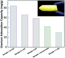
This series of monolith sorbents were screened with simulated seawater consisting of a high uranyl concentration (∼6 ppm) to identify candidate for marine testing. In Fig. 4, samples 1-0.75/5, 2-1.5/5, 3-2.0/5 (the first three bars) were powder samples with increasing ratios of AN to the crosslinker MBAAm. The uranium adsorption capacity increased with the increasing AN/MBAAm ratio and the highest adsorption capacity of 83.8 g-U per kg was obtained from the Sample 1-0.75/5, which contains 0.75 g MBAAm and 5 mL AN. This is expected based on the higher acrylonitrile content of the polymer. In order to evaluate the effect of particle sizes on the adsorption capacity, two control tests of Sample 1-0.75/5 with different particle sizes, were performed under identical conditions. As expected, the adsorption capacity decreased with the increasing particle size. This suggests a hindered transport through the particle. This could be due to lowered hydrophilicity, e.g., too much crosslinking that prohibits effective swelling of the polymer thus restricting access to the interior chelation sites. However, the adsorption capacity of just one large particle (∼15 mg) of Sample 1-0.75/5 was still higher than the JAEA non-porous irradiation-grafted non-woven polyethylene fabric (JAEA) conducted under the same exposure conditions [22.0 g-U per kg after 24 h].2b


image file: c5ra02131f-f4.tif
Fig. 4 Uranium adsorption capacities for monolith adsorbents in high uranyl concentration simulated seawater. Sample 1-several blocks: 0.1–0.2 mm blocks with a total weight of 15 mg; Sample 1-1 block: a block sample with a weight of 15 mg.

From the screening test, large particles of Sample 1-0.75/5, having the highest U adsorption capacity in the simulated seawater, were chosen as the candidates to evaluate the efficiency of uranium adsorption from natural seawater. This sample represents a balance between high capacity and achieving a stable, porous monolith. The results of the 21 and 42 day contact times along with a 42 day contact time from a reference sample supplied by the JAEA are shown in Fig. 5. The myriad of elements present in seawater is exhibited by the broad distribution of elements extracted by the adsorbent. Calcium, magnesium, and sodium, which are present at high concentrations in seawater, are also present in high concentrations in the adsorbents, at 37.9, 28.8, and 39.7 g kg−1, respectively, after 42 days. Potassium and strontium are much lower at 1.6 and 0.2 g kg−1, respectively. The heavy metals increase in concentration over time, as expected. While the JAEA adsorbent has higher capacities for the metals usually reported for amidoximes and seawater extractions, i.e., U, V, Fe, Cu and Ni, the concentrations of titanium, molybdenum, chromium, and manganese are higher for the cryogel-based adsorbent. The concentration difference for molybdenum is significant at 0.038 g kg−1 (0.394 mmol-Mo per kg-ads.) for the cryogel polymer and 0.005 g kg−1 (0.052 mmol-Mo per kg-ads.) for the JAEA sample after 42 days. The vanadium and uranium ratios are in line with previous reports where vanadium is preferentially adsorbed over uranyl and is observed in the adsorbent supplied by JAEA researchers and tested alongside the present cryogel-based polymer.7a,d This is due, in part, to the relative concentrations of vanadium to uranium in seawater. Similar trends are observed for iron. The large amounts of ions smaller than uranyl, such as iron, manganese and molybdenum could be due to pore constrictions in the swollen polymer. The swelling that occurs during the base pre-treatment prior to seawater contact could result in a size exclusion phenomena where the larger uranyl is rejected while the smaller metals can access the adsorption sites.


image file: c5ra02131f-f5.tif
Fig. 5 Metal ions adsorption capacities (mmol-M per kg-ads.) of Sample 1-0.75/5 and Japanese fiber in continuous-flow experiments in seawater (inset is a zoom of the metal capacities for Sr–Co).

Overall, the uranium adsorption capacity was low, 0.95 g-U per kg (4.0 mmol-U per kg-ads.), compared to that of JAEA adsorbent (1.8 g-U per kg; 8.6 mmol-U per kg-ads.). This can be understood through mass transport effects. Fig. 6 shows the cryogel polymer after seawater contact. The surface color is due to surface adsorption of the metal ions while the interior did not exhibit a color change, indicating poor mass transport through the particle. The co-extraction of the other transition metals, such as molybdenum and manganese, increase the value of the adsorbent for seawater extractions, even with the low uranium capacity.


image file: c5ra02131f-f6.tif
Fig. 6 Picture of cryogel polymer (Sample 1-0.75/5) after seawater contact.

Conclusions

A new monolithic adsorbent for uranium seawater mining was prepared via the simple cryogel method extending it to include organic monomers with organic/aqueous solvent mixtures as the structure templates without the use of an emulsion. These macroporous monoliths with different ratio of the crosslinker N,N′-methylenebisacrylamide and the functional monomer acrylonitrile were screened using simulated seawater, containing a high uranyl concentration (∼6 ppm) and the uranium adsorption results have shown that the adsorption capacities are strongly influenced by the density of the amidoxime groups, i.e., the ratio of the crosslinker and functional monomer. Mass-transport limitations were observed in seawater testing using filtered seawater from Sequim Bay, WA (USA). The uranyl adsorption capacity was low, hindered by mass-transport limitations due possibly to the lack of hydrogel formation and rigidity of the polymer framework. Efforts to increase the uranyl capacity are on-going; however, the molybdenum extraction presented interesting results as the capacity was significantly higher than that of the fibrous JAEA adsorbent.

Acknowledgements

This research was conducted at Oak Ridge National Laboratory (ORNL) and supported by the U.S. DOE Office of Nuclear Energy, under Contract no. DEAC05-00OR22725 with ORNL, managed by UT Battelle LLC. Seawater screening was performed at the Marine Sciences Laboratory in Sequim, WA, a division of Pacific Northwest National Laboratory, supported by the U.S. DOE Office of Nuclear Energy, under Contract no. DE-AC05-76RL01830.

Notes and references

  1. (a) J. Kim, C. Tsouris, R. T. Mayes, Y. Oyola, T. Saito, C. J. Janke, S. Dai, E. Schneider and D. Sachde, Sep. Sci. Technol., 2013, 48, 1–48 CrossRef PubMed; (b) G. Tian, S. J. Teat, Z. Zhang and L. Rao, Dalton Trans., 2012, 11579–11586 RSC; (c) J. L. Vivero-Escoto, M. Carboni, C. W. Abney, K. E. deKrafft and W. Lin, Microporous Mesoporous Mater., 2013, 180, 22–31 CrossRef CAS PubMed.
  2. (a) B. L. K. Somayajulu and E. D. Goldberg, Earth Planet. Sci. Lett., 1966, 1, 102–106 CrossRef CAS; (b) M. Gao, G. Zhu and C. Gao, Energy Environ. Focus, 2014, 3, 219–226 CrossRef PubMed.
  3. (a) T. Saito, S. Brown, S. Chatterjee, J. Kim, C. Tsouris, R. T. Mayes, L.-J. Kuo, G. Gill, Y. Oyola, C. J. Janke and S. Dai, J. Mater. Chem. A, 2014, 2, 14674–14681 RSC; (b) S. Xie, X. Liu, B. Zhang, H. Ma, C. Ling, M. Yu, L. Li and J. Li, J. Mater. Chem. A, 2015, 3, 2552–2558 RSC.
  4. (a) R. V. Davies, J. Kennedy, R. W. McIlroy, R. Spence and K. M. Hill, Nature, 1964, 203, 1110–1115 CrossRef PubMed; (b) I. Tabushi, Y. Kobuke and T. Nishiya, Nature, 1979, 280, 665–666 CrossRef CAS PubMed; (c) J. Górka, R. T. Mayes, L. Baggetto, G. M. Veith and S. Dai, J. Mater. Chem. A, 2013, 1, 3016–3026 RSC.
  5. T. Suzuki, K. Saito, T. Sugo, H. Ogura and K. Oguma, Anal. Sci., 2000, 16, 429–432 CrossRef CAS.
  6. (a) S. D. Alexandratos, Ind. Eng. Chem. Res., 2009, 48, 388–398 CrossRef CAS; (b) R. Rafiee, A. A. Babalou, A. Nilchi and M. K. R. Aghjeh, J. Appl. Polym. Sci., 2012, 126, 1069–1076 CrossRef CAS PubMed; (c) P. S. Barber, S. P. Kelley and R. D. Rogers, RSC Adv., 2012, 2, 8526–8530 RSC; (d) A. C. Sather, O. B. Berrymanb and J. Rebek, Chem. Sci., 2013, 4, 3601–3605 RSC; (e) M. Carboni, C. W. Abney, S. Liu and W. Lin, Chem. Sci., 2013, 4, 2396–2402 RSC.
  7. (a) J. Kim, C. Tsouris, Y. Oyola, C. J. Janke, R. T. Mayes, S. Dai, G. Gill, L.-J. Kuo, J. Wood, K.-Y. Choe, E. Schneider and H. Lindner, Ind. Eng. Chem. Res., 2014, 53, 6076–6083 CrossRef CAS; (b) T. Hirotsu, S. Katoh, K. Sugasaka, N. Takai, M. Senō and T. Itagaki, Ind. Eng. Chem. Res., 1987, 26, 1970–1977 CrossRef CAS; (c) H.-B. Pan, W. Liao, C. M. Wai, Y. Oyola, C. J. Janke, G. Tian and L. Rao, Dalton Trans., 2014, 10713–10718 RSC; (d) Y. Yue, X.-G. Sun, R. T. Mayes, J. Kim, P. F. Fulvio, Z.-A. Qiao, S. Brown, C. Tsouris, Y. Oyola and S. Dai, Sci. China: Chem., 2013, 56, 1510–1515 CrossRef CAS PubMed.
  8. (a) E. G. Witte, K. S. Schwochau, G. Henkil and B. Krebs, Inorg. Chim. Acta, 1984, 94, 323–331 CrossRef CAS; (b) N. Sahiner, H. Yu, G. Tan, J. He, V. T. John and D. A. Blake, ACS Appl. Mater. Interfaces, 2012, 4, 163–170 CrossRef CAS PubMed; (c) P. A. Kavaklı, N. Seko, M. Tamada and O. Güven, Sep. Sci. Technol., 2004, 39, 1631–1643 CrossRef; (d) X. Sun, C. Xu, G. Tian and L. Rao, Dalton Trans., 2013, 14621–14627 RSC; (e) C.-Z. Wang, J.-H. Lan, Q.-Y. Wu, Q. Luo, Y.-L. Zhao, X.-K. Wang, Z.-F. Chai and W.-Q. Shi, Inorg. Chem., 2014, 53, 9466–9476 CrossRef CAS PubMed.
  9. (a) M. Kanno, J. Nucl. Sci. Technol., 1984, 21, 1–9 CrossRef CAS PubMed; (b) K. Schwochau, Top. Curr. Chem., 1984, 124, 91–133 CAS; (c) D. Dojozan, M. H. Pournaghi-Azar and J. Toutounchi-Asr, Talanta, 1998, 46, 123–128 CrossRef CAS.
  10. (a) P. S. Barber, S. P. Kelley, C. S. Griggs, S. Wallace and R. D. Rogers, Green Chem., 2014, 16, 1828–1836 RSC; (b) K. J. Barlow, X. Hao, T. C. Hughes, O. E. Hutt, A. Polyzos, K. A. Turner and G. Moad, Polym. Chem., 2014, 5, 722–732 RSC; (c) S. Xie, R. W. Allington, F. Svec and J. M. J. Fréchet, J. Chromatogr. A, 1999, 865, 169–174 CrossRef CAS; (d) R. E. Moore, L. Licklider, D. Schumann and T. D. Lee, Anal. Chem., 1998, 70, 4879–4884 CrossRef CAS.
  11. (a) N. Seko, A. Katakai, S. Hasegawa, M. Tamada, N. Kasai, H. Takeda and T. Sugo, Nucl. Technol., 2003, 144, 274–278 CAS; (b) N. Kabay, A. Katakai and T. Sugo, Radiat. Phys. Chem., 1995, 46, 833–836 CrossRef CAS; (c) T. L. Prasad, P. K. Tewari and D. Sathiyamoorthy, Int. J. Nucl. Desalin., 2011, 4, 261–276 CrossRef; (d) K. Sekiguchi, K. Saito, S. Konishi, S. Furusaki, T. Sugo and H. Nobukawa, Ind. Eng. Chem. Res., 1994, 33, 662–666 CrossRef CAS.
  12. S. Das, A. K. Pandey, A. Athawale, V. Kumar, Y. K. Bhardwaj, S. Sabharwal and V. K. Manchanda, Desalination, 2008, 232, 243–253 CrossRef CAS PubMed.
  13. (a) Y. Yue, R. T. Mayes, J. Kim, P. F. Fulvio, X.-G. Sun, C. Tsouris, J. Chen, S. Brown and S. Dai, Angew. Chem., Int. Ed., 2013, 52, 13458–13462 CrossRef CAS PubMed; (b) K. Saito, T. Hori, S. Furusaki, T. Sugo and J. Okamoto, Ind. Eng. Chem. Res., 1987, 26, 1977–1981 CrossRef CAS.
  14. (a) N. E. Vrana, K. Matsumura, S. H. Hyon, L. M. Geever, J. E. Kennedy, J. G. Lyons, C. L. Higginbotham, P. A. Cahill and G. B. McGuinness, J. Tissue Eng. Regener. Med., 2012, 6, 280–290 CrossRef CAS PubMed; (b) I. F. Nata, T. M. Wu, J. K. Chen and C. K. Lee, J. Mater. Chem. B, 2014, 2, 4108–4113 RSC; (c) M. Ottens, G. Leene, A. A. C. M. Beenackers, N. Cameron and D. C. Sherrington, Ind. Eng. Chem. Res., 2000, 39, 259–266 CrossRef CAS.
  15. K. Okada, M. Nandi, J. Maruyama, T. Oka, T. Tsujimoto, K. Kondohc and H. Uyama, Chem. Commun., 2011, 47, 7422–7424 RSC.
  16. (a) M. Nasef and O. Güven, Prog. Polym. Sci., 2012, 37, 1597–1656 CrossRef CAS PubMed; (b) E. Jain and A. Kumar, J. Biomater. Sci., Polym. Ed., 2009, 20, 877–902 CrossRef CAS PubMed.
  17. (a) S. Vukovic, L. A. Watson, S. O. Kang, R. Custelcean and B. P. Hay, Inorg. Chem., 2012, 51, 3855–3859 CrossRef CAS PubMed; (b) S. P. Kelley, P. S. Barber, P. H. K. Mullins and R. D. Rogers, Chem. Commun., 2014, 50, 12504–12507 RSC.

This journal is © The Royal Society of Chemistry 2015
Click here to see how this site uses Cookies. View our privacy policy here.