Taihei Taniguchi,
Kyu-Hong Kyung and
Seimei Shiratori*
Grad. Sch. Sci. Tech., Keio Univ., 3-14-1 Hiyoshi, Kohoku-ku, Yokohama, Japan. E-mail: shiratori@appi.keio.ac.jp; Fax: +81-45-566-1602; Tel: +81-45-566-1602
First published on 9th December 2015
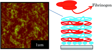
Anti-thrombus coatings using natural polymers with high biocompatibility were successfully fabricated by a Layer-by-Layer (LbL) method. The cationic and anionic components were chitin nanofibers and heparin, respectively. LbL multilayers were successfully fabricated by depositing different numbers of bilayers. Analyzing the damping waves of Quartz Crystal Microbalance (QCM) measurements showed that the film hardened as the number of bilayers deposited increased. Mass change measurements via QCM, scanning electron microscopy, atomic force microscopy, ellipsometry, and X-ray photoelectron spectroscopy were also carried out. The density of the chitin nanofiber layers increased as the number of bilayers increased. Simultaneously, the film stability in solution improved. The fabricated 30-bilayer film showed a fibrous structure on its surface and no structural disorder, even after 48 hours immersion in phosphate buffered saline (PBS), so high stability in PBS was demonstrated. The anti-thrombus properties of the film were also investigated by measuring the amount of fibrinogen adsorbed using QCM. This film not only inhibits fibrinogen adsorption, but those fibrinogen which do adsorb detach relatively easily (compared with fibrinogen adsorbed onto an uncoated QCM substrate). Furthermore, while the uncoated glass substrate adsorbed a large quantity of blood on its surface, the 30-bilayer coated glass substrate successfully inhibited blood adsorption and blood solidification.
Previous studies show that some materials, such as poly ethylene glycol (PEG) and poly acrylic acid, can be used to make coatings which suppress thrombus formation. PEG, a hydrophilic macromolecule with a long polymer chain, is effective at preventing fibrinogen adhesion because of the entropic and steric repulsive forces arising when the polymer molecules are compressed by protein molecules approaching the surface.5–9
Layer-by-Layer (LbL) assembly was first developed by Decher and coworkers in the early 1990s.10–13 The LbL assembly method has attracted much attention because of its versatility and convenience.13,14 LbL assembly can be achieved by the alternate immersion of certain materials into solutions of oppositely charged, water-soluble polymers;15–18 this alternately forms polyion complexes on the substrate surface to create ultrathin polymeric films. Moreover, LbL assembly allows the surface structure and thickness of a film to be controlled on micro- and nanoscales. There have been various applications of LbL assembly, including for antireflective and superhydrophobic coatings, dye release and drug delivery systems.19–25 We also reported that antithrombogenicity was achieved with polyelectrolyte multilayer thin film prepared from poly(vinyl alcohol)–poly(acrylic acid) (PVA–PAA) blends, deposited in alternate layers with poly(allylamine hydrochloride) (PAH).26
For medical applications, such as stents and artificial blood vessels, it is critical that the coating not only inhibit thrombus formation, but also be highly biocompatible and mechanically durable. Sheng Meng and coworkers developed chitosan–heparin LbL coatings which promoted endothelialization, and showed high biocompatibility and anticoagulation characteristics.27 Yao Shu and coworkers deposited LbL multilayers of chitosan and heparin on titanium substrates. The multilayers are beneficial to cytobiocompatibility.28 Also, Maria Bulwan and coworkers developed one-component chitosan-based ultrathin films fabricated using LbL technique, which may be potentially useful for many biomedical and environmental protection applications.29 Chitosan and heparin (HEP) is both biopolymers. Chitosan is a natural cationic polysaccharide, widely studied and used as a matrix for tissue engineering and drug release because of its good biocompatibility and biodegradability. Crab shells show high tensile strength and bendability,30,31 and thus chitin nanofibers (CHNFs) purified from crab shells and partially transformed from chitin to chitosan by deacetylation have also attracted much attention because of their high potential to show mechanical strength in addition to the advantageous characteristics of chitosan. HEP is biocompatible and widely used in medical applications, where it shows strong anti-thrombus characteristics. For example, it strongly inhibits sub-acute in-stent thrombus formation.32–34 There are many reports of anti-thrombus coatings using HEP prepared by LbL-assembly methods.28,35 However, these studies did not evaluate the film stability.
In the present research, to take advantage of the biocompatibility of the two polysaccharides, and combine the mechanical stability of CHNFs and anti-thrombus characteristics of HEP, new CHNFs/HEP LbL-assembly multilayers were deposited on glass substrates. Each polymer deposition step was evaluated by a Quartz Crystal Microbalance (QCM); the QCM was also used to determine fibrinogen adsorption and detachment abilities. QCM is often used to investigate protein adsorption, lipids and membranes, cells, DNA, and carbohydrates, and in applications within polymer physics.36,37 Furthermore, from differences in the rate of decay of the oscillation wave after turning off the voltage of the QCM, we evaluated the relative harnesses of the films. The films were also characterized by X-ray photoelectron spectroscopy (XPS), scanning electron microscopy (SEM), and atomic force microscopy (AFM). The anti-thrombus performance was evaluated indirectly measuring fibrinogen adsorption and detachment in PBS solution, and directly from the mass of blood that adhered to the coated and uncoated surfaces.
000–350
000 10 mM aqueous solution), poly(4-styrenesulfonic acid sodium salt) (PSS, Mw 70
000 10 mM aqueous solution) and phosphate buffered saline (PBS) were from Sigma-Aldrich Japan (Tokyo, Japan). Pig blood was purchased from Shibaura Zoki industry (Tokyo, Japan).
All experiments were conducted in air at ambient temperature and used ultra-pure water (Aquarius GS-500.CPW, Advantec, Tokyo, Japan, 18.2 MΩ); the pH values of the of dipping solutions for the LbL method were adjusted using sodium hydroxide and acetic acid.
:
120
:
60 wt% KOH/H2O/IPA), and rinsed twice by ultrasonication in water as above. The sequence for multilayer construction was as follows: the hydroxylated glass substrate was immersed in CHNFs solution for 15 min, which changes the surface charge to positive, and then rinsed three times in ultra-pure water for 1 min. Then the sample was exposed to negatively-charged HEP solution for 15 min, which changes the surface charge to negative again, followed by the same rinsing procedure and dried at natural condition. The cycle of immersing the substrate into CHNFs and HEP solutions alternately was repeated n times to obtain the desired film. To increase the anti-thrombus ability, we use heparin to the outermost layer, because heparin is the key material for the film to show anti-thrombus ability. The ultrathin films containing n alternating layers of CHNFs and HEP were defined as (CHNFs/HEP)n.
LbL assembly by alternating electrostatic adsorption of CHNFs and HEP occurs as illustrated schematically in Fig. 2. Fig. 3 shows the adsorbed mass of fibrinogen to the QCM, which was calculated by the frequency shift decreased with increasing amount of polymer adsorbed on the surface of QCM substrate, plotted against the steps of the CHNFs/HEP LbL assembly. The stepwise polymer depositions are shown from the decrease in frequencies of QCM as assembly proceeds. The total thicknesses of the prepared CHNFs/HEP films were 73.7 nm for 10 bilayers and 350.2 nm for 30 bilayers.
![]() | ||
| Fig. 2 Schematic illustration of LbL self-assembly process with oppositely-charged CHNFs and HEP polyelectrolyte multilayers on a glass substrate. | ||
When the deposition time was 3 minutes for each layer, a homogeneous LbL film was not formed. We consider that such a short adsorption time resulted in unstable and slow adsorption during the deposition (ESI S2†). Sufficient adsorption time (such as 15 minutes) is required for stable deposition.
AFM images of the surface structures of (CHNFs/HEP)n films with n = 10, 20, 30 and 50 are shown in Fig. 4. Though the top views of 10-bilayer and 20-bilayer films have less fibrous structure, the 30-bilayer film showed a complex, intertwined, fibrous structure, derived from CHNFs. Also about 50 bilayer, the groups of the fiber structure can also be confirmed extensively, which is also indicated by the RMS values. Our suggested reasons for this fibrous structure appearing on the top of the films containing more bilayers are as follows: in the early stages of LbL adsorption, the surface of the film has a nearly flat structure (because there has been less adsorption); this means that it also has a relatively small specific surface area, and a low amount of electrostatic charge. However, as the number of bilayers increases, the increasing mass of material adsorbed makes the surface rougher, increasing the specific surface area and meaning that there is more charge on this surface than on the earlier-stage surface. This observation is consistent with the RMS values measured by AFM (10 bilayer: 7.53 nm, 20 bilayer: 10.4 nm, 30 bilayer: 12.4 nm, 50 bilayer: 39.9 nm). This larger quantity of electrostatic charge causes numerous CHNFs to absorb onto the film. Unless otherwise noted, all subsequent experiments used 30-bilayer films.
![]() | ||
| Fig. 4 AFM images of (CHNFs/HEP) multilayers. (a) and (e) 10 bilayer, (b) and (f) 20 bilayer, (c) and (g) 30 bilayer, (d) and (h) 50 bilayer. | ||
The XPS spectra of (CHNFs/HEP)30 LbL films are shown in Fig. 5. Sulfone group peaks are observed, confirming that HEP exists on the top of the film. The two peaks are derived from the S 2s and S 2p orbitals of the sulfone. It is important that the sulfone groups of HEP are located on the surface of the films, because they are expected to play an important role in the antithrombogenic activity of the multilayer films. QCM results that are related to the film density and hardness are shown in Fig. 6. These damping waves are for non-coated, buffer only coated, the 30-bilayer coated, the 50-bilayer coated, cast film of CHNFs, respectively. The damping time for each QCM wave becomes shorter for the high viscoelasticity films, because higher viscoelasticity films absorb more vibration than lower films.42–44 For this measurement, we evaluated the films by the simple QCM-D system used a digital storage oscilloscope and UNIX programs for analyzing the 2 × 10⁶ (2 M) plots stored in the oscilloscope. We used Cygwin UNIX for Windows for large-data-set analysis techniques to obtain QCM-D frequency information and for the determination of viscoelasticities of stacked film made of soft materials.44
Therefore, observing the damping behavior of the vibration of the quartz which coated the film, relative viscosity of the film, which lead to the hardness, is easily found. The results are shown in Fig. 6. Non-coated QCM, which is metal surface, is the hardest, therefore its damping time is the longest. As shown in the figure, as the number of bilayers increased, the damping time of the QCM wave became longer. The values of the dissipation of buffer only coater, 30 bilayer coated and 50 bilayer coated (D × 106 where D = 1/πfτ is dissipation factor of the films; f is characteristic frequency of the quartz (10 MHz) and tau is the relaxation time of the decaying amplitude) was 6.38 × 10⁻⁴, 6.47 × 10⁻⁴ and 5.99 × 10⁻⁴, respectively. This means that the LbL films in our study became lower viscoelasticity as the number of bilayers increased. We consider that this phenomenon was caused by accretion of high mechanical strength CHNFs into the film as the number of bilayers increased and imply that the films with more bilayers should have higher stability due to the high mechanical strength of CHNFs. By CNHFs first adsorbing onto the PDDA/PSS buffer layer, the film became hard, so the uncoated (buffer coated) film is relatively high viscoelastic and reveals lowest oscillation time. The AFM images in Fig. 4 also show a more extensive nanofiber structure as the number of bilayers increases – this would result in increased film strength. Hardness of CHNFs was demonstrated by the damping wave of the cast film of CHNFs. The damping time of the cast film of CHNFs is longer than that of PDDA/PSS polymer coating and the 30 bilayers LbL coating. However, compared with 50 bilayers coatings, the cast film of CHNFs was found to be soft because of the short damping times. One reason of this is the mass difference of adsorbed polymer. The weight of 50 bilayer (CHNFs/HEP) LbL coating was 4860 ng, which was estimated from QCM date of Fig. 3, while that of the cast film of CHNFs was 7365 ng. Therefore, the cast film of CHNFs, which is larger amount of adsorbed mass, became softer than 50 bilayer LbL coating. Moreover, which is more important, the other reason is the adsorbing force between the layer materials. The cast film of CHNFs was adsorbed onto the QCM surface by van der Waals force, on the other hand, the 50 bilayer LbL coating was formed by the electrostatic force between CHNFs and HEP. In other words, it may suggest that electrostatic interaction between CHNFs and HEP is the main difference that improves coating “hardness” as opposed to CHNFs as a cast film. For this point of view, we tried to fabricate the cast film by CHNFs and HEP mixed solution, however, the polyion complex formed precipitation on substrate and the numerous aggregated complex particles did not form uniform film. We consider that LbL method is very effective to form uniform film by CHNFs and HEP and the formed (CHNFs/HEP) LbL coating was harder than that of the cast film of CHNFs because the electrostatic force is stronger than the van der Waals force.
From these experimental results and considerations, we concluded that the stability of (CHNFs/HEP) LbL film are caused by the usage of CHNFs and LbL fabrication method based on electrostatic force.
SEM images after immersion of the (CHNFs/HEP)30 in PBS are shown in Fig. 7. As shown in the figure, fibrous structures derived from CHNFs are not degraded by immersion in PBS. This indicates that the films are durable enough to be potentially useful in vivo. Even after immersion for 48 h, there is no damage to the CHNFs, although the fibrous structure swelled a little with the diameter of CHNFs increasing from ∼48 nm before immersion to ∼69 nm after 48 h immersion, which was measured form SEM image and by using image-J software. This stability probably results from both the strength of the original CHNFs (purified from hard crab shell) and the complicated entanglement of the fibers.
![]() | ||
| Fig. 7 SEM images of the (CHNFs/HEP)30 multilayer film after immersion in PBS for: (a) 0 h, (b) 3 h, (c) 6 h, and (d) 48 h. | ||
The masses of fibrinogen adsorbed onto different surfaces are shown in Fig. 8. The amount of fibrinogen adsorbed on the LbL film was less than half that adsorbed on a non-coated QCM substrate. Also, while it took 2 min 30 s for the non-coated QCM substrate to acquire 80% of its final mass of adsorbed fibrinogen, it took 32 min for the (CHNFs/HEP)30 film-coated QCM substrate. This fibrinogen-resistance property is thought to be caused by the negatively charged sulfone groups of HEP, which show anticoagulation ability. The sulfate group of heparin is highly negatively charged, and we consider that fibrinogen which mainly has negative charge likely to repel by the negative charge of heparin by electrostatic interaction, and thus result in the lower adsorption of fibrinogen on to the QCM electrode which were covered by (CHNFs/HEP)30 film.
Moreover, Fig. 9 shows the fibrinogen detachment from the LbL films when they are rinsed (after being dipped for 10 min in fibrinogen solution). The fibrinogens adsorbed onto the LbL film are detached more easily than those adsorbed onto the non-coated QCM substrate; this implies that the CHNFs/HEP LbL films can not only reduce fibrinogen adsorption but also weaken the interaction between the fibrinogen and the surface.
Finally the anti-thrombus property of the multilayer film was examined. The results are shown in Fig. 9. As described in the section of “Blood Anticoagulation testing”, pig blood was prepared for the anticoagulation test. 80 μl of blood were cast both onto non-coated glass and film-coated glass (1 × 1 cm) substrates simultaneously; the substrates were then incubated in air at 37 °C for 15 min. After 15 min, the each substrate was rinsed vigorously using a plastic syringe with 10 ml of ultra-pure water. The results are shown in Fig. 10, while much blood adhered to the non-coated substrate, on the other hand, only a small amount of blood (less than 0.5 μl) adhered to the LbL-coated substrate. From these results, we can confirm that the CHNFs/HEP LbL multilayers have anti-thrombus properties.
Footnote |
| † Electronic supplementary information (ESI) available. See DOI: 10.1039/c5ra01853f |
| This journal is © The Royal Society of Chemistry 2015 |